EM · Endocrine emergencies
Adrenal crisis (Addisonian crisis)
Also known as Addisonian crisis · Acute adrenal insufficiency · Adrenal emergency · Glucocorticoid crisis
Adrenal crisis (Addisonian crisis) is the acute, life-threatening state of cortisol deficiency producing vasodilatory shock refractory to fluid, abdominal pain, vomiting and the classic biochemistry of hyponatraemia, hyperkalaemia and hypoglycaemia. The triggers are an infection, surgery, trauma, sepsis and the abrupt withdrawal of chronic steroids. The Fellowship-critical management is hydrocortisone 200 mg intravenously as a stat dose then 100 mg intravenously every six hours, aggressive 0.9 per cent saline 1 L rapidly, and glucose if hypoglycaemic — fludrocortisone is not needed acutely because hydrocortisone has mineralocorticoid activity at the high doses. Hydrocortisone is never delayed for the cortisol result: the blood is drawn but treatment is immediate. The differential is septic shock, the upper gastrointestinal bleed and diabetic ketoacidosis. ACEM-primary, globally tagged.
On this page & tools
Your progress
Saved locally on this device.
5 MCQs with explanations
Target exams
Red flags
Related topics
- Sepsis and septic shock — the emergency department approach
- DKA, HHS and hypoglycaemia
- Upper gastrointestinal bleed
- Thyroid emergencies — thyroid storm and myxoedema coma
- Fluid resuscitation in the emergency department
- Electrolyte emergencies — potassium and sodium
- Cardiogenic shock in the emergency department
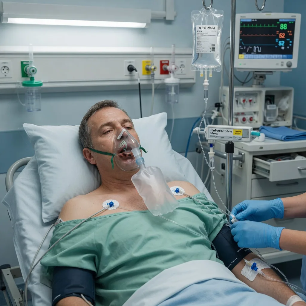
Adrenal crisis (the Addisonian crisis) is the acute, life-threatening state of cortisol deficiency. The cortisol deficit removes vascular tone and the permissive effect on catecholamines, producing a vasodilatory shock that is refractory to fluid; when the aldosterone is also deficient (primary adrenal failure), renal sodium loss and potassium retention compound the volume depletion. The Fellowship candidate must recognise the syndrome from its bedside and biochemical fingerprint — refractory hypotension with abdominal pain and vomiting, hyponatraemia, hyperkalaemia and hypoglycaemia — then act immediately with hydrocortisone and fluid, never waiting for the confirmatory cortisol. The crisis is reversible in minutes to hours when treated correctly, and lethal within hours when missed.[1][2][4]
Definition and classification
Adrenal crisis is an acute, severe insufficiency of cortisol (with or without aldosterone) that produces haemodynamic compromise — hypotension that is unresponsive to fluid — together with the metabolic derangements of cortisol and aldosterone deficit. It is graded in practice by the haemodynamic and conscious-level state rather than by a single number, because the emergency is treated on clinical suspicion before any confirmatory test returns.[2]
Two anatomical classifications carry management weight. Primary adrenal insufficiency (Addison disease) is failure of the adrenal cortex itself: cortisol and aldosterone are both deficient, adrenocorticotrophic hormone (ACTH) rises in response, and the patient develops the hyperpigmentation of chronic ACTH excess together with hyperkalaemia from aldosterone loss. Secondary adrenal insufficiency is pituitary or hypothalamic failure of ACTH: cortisol is deficient but aldosterone is preserved (the renin–angiotensin system is intact), so potassium is normal and there is no hyperpigmentation; hyponatraemia still occurs, from cortisol-driven water retention and the loss of the tonic suppression of antidiuretic hormone. The commonest cause of adrenal crisis in contemporary practice is neither classical entity but glucocorticoid-induced adrenal suppression — the chronic exogenous steroid that is withdrawn or faced with an acute stress.[1][3]
Epidemiology and risk factors
In patients with established adrenal insufficiency, adrenal crisis occurs at a rate of roughly six to eight events per hundred patient-years, and around half of all such patients will suffer at least one crisis in their lifetime; the precipitant is usually an infection, a gastrointestinal illness with vomiting, or a surgical or procedural stress that outstrips the patient's glucocorticoid reserve.[2][4] Mortality per crisis sits between 0.5 and 7 per cent and climbs in the elderly and in those presenting in septic shock, with the cause of death usually refractory vasodilatory shock or an untreated precipitant rather than the hormone deficit itself.[4]
The risk profile is the key to the bedside suspicion. The patient may carry a known diagnosis of Addison disease, congenital adrenal hyperplasia, or a pituitary disorder; may be on chronic glucocorticoids (oral, inhaled for asthma or COPD, or long-acting injections) stopped abruptly within the preceding days to weeks; may have an autoimmune diathesis; or may have metastatic disease, tuberculosis, or HIV with cytomegalovirus involvement of the adrenals. Pregnancy, the post-partum period, and recent surgery or trauma are the acute stresses that uncover a marginal reserve.[1][3]
Pathophysiology
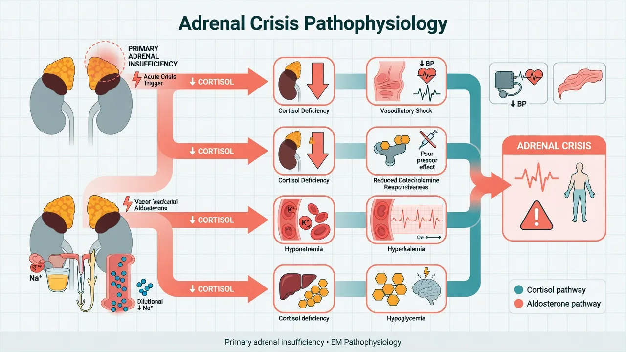
Cortisol is the body's principal stress hormone and its principal permissive agent for vascular tone. In health it is released in surges against any physiological stress, sustaining the catecholamine response, maintaining vascular smooth-muscle responsiveness, suppressing the inflammatory cytokine cascade, and supporting hepatic gluconeogenesis. When cortisol fails acutely, vascular tone collapses: the vessels become unresponsive to endogenous and exogenous catecholamines, producing a vasodilatory shock that is refractory to fluid resuscitation — the haemodynamic hallmark of the crisis. Aldosterone deficit (in primary insufficiency) removes the renal sodium-retaining, potassium-excreting drive, deepening the volume depletion and producing the hyperkalaemia.[2]
The biochemistry follows directly. Hyponatraemia arises from two mechanisms — aldosterone-driven sodium loss in primary disease, and cortisol-driven failure to suppress antidiuretic hormone (cortisol normally tonically inhibits vasopressin release), causing water retention. Hyperkalaemia is present only in primary insufficiency, where aldosterone deficit reduces distal tubular potassium secretion. Hypoglycaemia reflects the loss of cortisol's gluconeogenic and anti-insulin action, and is most pronounced in children, the fasting patient, and those with coexisting sepsis. A metabolic acidosis with a raised lactate follows the poor perfusion. [1]
[1]Aetiology — the three compartments and the contemporary iatrogenic crisis
The aetiology of adrenal crisis is best held in the Fellowship mind as three compartments, because the compartment predicts the biochemistry, the bedside signs and the precipitant. Primary adrenal insufficiency is destruction of the adrenal cortex itself — cortisol and aldosterone both fail, ACTH rises, and the patient carries the hyperpigmentation, the hyperkalaemia and the salt craving of chronic Addison disease. Secondary adrenal insufficiency is hypothalamic–pituitary failure of corticotrophin (ACTH) release — cortisol fails but aldosterone is preserved (the renin–angiotensin–aldosterone axis is intact), so potassium is normal and there is no pigmentation; hyponatraemia still occurs, from cortisol-driven water retention and the loss of the tonic suppression of vasopressin. Tertiary refers to the hypothalamic (CRH) level and is clinically folded into secondary. The single commonest precipitant of crisis in the modern developed-world emergency department is none of the classical entities but iatrogenic glucocorticoid suppression — the chronic oral, inhaled or injected steroid withdrawn abruptly or overwhelmed by an acute stress.[1][3][5][6]
Primary adrenal insufficiency — autoimmune destruction is the modern majority
In the developed world, autoimmune Addison disease accounts for the majority of primary adrenal insufficiency — the immune destruction of the adrenal cortex, often as part of autoimmune polyglandular syndrome type 2 with type 1 diabetes, autoimmune thyroid disease, pernicious anaemia and vitiligo. The onset is insidious, the cortex is destroyed over months to years, and the patient presents in crisis only when an intercurrent illness outstrips the residual reserve — or, occasionally, when the chronic disease is first diagnosed in the resuscitation bay. Tuberculosis is the commonest cause worldwide and the dominant cause in endemic regions — the caseating granulomatous destruction of both glands, which must be over 90 per cent destroyed before clinical failure appears, because the adrenal reserve is vast. HIV/AIDS with cytomegalovirus adrenalitis, disseminated fungal infection (histoplasmosis, paracoccidioidomycosis) and metastatic disease (lung, breast, melanoma, renal — the adrenals are a common metastatic site, though clinical failure is uncommon) are the infiltrative causes. Adrenoleukodystrophy causes adrenal failure in young males; congenital adrenal hyperplasia (21-hydroxylase deficiency) is the neonatal and paediatric cause; bilateral adrenalectomy for Cushing disease or bilateral pheochromocytoma produces an instant, absolute primary insufficiency.[1][3][6]
Bilateral adrenal haemorrhage and infarction — the anticoagulated and the septicaemic
Bilateral adrenal haemorrhage is a surgical–medical cause of sudden bilateral cortical destruction and an under-recognised emergency. It occurs in three settings: the anticoagulated patient (especially in the first weeks of heparin or warfarin therapy, and in heparin-induced thrombocytopenia, where adrenal vein thrombosis produces haemorrhagic infarction), the postoperative or trauma patient (major surgery, burns, the coagulopathy of massive transfusion), and the septicaemic patient — classically meningococcaemia, where the Waterhouse–Friderichsen syndrome is the bilateral adrenal haemorrhage of fulminant meningococcal or pneumococcal sepsis with purpura, disseminated intravascular coagulation and rapid progression to vasodilatory shock. The patient with new abdominal or flank pain, a falling haemoglobin and unexplained shock in the context of anticoagulation or sepsis demands a cortisol level and empirical hydrocortisone, because the bilateral haemorrhage can destroy both glands within hours.[4][6]
Secondary adrenal insufficiency — the pituitary and the withdrawn steroid
Secondary adrenal insufficiency is cortisol deficit with preserved aldosterone, driven by ACTH lack. The commonest cause in contemporary practice is exogenous glucocorticoid suppression — the patient on chronic oral prednisolone (above the equivalent of prednisolone 5 mg daily for more than three weeks), high-dose inhaled corticosteroids (especially fluticasone, potent and long-acting), or depot steroid injections whose hypothalamic–pituitary–adrenal axis recovers over months. The suppression persists for up to a year after cessation, and the crisis is precipitated by an acute stress — an infection, a surgical procedure, a gastroenteritis with vomiting — or by the act of withdrawal itself; the axis cannot mount the cortisol surge the stress demands, and the patient collapses. The structural causes are the pituitary mass and its catastrophes: pituitary apoplexy (the haemorrhagic infarction of a pituitary adenoma — sudden severe headache, ophthalmoplegia, visual field defect, meningismus and shock); Sheehan syndrome (post-partum pituitary necrosis from profound hypovolaemia at delivery, presenting in the days to weeks post-partum with failure to lactate, amenorrhoea, hyponatraemia and progressive hypotension); pituitary surgery and radiation, craniopharyngioma, infiltrative disease (sarcoidosis, lymphocytic hypophysitis, haemochromatosis) and traumatic brain injury.[1][6]
Primary (Addison)
- Destruction of the adrenal cortex — cortisol AND aldosterone deficient; ACTH high from the loss of feedback
- Autoimmune is number one in the developed world; tuberculosis number one globally; bilateral haemorrhage, metastases, congenital adrenal hyperplasia, adrenoleukodystrophy, adrenalectomy
- Hyperpigmentation of the palmar creases, buccal mucosa, gums, scars and areolae; salt craving and weight loss — the chronic ACTH-driven melanocyte stimulation
- Hyponatraemia AND hyperkalaemia (the aldosterone deficit), hypoglycaemia, metabolic acidosis — the hyperkalaemia is the discriminating feature
- Needs fludrocortisone in the maintenance phase (aldosterone is permanently lost); hydrocortisone alone provides mineralocorticoid cover only at the stress doses
- Crisis from any intercurrent stress; the reserve is gone once 90 per cent of the cortex is destroyed
Secondary (pituitary)
- ACTH deficiency — cortisol deficient, aldosterone PRESERVED (the renin–angiotensin axis is intact)
- Steroid withdrawal or suppression is number one; pituitary apoplexy, Sheehan syndrome, tumour, surgery, radiation, infiltrative disease, traumatic brain injury
- NO hyperpigmentation (ACTH is low, not high); may carry pallor, loss of axillary and pubic hair, amenorrhoea, the small-volume pituitary features
- Hyponatraemia (cortisol-driven water retention) but POTASSIUM IS NORMAL; hypoglycaemia; no hyperkalaemia — the discriminating feature
- Does NOT need fludrocortisone even in maintenance — aldosterone is intact; hydrocortisone replacement alone
- May have coexisting thyroid failure — NEVER start thyroid hormone before cortisol in panhypopituitarism, because it precipitates crisis
SICK DAY — the stress-dose steroid rules in one breath
SICKDAY
Minor illness (fever, upper respiratory infection, dental work) — double the oral hydrocortisone for two to three days; the patient self-manages and carries the card
Moderate stress (fracture, significant infection, procedure under sedation, vomiting with inability to absorb) — hydrocortisone 50 to 100 mg intramuscularly or intravenously; the emergency self-injection kit
Major stress (surgery, sepsis, major trauma, the actual crisis) — hydrocortisone 200 mg IV stat then 100 mg every 6 hours, tapered over one to two days as the stress resolves
Every adrenal-insufficient patient carries a parenteral hydrocortisone emergency injection kit (100 mg IM) and a steroid emergency card — and the family is taught to use it
Never stop chronic glucocorticoids abruptly; taper over weeks to months, and stress-dose through any illness or procedure
MedicAlert bracelet and the steroid emergency card carried at all times; the unconscious patient is identified by the alert
Vomiting is the danger sign — oral steroids are not absorbed; switch to the parenteral route at the first vomit
Clinical presentation
The presentation is the combination of shock with the gastrointestinal and metabolic features of cortisol deficit. The patient is hypotensive — often profoundly, with a systolic below 90 — and the hypotension does not respond adequately to a fluid bolus. Nausea and vomiting are near-universal and accelerate the crisis by preventing oral steroid intake; abdominal pain is common and may be severe enough to mimic an acute abdomen or a perforated viscus, occasionally triggering a misleading surgical referral. Lethargy, confusion and progressive coma reflect the hyponatraemia, the hypoglycaemia and the loss of cerebral perfusion. Fever may accompany the precipitating infection, but hypothermia is also seen in the shocked patient.[2][4]
The chronic clues point to long-standing primary Addison disease: hyperpigmentation of the palmar creases, the buccal mucosa, the gums, recent scars and the areolae, produced by chronic ACTH-driven melanocyte stimulation; salt craving; weight loss; and a preceding history of fatigue, postural dizziness and intolerance of minor illness. A history of chronic oral, inhaled or injected steroid use — or a recent taper — is the single most important historical clue in the emergency setting, because glucocorticoid-induced suppression is now the commonest precipitant of crisis in the developed world.[1][3]
Differential diagnosis
Adrenal crisis enters the differential of any undifferentiated shock, and several mimics share its biochemistry or its presentation. The distinction is made at the bedside by the cortisol-deficient fingerprint — the combination of refractory hypotension with hyponatraemia, hyperkalaemia and hypoglycaemia — and by the history of steroid use or chronic Addison disease. [1]
Septic shock
- Fever, source (urine, chest, line), warm vasodilated shock, raised lactate; may coexist with and trigger an adrenal crisis
- Cortisol normal or appropriately high; biochemistry of sepsis rather than cortisol deficit
- Fluids, early antibiotics within the hour, vasopressors, source control; hydrocortisone only if refractory shock or known adrenal insufficiency
- The commonest precipitant of a true adrenal crisis — always look for sepsis behind it
Diabetic ketoacidosis
- Known diabetic, polyuria and polydipsia, Kussmaul breathing, ketotic breath; glucose typically high
- Hyperglycaemia with ketones above 3 mmol/L, metabolic acidosis, pH below 7.3; potassium may be high but glucose is high not low
- Fluid, fixed-rate insulin 0.1 units/kg/h, potassium; not steroids
- Hypoglycaemia distinguishes the adrenal crisis from DKA, where glucose is high
Upper gastrointestinal bleed
- Melaena or haematemesis, pallor, collapse; a known Addison patient on steroids may also bleed
- Anaemia, raised urea-to-creatinine ratio; cortisol not the driver unless steroid intake is interrupted
- Resuscitate, transfuse, endoscopy; continue the usual steroid cover
- A GI bleed can precipitate crisis by interrupting oral steroid absorption — both may coexist
Acute abdomen (perforation, pancreatitis)
- Severe abdominal pain with guarding or rigidity; the adrenal crisis itself causes pain and can mimic this
- Raised amylase or lipase, pneumoperitoneum on imaging; the biochemistry of cortisol deficit is absent
- Surgical or pancreatitis pathway; steroids only if steroid-dependent
- Do not send an adrenal-crisis patient with abdominal pain to theatre before the hydrocortisone is given
Investigations and diagnostic targets
The emergency investigations are sent in parallel with — never before — the first dose of hydrocortisone. A venous blood gas demonstrates the metabolic acidosis and the perfusion deficit, and gives an immediate potassium and glucose. Urea and electrolytes show the hyponatraemia with, in primary disease, a hyperkalaemia; urea is raised from volume depletion. A bedside glucose is checked immediately and repeated — hypoglycaemia is common, may be profound, and is reversed within a minute of intravenous dextrose. A full blood count, coagulation, group and save (or crossmatch if bleeding or surgery is plausible), a blood culture, urine culture and a chest radiograph seek the precipitant. An ECG is mandatory — the hyperkalaemia may produce peaked T waves and widening QRS, and the older patient may carry a silent myocardial infarction as the trigger. A β-hCG is checked in any woman of reproductive age, both because pregnancy is a stressor and because the management is modified.[1][3]
The single diagnostic sample — a random serum cortisol and a plasma ACTH — is drawn immediately before the first dose of hydrocortisone, because exogenous hydrocortisone will confound every later measurement. A high random cortisol in a sick patient effectively excludes adrenal insufficiency; a low cortisol with a high ACTH confirms primary disease, and a low cortisol with a low or inappropriately normal ACTH confirms secondary disease. A short Synacthen (250 microgram ACTH) stimulation test is the confirmatory investigation for adrenal reserve, but it is a ward or endocrine-unit test performed after stabilisation, never an emergency test — its result cannot be allowed to delay treatment. The imaging of choice when secondary (pituitary) insufficiency is suspected is a pituitary MRI, again performed after stabilisation.[1][3]
The adrenal crisis at a glance
Immediate management and resuscitation
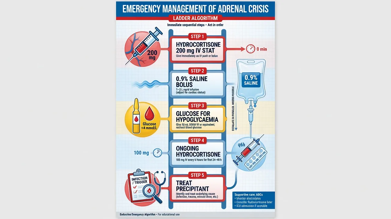
The resuscitation runs in parallel with the diagnostic sampling and the first dose of hydrocortisone. Secure the airway and give high-flow oxygen to the hypoxaemic or shocked patient; establish two large-bore intravenous cannulae; attach cardiac monitoring (the hyperkalaemia threatens the rhythm); and obtain the venous gas, urea and electrolytes, glucose, full blood count, cultures and the cortisol–ACTH sample. A urinary catheter is placed to track the perfusion response.[1][2]
Fluid is given aggressively: 0.9 per cent saline, 1 L rapidly, repeated to restore the perfusion — the patient may be several litres behind from vomiting, aldosterone-driven sodium loss and vasodilatory third-spacing. The rate is moderated in the elderly and in cardiac or renal impairment, and the sodium is watched because an overly rapid correction of a longstanding hyponatraemia risks the osmotic demyelination syndrome. Hypoglycaemia is treated immediately with glucose — 50 mL of 50 per cent dextrose (25 g) into a large vein in the adult, or the age-appropriate equivalent in a child — and rechecked, because the cortisol deficit sustains a tendency to recurrent hypoglycaemia until the hydrocortisone is established. Hyperkalaemia usually resolves with fluid, glucose and insulin-free correction of the cortisol deficit, and rarely needs the standard hyperkalaema pathway, but the ECG is treated on its merits with calcium gluconate if there is conduction disturbance.[1][3]
[1]The first 60 minutes of adrenal crisis in one breath
Recognise the syndrome — refractory hypotension with abdominal pain, hyponatraemia, hyperkalaemia and hypoglycaemia in a patient with known Addison disease, chronic steroids, or an autoimmune or pituitary history. Draw a random cortisol and ACTH, then give hydrocortisone 200 mg intravenously as a stat dose. Run in 0.9 per cent saline 1 L rapidly, repeated to clinical end-points, moderating the rate in the elderly and cardiac patient. Give glucose 25 g intravenously (50 mL of 50 per cent) for the hypoglycaemic patient and recheck. Send the blood gas, urea and electrolytes, full blood count, cultures, ECG and chest radiograph; place a urinary catheter. Find and treat the precipitant — the infection, the bleed, the infarct. Admit to high-dependency or intensive care. Fludrocortisone is not needed acutely. [1]
Adrenal crisis — the parallel resuscitation in the first 60 minutes
0 min — recognise and draw
Recognise the syndrome: refractory hypotension with abdominal pain, nausea, hyponatraemia, hyperkalaemia and hypoglycaemia in a patient with known Addison disease, chronic oral or inhaled steroids, a recent taper, an autoimmune or pituitary history, or anticoagulation with new abdominal pain. Draw the random cortisol and ACTH into a single tube IMMEDIATELY — before any hydrocortisone — but do not wait for the result.
0 to 5 min — hydrocortisone 200 mg IV stat
Give hydrocortisone 200 mg intravenously as a stat dose on suspicion, empirically, the moment the cortisol tube is drawn. This is the single most time-critical and most defensible act in the resuscitation. Establish two large-bore cannulae, give high-flow oxygen to the hypoxaemic or shocked, and attach cardiac monitoring (the hyperkalaemia threatens the rhythm).
0 to 30 min — aggressive 0.9 per cent saline and glucose
Run 0.9 per cent saline 1 L rapidly, repeated to restore perfusion — the patient may be several litres behind from vomiting, aldosterone-driven sodium loss and vasodilatory third-spacing. Moderate the rate in the elderly, the cardiac and the renal. Check and treat hypoglycaemia immediately with glucose 25 g intravenously (50 mL of 50 per cent) into a large vein, and recheck.
0 to 60 min — the parallel workup
Send the venous gas, urea and electrolytes, glucose, full blood count, coagulation, group and save, blood and urine cultures, and the beta-hCG in women of reproductive age. Perform the ECG (treat hyperkalaemic conduction disturbance with calcium gluconate on its merits) and the chest radiograph. Place a urinary catheter to track the perfusion response.
Hour 1 — start the continuing infusion
Begin hydrocortisone 100 mg intravenously every six hours (or a continuous infusion of 10 mg per hour, around 200 mg per 24 hours). Do NOT give fludrocortisone acutely — the high-dose hydrocortisone saturates the mineralocorticoid receptor. Admit to high-dependency or intensive care for the first 24 hours of intravenous hydrocortisone, fluid and electrolyte monitoring.
Hours 1 to 24 — find and treat the precipitant
The precipitant — an infection, a gastrointestinal bleed, a myocardial infarct, a pulmonary embolus, a surgical stress — is usually what kills the patient, not the hormone deficit. Start the antibiotics within the hour for the septic source, the transfusion and endoscopy for the bleed, the antiplatelet and the reperfusion for the infarct. Track the sodium (avoid a rise over 8 to 10 mmol per litre in 24 hours), the potassium (it falls as the mineralocorticoid activity returns) and the glucose (the cortisol deficit sustains recurrent hypoglycaemia).
Stabilisation and taper
Once haemodynamically stable, the vomiting settled and the precipitant controlled, taper the intravenous hydrocortisone over one to two days to the oral maintenance dose (hydrocortisone 15 to 25 mg per day in divided doses; add fludrocortisone 50 to 100 micrograms daily in primary disease). Educate on the sick-day rules, issue the emergency hydrocortisone injection kit and the MedicAlert, and arrange the endocrine follow-up and the confirmatory short Synacthen test.
Definitive management — hydrocortisone, fluid and glucose
The definitive therapy is glucocorticoid replacement, fluid resuscitation and the correction of hypoglycaemia, with the precipitant treated in parallel.[1][2][3]
Hydrocortisone is the first-specific drug and is given immediately. The standard emergency regimen is hydrocortisone 200 mg intravenously as a stat dose, followed by 100 mg intravenously every six hours (an equivalent is a continuous infusion of 10 mg per hour). Hydrocortisone is chosen over dexamethasone because it has both glucocorticoid and, at these doses, mineralocorticoid activity; dexamethasone is reserved for the rare scenario in which a subsequent short Synacthen test must not be confounded, because it does not cross-react with the cortisol assay. The high-dose hydrocortisone promptly restores vascular tone and catecholamine responsiveness, often producing a dramatic improvement in the blood pressure within an hour, and corrects the aldosterone deficit by saturating the mineralocorticoid receptor — which is why fludrocortisone is not required in the acute phase.[1][3]
Fludrocortisone (50 to 100 micrograms once daily by mouth) is added only once the patient has stabilised and is moving to oral therapy, because its onset of action is in hours to days and the high-dose intravenous hydrocortisone already provides full mineralocorticoid cover. Giving fludrocortisone acutely adds nothing and can cause fluid overload in a patient receiving aggressive saline. [1]
Fluid and electrolytes. 0.9 per cent saline continues at a rate titrated to the haemodynamics and the sodium, with the potassium monitored and replaced only once it has normalised under hydrocortisone — the early hyperkalaemia typically resolves as the aldosterone activity is restored. Hypoglycaemia is treated with intravenous glucose and rechecked. The precipitant — an infection, a bleed, an infarct, a surgical stress — is identified and managed in parallel, because it is usually what kills the patient, not the hormone deficit.[4]
Tapering to maintenance. Once the patient is haemodynamically stable, the vomiting has settled, and the precipitant is controlled, the intravenous hydrocortisone is tapered over one to two days to the oral maintenance dose — typically hydrocortisone 15 to 25 mg per day in divided doses, with fludrocortisone 50 to 100 micrograms daily added in primary disease. The patient is educated on the sick-day rules and discharged with an emergency hydrocortisone injection kit and a MedicAlert alert.[1]
Hydrocortisone dosing across the phases
Steroid stress-dose rules and prevention
Prevention is the highest-yield intervention because most crises are foreseeable. Any patient with known adrenal insufficiency or chronic glucocorticoid use must have their steroid dose increased in the face of physiological stress — the sick-day rules. For a minor illness (a fever, an upper respiratory infection), the oral hydrocortisone is doubled for two to three days. For a moderate stress (a fracture, a significant infection, a procedure under sedation), the patient takes hydrocortisone 50 to 100 mg per day orally or intramuscularly. For a major stress (surgery, severe sepsis, major trauma), the patient receives hydrocortisone 100 mg intravenously every six to eight hours, tapered over one to two days as the stress resolves. Every such patient should carry a steroid emergency card, a MedicAlert bracelet, and a parenteral hydrocortisone emergency injection kit for self-administration in a crisis, with their family taught to use it.[1][2][3]
The corollary for the emergency clinician is that any steroid-dependent patient presenting with an acute illness, vomiting, or a procedure must not have their steroids withheld — the most common iatrogenic precipitant of crisis in hospital is the failure to prescribe the stress-dose glucocorticoid when the patient is fasted, nil-by-mouth, or transferred between wards. [1]
Complications and pitfalls
Death from refractory vasodilatory shock is the feared outcome, driven by delay in recognising the syndrome or in giving hydrocortisone; a hypoglycaemic brain injury may follow an unrecognised and untreated low glucose; and the rapid correction of a longstanding hyponatraemia may precipitate the osmotic demyelination syndrome. The precipitant itself — a sepsis, a myocardial infarction, a gastrointestinal bleed — carries its own morbidity and is often the actual cause of death.[4]
The recurring pitfalls are the inverse of the protocol. The first is delaying hydrocortisone for the cortisol result — the single most dangerous error, because the cortisol takes hours and the patient may not have them. The second is treating the crisis with the maintenance dose instead of the high-dose stress regimen: a patient on 20 mg of oral hydrocortisone a day who is given only 20 mg intravenously has not been resuscitated. The third is giving fludrocortisone acutely and expecting a mineralocorticoid effect within minutes, when hydrocortisone already provides it. The fourth is missing the precipitant: a crisis without an obvious trigger demands a search for sepsis, a bleed, an infarct or a pulmonary embolus. The fifth is failing to issue the sick-day rules and the emergency injection kit at discharge, guaranteeing a recurrence.[1][2]
Prognosis and disposition
Treated promptly, an adrenal crisis reverses within hours: the blood pressure responds to hydrocortisone and fluid, the biochemistry corrects, and the conscious level clears. The patient is admitted to a high-dependy or intensive-care bed for the first 24 hours of intravenous hydrocortisone, fluid and electrolyte monitoring, then to a ward as the regimen is tapered. Mortality is driven by the precipitant and the timeliness of treatment, and remains significant — crisis is, as the literature notes, still a deadly event in the modern era.[4] Discharge follows education on the sick-day rules, the provision of an emergency hydrocortisone injection kit, MedicAlert registration, and endocrine follow-up.
Special populations
The steroid-dependent patient is the highest-risk group in the emergency department — every acute illness, fasted state or procedure demands an explicit stress-dose glucocorticoid prescription. Pregnancy increases the cortisol requirement, and crisis in pregnancy threatens both mother and fetus, mandating early hydrocortisone and obstetric involvement. Children present more often with hypoglycaemia and seizures, and the hydrocortisone is weight-adjusted (a common regimen is hydrocortisone 50 mg per square metre per day intravenously, with a stat bolus of 2 mg per kilogram in the crisis). The septic patient with refractory vasodilatory shock may have a relative adrenal insufficiency, and the older trial-derived practice of stress-dose hydrocortisone in septic shock remains reserved for those whose shock is refractory to fluid and vasopressor — distinct from the known adrenal-insufficiency patient, in whom hydrocortisone is mandatory and immediate.[1][3][4]
Evidence and regional guidelines
The contemporary framework is the Endocrine Society 2016 clinical practice guideline (Bornstein and colleagues), which sets the emergency regimen of hydrocortisone 200 mg intravenously as a stat then 100 mg every six hours, the aggressive 0.9 per cent saline, the empiric-treatment-before-results principle, and the sick-day stress-dose rules for prevention.[1] The European consensus (Husebye and colleagues, 2014) and the Allolio review (2015) align on the same doses and emphasise patient education, the emergency injection kit and the prevention of recurrence as the determinants of long-term outcome.[2][3] The Puar review (2016) quantifies the persistent mortality and reinforces that crisis remains a deadly event when delayed or missed.[4] The Adrenal Crisis Campaign of the patient-advocacy bodies operationalises these into the steroid emergency card and the school-and-travel sick-day pack.
The Fellowship candidate must separate the known adrenal-insufficiency patient, in whom hydrocortisone is mandatory and immediate on suspicion, from the septic-shock patient with relative adrenal insufficiency, in whom stress-dose hydrocortisone is reserved for the refractory case. The evidence base for the latter is a three-trial arc the candidate should be able to summarise, because it is the recurring viva question on the boundary between the sepsis pathway and the adrenal pathway.[7][8][9]
Annane et al — low-dose hydrocortisone in septic shock (JAMA 2002)
JAMA
PMID 12186604
Key finding
A multicentre randomised trial of 299 patients with septic shock and relative adrenal insufficiency (a cortisol rise of 9 micrograms per decilitre or less on the short Synacthen test), comparing low-dose hydrocortisone (50 mg intravenously every 6 hours for 7 days) plus fludrocortisone against placebo. 28-day mortality fell from 63 per cent in the non-responders on placebo to 53 per cent on hydrocortisone (p=0.02), with no benefit in the responders.
Practice change
Established that the septic-shock patient with an impaired cortisol response (relative adrenal insufficiency) benefits from low-dose stress hydrocortisone — the rationale for testing the axis in refractory vasodilatory shock. This is distinct from the known adrenal-insufficiency patient, in whom hydrocortisone is never contingent on a test.
CORTICUS — hydrocortisone for septic shock (NEJM 2008)
New England Journal of Medicine
PMID 18184957
Key finding
A multicentre randomised trial of 499 patients with septic shock of any severity, comparing hydrocortisone 50 mg intravenously every 6 hours for 5 days then tapered against placebo. No mortality benefit at 28 days overall (34.9 per cent vs 31.9 per cent, p=0.51), and no benefit even in the non-responders to Synacthen — contradicting Annane. Hydrocortisone did reverse shock faster but was associated with more superinfections.
Practice change
Retreated from the universal hydrocortisone-for-septic-shock position: stress-dose hydrocortisone is reserved for the vasopressor-refractory septic shock, not given routinely. The known adrenal-insufficiency patient remains in a separate, mandatory-treatment category.
APROCCHSS — hydrocortisone plus fludrocortisone in septic shock (NEJM 2018)
New England Journal of Medicine
PMID 29490185
Key finding
A multicentre randomised trial of 1,241 patients with septic shock on vasopressors for at least 6 hours, comparing hydrocortisone 200 mg per day plus fludrocortisone 50 micrograms per day for 7 days against placebo in a factorial design. 90-day mortality fell from 49.1 per cent in the placebo group to 43.0 per cent in the hydrocortisone-plus-fludrocortisone group (p=0.03), with more superinfection but no increase in mortality.
Practice change
Rehabilitated the hydrocortisone-plus-fludrocortisone combination for the vasopressor-refractory septic shock, and is the contemporary basis for the stress-dose regimen in that selected population — again distinct from the known adrenal-insufficiency emergency, where the same doses are mandatory and immediate on suspicion.
ANZ practice note. The Australian and New Zealand endocrine and emergency pathways follow the Endocrine Society and European consensus doses — hydrocortisone 200 mg intravenously as a stat then 100 mg every six hours (or a 10 mg per hour infusion), with 0.9 per cent saline 1 L rapidly and glucose for hypoglycaemia, and the cortisol–ACTH drawn before the first dose but never delaying it. Addison's Disease Self-Help Group Australia and the equivalent New Zealand resources distribute the steroid emergency card and the parenteral hydrocortisone injection kit, and the sick-day rules are the standard discharge prescription for every steroid-dependent patient. [1]
High-yield Fellowship pearls — the recurring viva themes
[1] [1] [1] [1] [1] [1] [1] [1] [1] [1]Infection (the number-one precipitant)
- The commonest trigger of crisis in the known adrenal-insufficient patient — gastroenteritis with vomiting (which stops the oral steroid absorption), pneumonia, urinary sepsis
- The fever raises the cortisol demand; the vomiting prevents the oral replacement; the patient slides into crisis within hours
- Treat the infection (antibiotics, source control) AND stress-dose the steroid in parallel — the antibiotic alone will not cover the cortisol deficit
Surgery and procedural stress
- Surgery, fractures, labour and delivery, dental work and endoscopy all raise the cortisol requirement above the maintenance dose
- The pre-operative assessment MUST identify the steroid-dependent patient and prescribe the stress-dose regimen on the drug chart before the fast
- The recurrent failure is the patient admitted for surgery whose chronic steroid is withheld because they are nil-by-mouth
Trauma and major illness
- Major trauma, burns, myocardial infarction, pulmonary embolism, stroke — any physiological stress that outstrips the reserve
- The crisis may be the first presentation of previously unrecognised adrenal insufficiency — refractory shock in the trauma bay demands a cortisol level
- Stress-dose empirically in the known patient; draw the level and treat on suspicion in the unknown
Steroid withdrawal
- The abrupt cessation of chronic glucocorticoids (oral, inhaled, depot) — the suppressed HPA axis cannot mount the basal, let alone the stress, output
- The suppression persists for up to a year after cessation; the patient is vulnerable long after the last dose
- Taper slowly, stress-dose through illness, and never assume the patient who stopped their steroids months ago is safe
TRIPLE — the three legs of the crisis resuscitation
TRIPLE
Hydrocortisone 200 mg IV stat then 100 mg every 6 hours — immediately on suspicion, never waiting for the cortisol
0.9 per cent saline 1 L rapidly, repeated to clinical end-points, with glucose for the hypoglycaemia
Cultures, ECG, chest radiograph, beta-hCG — the infection, the bleed, the infarct is what usually kills the patient
Correct the hyponatraemia slowly — no more than 8 to 10 mmol per litre in 24 hours — to avoid the osmotic demyelination
No fludrocortisone acutely — the high-dose hydrocortisone provides the mineralocorticoid cover; add it once stable and oral
Sick-day rules, the emergency hydrocortisone injection kit, the MedicAlert bracelet, and the endocrine follow-up before discharge
SAQs — exam practice
SAQ — Addisonian crisis with hyperkalaemia and ECG changes
10 minutes · 10 marks
A 52-year-old woman with known autoimmune Addison disease (hydrocortisone 20 mg/day, fludrocortisone 100 microgram/day) is brought to the emergency department by her husband after two days of vomiting and diarrhoea. She stopped her oral hydrocortisone 36 hours ago because she could not keep it down. On arrival she is drowsy (GCS 13), profoundly hypotensive at BP 74/44, HR 118, RR 26, SpO2 94 per cent on room air, with pigmented palmar creases and buccal mucosa. The venous gas shows pH 7.28, glucose 2.4 mmol/L, sodium 122 mmol/L, potassium 7.6 mmol/L, lactate 4.2 mmol/L. The ECG shows peaked T waves and a QRS width of 130 ms.
SAQ — Etomidate-induced adrenal suppression in the intubated septic patient
10 minutes · 10 marks
A 66-year-old man with community-acquired pneumonia and septic shock is intubated in the emergency department using etomidate 0.3 mg/kg and suxamethonium 1.5 mg/kg. Twelve hours later in the intensive care unit he remains on noradrenaline 0.4 microgram/kg/min and vasopressin 0.04 units/min, with a MAP of 58, lactate rising from 3.2 to 5.8 mmol/L, and no response to a further 30 mL/kg crystalloid bolus. A random cortisol drawn at the bedside is 280 nmol/L (inappropriately low for a critically ill patient in vasodilatory shock). The team asks whether etomidate caused the adrenal suppression and whether he should receive stress-dose hydrocortisone.
Exam pearls
- Hydrocortisone 200 mg IV stat, then 100 mg IV every 6 h — give immediately on suspicion, never wait for the cortisol.
- Draw the random cortisol and ACTH first, then treat — a high later result is reassuring, a low confirms, but treatment is unconditional.
- Fludrocortisone is not needed acutely — hydrocortisone at 200 mg saturates the mineralocorticoid receptor; fludrocortisone is for the maintenance phase.
- Refractory hypotension plus hyponatraemia, hyperkalaemia and hypoglycaemia = think Addison — the hyperpigmentation of the palmar creases and buccal mucosa is the chronic clue.
- The commonest precipitant in the developed world is the abrupt withdrawal of chronic exogenous steroids — ask every shocked patient about a steroid card, inhalers, and a recent taper.
- 0.9% saline 1 L rapidly, repeated to end-points; watch the sodium to avoid osmotic demyelination in a longstanding hyponatraemia.
- Find and treat the precipitant — the infection, the bleed, the infarct is what usually kills the patient, not the hormone deficit.
- Stress-dose every steroid-dependent patient who is acutely ill, fasted or undergoing a procedure — the commonest iatrogenic cause of in-hospital crisis is a withheld glucocorticoid.
- Primary vs secondary: hyperpigmentation and hyperkalaemia indicate primary (Addison); their absence with other pituitary signs indicates secondary, where potassium is normal. [1]
Red flags
[1]References
- [1]Bornstein SR, Allolio B, Arlt W, et al. Diagnosis and Treatment of Primary Adrenal Insufficiency: An Endocrine Society Clinical Practice Guideline J Clin Endocrinol Metab, 2016.PMID 26760044
- [2]Allolio B Extensive expertise in endocrinology. Adrenal crisis Eur J Endocrinol, 2015.PMID 25288693
- [3]Husebye ES, Allolio B, Arlt W, et al. Consensus statement on the diagnosis, treatment and follow-up of patients with primary adrenal insufficiency J Intern Med, 2014.PMID 24330030
- [4]Puar TH, Stikkelbroeck NM, Smans LC, Zelissen PM, Hermus AR Adrenal Crisis: Still a Deadly Event in the 21st Century Am J Med, 2016.PMID 26363354
- [5]Hahner S, Spinnler C, Fassnacht M, et al. High incidence of adrenal crisis in educated patients with chronic adrenal insufficiency: a prospective study J Clin Endocrinol Metab, 2015.PMID 25419882
- [6]Bancos I, Hahner S, Tomlinson J, Arlt W Diagnosis and management of adrenal insufficiency Lancet Diabetes Endocrinol, 2015.PMID 25098712
- [7]Annane D, Sebille V, Charpentier C, et al. Effect of treatment with low doses of hydrocortisone and fludrocortisone on mortality in patients with septic shock JAMA, 2002.PMID 12186604
- [8]Sprung CL, Annane D, Keh D, et al. Hydrocortisone therapy for patients with septic shock N Engl J Med, 2008.PMID 18184957
- [9]Annane D, Renault A, Brun-Buisson C, et al. Hydrocortisone plus Fludrocortisone for Adults with Septic Shock N Engl J Med, 2018.PMID 29490185