EM · Renal colic and nephrolithiasis
Renal colic and nephrolithiasis
Also known as Renal colic · Ureteric colic · Nephrolithiasis · Kidney stone · Urolithiasis · Pyonephrosis
Renal colic — the acute ureteric obstruction by a stone producing severe colicky flank pain radiating to the groin, with nausea, vomiting and haematuria; investigated by urinalysis, urine culture, U&E and creatinine, and CT KUB (the gold standard), with ultrasound reserved for pregnancy, children and recurrent known stone-formers; managed with diclofenac 75 mg IM (NSAID first-line) and morphine 5 mg IV for refractory pain, an antiemetic, medical expulsive therapy (tamsulosin 0.4 mg nocte for distal stones 5 to 10 mm), a stone filter and outpatient urology follow-up — with the infected obstructed system (pyonephrosis) distinguished as the urological emergency needing urgent decompression by ureteric stent or percutaneous nephrostomy and intravenous antibiotics. ACEM-primary, globally tagged.
On this page & tools
Your progress
Saved locally on this device.
8 MCQs with explanations
Target exams
Red flags
Related topics
- Acute abdominal pain — the emergency department approach
- Testicular torsion
- Ectopic pregnancy
- Acute appendicitis
- Abdominal aortic aneurysm (ruptured and intact)
- Biliary disease — biliary colic, acute cholecystitis and ascending cholangitis
- Point-of-care ultrasound: biliary and renal (and the incidental AAA)
Renal colic is the severe pain produced when a calculus lodges in and acutely obstructs the ureter, generating proximal urinary stasis, ureteric spasm and a rise in capsular pressure. The Fellowship candidate must hold two quite different patients in mind at once: the otherwise well, afebrile patient with an uncomplicated ureteric stone who needs analgesia and safe discharge with a stone filter and urology follow-up, and the septic patient with an infected obstructed system (pyonephrosis) who needs urgent surgical decompression within hours. Confusing the two — discharging the febrile obstructed patient as "simple colic" — is the classic Fellowship pitfall and a cause of renal loss and death. The emergency task is to make the diagnosis, exclude the lethal mimics (the abdominal aortic aneurysm, the testicular torsion, the ectopic pregnancy), provide analgesia with a non-steroidal agent before reaching for an opioid, identify the infected or solitary-kidney patient who cannot go home, and arrange safe disposition.[1][3]
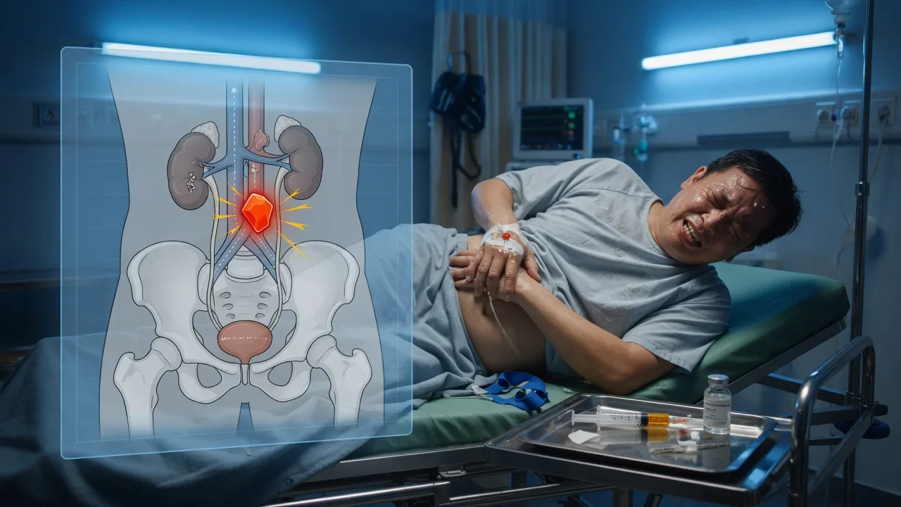
Definition and classification
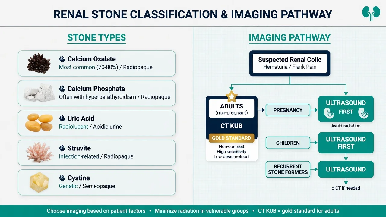
Nephrolithiasis (urolithiasis) is stone formation anywhere in the urinary tract. Renal colic is the clinical syndrome of acute ureteric obstruction by a stone — the term "colic" denoting the wave-like, building-and-fading character of the pain, not the lesser meaning of intestinal cramp. Stones are classified by composition: calcium oxalate is by far the commonest (about 70 to 80 per cent), followed by calcium phosphate, uric acid (radiolucent, in gout and acidic concentrated urine), struvite (magnesium-ammonium-phosphate, the infection stone formed by urease-splitting organisms such as Proteus, forming large staghorn calculi), and cysteine (the inherited tubular transport defect). Stones are also classified by location — renal, or ureteric at the proximal ureter, mid-ureter, or the vesicoureteric junction (VUJ), the narrowest point and the commonest site of impaction. [1]
Calcium oxalate
~70–80 % — the commonest
- The single commonest stone — 70 to 80 per cent of all calculi
- Radiopaque; the spiky envelope or dumbbell crystal on microscopy
- Driven by hypercalciuria, hyperoxaluria and hypocitraturia; high-oxalate foods (spinach, nuts, tea, chocolate)
- Check the serum calcium — hyperparathyroidism is the treatable driver
Calcium phosphate
~5–10 %
- Radiopaque; forms in a persistently alkaline urine
- Distal (type 1) renal tubular acidosis and medullary sponge kidney
- Read the venous gas for the acidosis; check the urinary pH (over 5.5)
- Often a component of mixed calcium stones
Uric acid
~5–10 % — the acidic-urine stone
- RADIOLUCENT — invisible on a plain KUB, seen only on CT or ultrasound
- Forms when the urine pH is persistently below 5.5 (acidic, concentrated urine)
- Gout, hyperuricaemia, myeloproliferative disease, chronic diarrhoea or ileostomy
- TREATABLE by alkalinising the urine (potassium citrate) to a pH above 6.5 — the one stone you can dissolve medically
Struvite
the infection stone
- Magnesium-ammonium-phosphate; formed by urease-splitting organisms (Proteus, Klebsiella, Pseudomonas, Providencia)
- Forms large staghorn calculi that fill the renal pelvis and destroy the kidney
- A persistently alkaline urine (pH above 7.2) with recurrent UTIs is the fingerprint
- Drain the system, culture the urine, and refer for complete surgical clearance — fragments regrow
Cystine
hereditary
- Autosomal recessive cystinuria — the inherited proximal-tubule amino-acid transport defect
- Hexagonal crystals; faintly radiopaque; presents in children and young adults with recurrent stones
- Suspect in any first presentation under 30 with a family history
- Lifelong management — high fluid intake, urinary alkalinisation (potassium citrate) and tiopronin or captopril
Drug-related
rare
- Indinavir and atazanavir stones (radiolucent, in HIV therapy)
- Triamterene, sulfadiazine, ephedrine and guaifenesin stones
- Ammonium acid urate (laxative abuse, ileostomy)
- Diagnosed by stone analysis and a careful drug history
Epidemiology and risk
The lifetime risk of a stone is roughly 10 to 15 per cent in Western populations, with men affected roughly twice as often as women, and the peak incidence between the ages of 20 and 50. Australia sits in the stone belt — the hot, arid climate drives chronic dehydration and concentrates urine, so prevalence is among the highest in the world, matched by the Middle East and the southern United States. After a first stone, recurrence is roughly 50 per cent at 10 years, so every first stone-former deserves an outpatient metabolic workup (serum calcium, urate, and a 24-hour urine where indicated). [1]
The risk factors for stone formation
STONE
Heat, dehydration, concentrated urine — the Australian stone belt
UTI with urease-splitting organisms (Proteus) → struvite; obstruction; cystic kidneys
High animal protein, high sodium, low fluid intake; high oxalate (spinach, nuts, tea)
Hyperparathyroidism, gout/hyperuricaemia, renal tubular acidosis, Crohn's/ileal disease
Cystinuria, primary hyperoxaluria, family history, medullary sponge kidney
Pathophysiology
Stone formation begins when urine becomes supersaturated with a stone-forming salt — calcium oxalate, uric acid, cysteine. Dehydration, low urinary volume, and dietary or metabolic excess drive supersaturation; crystals nucleate, aggregate, and grow into a stone within the renal collecting system. Most stones lie silently in the kidney until they migrate into the ureter, where they lodge at one of the three natural narrowings — the pelviureteric junction, the pelvic brim where the ureter crosses the iliac vessels, and the vesicoureteric junction (narrowest, commonest site of impaction). [1]
[1]The pain localisation follows the embryological origin of the ureter: visceral afferents from T11 to L2 produce flank and loin pain when the stone is in the proximal ureter; as it descends past the pelvic brim the pain radiates via the genitofemoral nerve (L1–L2) to the groin, testis or labia; and a stone lodged at the vesicoureteric junction produces urethral, perineal or testicular pain with urinary urgency, frequency and strangury — the latter easily mistaken for cystitis or torsion. [1]
[1]Clinical presentation
The classic presentation is unmistakable: sudden-onset, severe, colicky flank pain that builds to a peak over minutes, lasts hours, radiates from loin to groin as the stone descends, and is accompanied by nausea and vomiting. The patient is characteristically unable to find a comfortable position and writhes around the bed — the single most useful behavioural clue at the bedside, and the opposite of the patient with peritonitis who lies perfectly still. [1]
ANZ practice note. Australasian emergency practice gives an NSAID before an opioid for renal colic — diclofenac 75 mg intramuscularly (or 100 mg rectally) is the default first agent, with morphine 5 mg intravenously reserved for refractory or severe pain. This reflects the Holdgate systematic review and local Cochrane evidence that NSAIDs achieve equivalent or superior analgesia with fewer rescue doses and side effects.[1] A recent Australasian randomised trial reaffirmed the superiority of intramuscular diclofenac over intravenous tramadol for acute renal colic.[4] Tamsulosin 0.4 mg nocte is offered for distal ureteric stones of 5 to 10 mm.
Haematuria — microscopic in roughly 85 to 90 per cent and occasionally macroscopic — supports the diagnosis, but its absence does not exclude a stone. A stone at the vesicoureteric junction produces a misleading constellation of lower urinary tract symptoms — urgency, frequency, dysuria and strangury — easily mistaken for cystitis or, in the male, for prostatitis. Fever, rigors, hypotension or a frankly septic appearance are not features of uncomplicated colic and signal an infected obstructed system that requires a fundamentally different, urgent pathway. [1]
The stone at the bedside
Differential diagnosis
Loin pain radiating to the groin overlaps with several lethal and several benign mimics, and the first task is to exclude the abdominal aortic aneurysm, the testicular torsion, and the ectopic pregnancy before attributing the picture to a stone. Crucially, the presence of a stone on imaging does not exclude a concomitant lethal mimic — an older patient may have a small incidental renal calculus and a leaking aneurysm at the same time. [1]
Abdominal aortic aneurysm (leak/rupture)
- Older male, vasculopath; syncope with back/flank pain
- Pulsatile abdominal mass; hypotension; the lethal mimic of colic
- CT KUB may show the aneurysm incidentally — palpate the aorta on every older "colic" patient
- A patient may have BOTH an incidental stone and a ruptured AAA
Testicular torsion
- Sudden groin/testicular pain in a young male — identical referral pattern
- Examine the genitalia on EVERY male with apparent renal colic
- High-riding, transverse, exquisitely tender testis; absent cremasteric reflex
- Six-hour window to detorsion — never attribute groin pain to colic without examining the testis
Ectopic pregnancy
- Woman of reproductive age with lower abdominal pain radiating to shoulder tip
- β-hCG on EVERY woman — do not be reassured by a "stone" history
- Tender adnexa, vaginal bleeding, signs of rupture: tachycardia, hypotension
- Pelvic ultrasound is diagnostic
Acute pyelonephritis
- Fever, rigors, systemic illness DOMINANT (pain is constant, not colicky)
- Marked loin tenderness; leucocytes and nitrites on dipstick
- No ureteric stone on CT KUB (may have hydronephrosis)
- Bilateral or in a transplanted kidney is a urological emergency if obstructed
Acute appendicitis
- Central pain migrating to right iliac fossa over 12–24 h (slower onset)
- Anorexia, low-grade fever, McBurney point tenderness
- Inflammatory markers raised; CT shows appendiceal inflammation
- Right-sided colic may mimic appendix pain — examine carefully
Biliary colic
- Post-prandial right upper quadrant pain radiating to right scapula
- Onset 1–2 h after food; lasts 1–6 h; self-limiting
- Ultrasound shows gallstones; liver function tests may be mildly abnormal
- Afebrile between attacks; not genitourinary symptoms
Mesenteric ischaemia
- Severe abdominal pain disproportionate to examination findings
- Atrial fibrillation, vascular disease, hypoperfusion
- Lactate raised; metabolic acidosis; CT angiography diagnostic
- Pain out of proportion is the clue — colic is tender, ischaemia is silent initially
Herpes zoster (radicular pain)
- Burning unilateral dermatomal pain preceding the rash
- T7–T12 distribution; reproduced by light touch (allodynia)
- Normal urinalysis and CT
- Vesicular rash appears days later in the affected dermatome
Other considerations include renal papillary necrosis (in sickle cell trait, analgesic abuse, diabetes — sloughed papilla obstructs the ureter), renal infarction (atrial fibrillation, severe flank pain with haematuria and a raised lactate dehydrogenase), retroperitoneal fibrosis, and ureteric transitional cell carcinoma with clot colic. A pregnancy test is mandatory in every woman of reproductive age — a right-sided ectopic and a right-sided ureteric stone present identically. [1]
Bedside assessment
Begin with airway, breathing and circulation — the septic patient with pyonephrosis may be in established septic shock within the first hour. Attach monitoring, establish intravenous access, and give analgesia early: pain control is both humane and diagnostic, because the patient who settles on an NSAID has the classic analgesic response of ureteric colic. A focused history establishes the onset, character, radiation and duration of the pain, associated urinary symptoms, prior stone history, fever or rigors, and the vascular and reproductive risk factors (age, hypertension, smoking for AAA; last menstrual period for pregnancy). [1]
Examination looks for loin tenderness (mild, in keeping with capsular distension), assesses the abdomen for peritonism (which is NOT colic and demands another diagnosis), and crucially palpates the abdominal aorta in every older patient. Examine the genitalia in every male — a high-riding, tender, transverse testis with an absent cremasteric reflex is torsion until proven otherwise, and the six-hour window to detorsion cannot be missed while attributing the pain to a stone. An explicit screen for organ dysfunction — temperature, blood pressure, respiratory rate, oxygen saturation, conscious level, urine output and a venous lactate — separates the well patient who can be discharged from the septic patient who needs urgent decompression. [1]
Investigations
Send blood for urea and electrolytes with creatinine (baseline renal function — obstruction causes a post-renal acute kidney injury), full blood count, C-reactive protein, and a venous lactate (the septic marker in the infected obstructed system). Check a β-hCG in every woman of reproductive age, regardless of contraception history. Urinalysis is central: blood (haematuria) supports a stone, while leucocytes and nitrites suggest infection and, in the obstructed kidney, transform the case into a urological emergency. Send a urine culture in any febrile or septic patient. A coagulation screen is worthwhile if intervention (stent, nephrostomy, surgery) is anticipated. [1]
Ultrasound is the first-line imaging in pregnancy, in children, and in the recurrent known stone-former where radiation should be minimised. It demonstrates hydronephrosis and renal stones (with the characteristic "twinkling artefact" on colour Doppler), but it visualises the ureter poorly and misses many ureteric stones; in pregnancy it is the screening test, with magnetic resonance urography as the second line when the diagnosis remains unclear. A plain KUB radiograph captures only radiopaque stones (about 80 per cent of calcium-containing stones; uric acid and cysteine stones are radiolucent) and is reserved for follow-up of a known radiopaque stone, not for the initial diagnosis. [1]
[1]Imaging strategy at a glance
The ED first 60 minutes
Suspected renal colic — the ED first 60 minutes
0 to 10 minutes — triage, analgesia and the danger screen
ABCDE, full monitoring, two large-bore cannulae. Screen for the dangerous patient first — a fever, hypotension, a raised lactate or anuria in a solitary kidney divert to the urgent-decompression pathway, not the colic pathway. Give diclofenac 75 mg IM (or 100 mg PR) immediately — the NSAID is the first drug, not the opioid. Examine the genitalia in every male; palpate the aorta in every older patient.
10 to 20 minutes — the focused history and the bloods
Onset, character, radiation (loin to groin), prior stones, fever or rigors, last menstrual period in every woman. Send the U&E with creatinine, the FBC, the CRP, the venous lactate, and a beta-hCG in every woman of reproductive age. Dipstick the urine — blood supports a stone; leucocytes and nitrites in an obstructed kidney transform the case into pyonephrosis.
20 to 40 minutes — the CT KUB (or ultrasound in pregnancy and childhood)
Non-contrast CT KUB is the adult first-line test — 95 to 98 per cent sensitive, defines the size, site and density, grades the hydronephrosis and excludes the lethal mimics (AAA, appendicitis, diverticulitis). Reserve ultrasound for pregnancy, children and the recurrent known stone-former; MR urography is the second line in pregnancy.
40 to 60 minutes — the disposition split
Read the stone size, the obstruction, the infection markers and the renal function. The uncomplicated stone (under 10 mm, afebrile, controlled pain, normal creatinine) is discharged on an NSAID, a PPI, tamsulosin for the 5 to 10 mm distal stone, a stone filter and return precautions. The infected or obstructed solitary kidney, the rising creatinine, or the uncontrolled pain is admitted — the septic obstructed system is decompressed within hours, not treated with antibiotics alone.
Immediate management and resuscitation
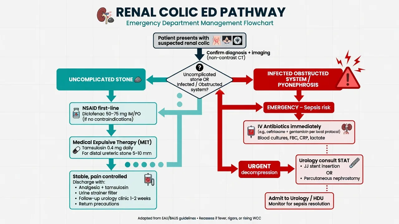
The priority is analgesia, and the analgesic ladder has a non-steroidal agent on the bottom rung. Give diclofenac 75 mg intramuscularly (or 100 mg rectally) as the first-line agent — it blocks the prostaglandin-mediated ureteric spasm and reduces the renal capsular pressure that drives the pain. The Holdgate systematic review established that NSAIDs achieve equivalent or superior analgesia to opioids in acute renal colic, with fewer rescue doses and fewer adverse effects, and a recent randomised trial confirmed the superiority of intramuscular diclofenac over intravenous tramadol.[1][4] Reserve morphine 5 mg intravenously, titrated, for refractory or severe pain, or use fentanyl 50 to 100 micrograms intravenously when the intramuscular route is contraindicated or rapid titration is required. Add an antiemetic — metoclopramide 10 mg intravenously or ondansetron 4 mg intravenously — for the nausea and vomiting that accompany the visceral afferent stimulation.
Caution in renal impairment. An NSAID is contraindicated when there is established acute kidney injury, in the dehydrated or septic patient, in solitary-kidney obstruction, and in pregnancy beyond 28 weeks (premature closure of the ductus arteriosus and fetal renal effects). In these settings use paracetamol 1 g intravenously and an opioid titrated to pain, and escalate to urology for definitive relief of the obstruction. [1]
Give intravenous fluids only to correct dehydration from vomiting or to maintain euvolaemia in the septic patient — routine aggressive hydration does not improve stone passage and may worsen the pain by further distending the renal capsule. If the patient is febrile or shows any sign of infection in an obstructed system, give broad-spectrum antibiotics immediately (ceftriaxone 2 g intravenously plus gentamicin, or piperacillin-tazobactam 4.5 g intravenously) after taking blood and urine cultures, and activate the urgent urology pathway — antibiotics alone will not cure an obstructed infected kidney. [1]
[1]Definitive management and disposition
The disposition splits cleanly along the line of infection, obstruction and pain control. [1]
The uncomplicated ureteric stone — discharge pathway
A patient with a stone of 10 mm or less, no fever, no evidence of infection, controlled pain, normal renal function, and single-kidney function preserved is managed as an outpatient. Provide: [1]
- Discharge analgesia: diclofenac 50 mg orally three times daily (with a proton-pump inhibitor) or naproxen 500 mg orally twice daily, with paracetamol and oral morphine for breakthrough pain.
- Medical expulsive therapy (MET): tamsulosin 0.4 mg nocte for 4 to 6 weeks for a distal ureteric stone between 5 and 10 mm. α-blockers relax the smooth muscle at the vesicoureteric junction and modestly increase the spontaneous passage rate and shorten the time to passage. MET is not indicated for proximal stones, stones under 5 mm (which pass anyway), or stones over 10 mm (which will not pass).[2]
- A stone filter (urine strainer) to catch the passed stone for analysis.
- Return precautions in writing: new fever or rigors, uncontrolled pain, intractable vomiting, or a marked reduction in urine output.
- Urology or general-practitioner follow-up in 2 to 4 weeks with repeat imaging to confirm passage.
The urological emergency — the infected obstructed system
The infected obstructed kidney (pyonephrosis) is the single most dangerous renal-colic scenario and a Fellowship favourite. The diagnosis is made when a patient with an obstructing stone has fever, rigors, leucocytosis, raised lactate, hypotension or any systemic sign of sepsis. Management is not antibiotics alone — the raised intraluminal pressure prevents antibiotic penetration of the urine and renal parenchyma, and the patient develops bacteraemia and septic shock. The definitive treatment is urgent surgical decompression of the obstructed upper tract within hours, by: [1]
- Percutaneous nephrostomy (the radiologically-placed drain, preferred in the haemodynamically unstable or septic patient), or
- Retrograde ureteric stent (placed cystoscopically in theatre). [1]
Decompression is combined with broad-spectrum intravenous antibiotics (ceftriaxone 2 g plus gentamicin, or piperacillin-tazobactam 4.5 g), fluid resuscitation titrated to mean arterial pressure and lactate, and admission to a monitored bed — high-dependency or intensive care for the patient with septic shock. The stone itself is dealt with electively once the sepsis has resolved. The same urgency applies to anuria in a patient with a solitary kidney, a renal transplant, or bilateral obstructing stones — these are emergencies because bilateral (or single-functioning-kidney) obstruction produces acute kidney injury rapidly and threatens renal loss. [1]
Definitive stone removal — extracorporeal shock-wave lithotripsy, ureteroscopy with laser lithotripsy, or percutaneous nephrolithotomy — is decided later by urology and depends on stone size, site and composition. Emergency indications for non-elective urology referral (within hours) are: infected obstructed system, anuria in a solitary or transplanted kidney or bilateral obstruction, intractable pain despite analgesia, and a rising creatinine with obstruction. Urgent (within days) referral applies to large stones over 10 mm, persistent obstruction, and failure of medical expulsive therapy. [1]
Special populations
Pregnancy: symptomatic stones are the commonest non-obstetric cause of admission in pregnancy. Ultrasound is the first-line imaging test; MR urography is second-line when the diagnosis remains unclear, and low-dose CT KUB is reserved for the refractory diagnostic dilemma in the third trimester. Avoid NSAIDs after 28 weeks (premature ductus arteriosus closure, oligohydramnios) — use paracetamol and opioid analgesia. Ureteric stenting or percutaneous nephrostomy is the definitive temporising measure when obstruction or infection complicates pregnancy. Children are imaged with ultrasound first, reserving CT for the equivocal case. The elderly patient with apparent colic has an abdominal aortic aneurysm excluded first — palpate the aorta, and image before attributing the pain to a stone. The patient with a solitary kidney or transplant is admitted with a lower threshold, because loss of the single functioning kidney is catastrophic. The anticoagulated patient bleeds more readily from the trauma of stone passage and from any intervention — coordinate anticoagulation reversal with urology. [1]
Complications and pitfalls
Complications of an obstructing stone include post-renal acute kidney injury (especially in the solitary or bilaterally obstructed kidney), pyonephrosis with progression to urosepsis and septic shock, permanent renal parenchymal loss from prolonged obstruction, and the long-term consequences of recurrent stones (chronic kidney disease, the infected staghorn calculus destroying the kidney). The common pitfalls invert good care. Discharging the febrile obstructed patient as simple colic misses pyonephrosis and may cost a kidney or a life. Missing the abdominal aortic aneurysm in the older patient with flank pain — the patient may have both an incidental stone and a leaking aneurysm. Failing to examine the genitalia in the male misses testicular torsion, which presents identically and has a six-hour window to detorsion. Relying on the absence of haematuria to exclude a stone — up to 15 per cent of confirmed stones have no blood on dipstick. Over-hydrating the well patient does not aid stone passage and may worsen the pain. Giving an NSAID to the dehydrated, septic or solitary-kidney patient worsens the acute kidney injury. Delaying drainage in pyonephrosis while awaiting cultures or a urology review in the morning kills patients — organ dysfunction mandates decompression within hours. [1]
Prognosis and disposition
The spontaneous passage rate depends on size: stones under 5 mm pass in roughly 80 per cent of cases, stones of 5 to 10 mm in about 50 per cent (improved modestly by tamsulosin for distal stones), and stones over 10 mm in under 20 per cent. The uncomplicated patient is discharged with analgesia, a stone filter and return precautions, and passes the stone at home in most cases. The infected obstructed patient is admitted for urgent decompression and intravenous antibiotics and, in septic shock, to intensive care — mortality in urosepsis from pyonephrosis is determined by the timeliness of source control, not the choice of antibiotic. A patient with a single functioning kidney, bilateral obstruction, anuria, intractable pain or a rising creatinine is admitted for urgent urological decompression. [1]
Evidence and regional guidelines
The contemporary evidence base for emergency management rests on three pillars. The Holdgate systematic review (BMJ 2004) established that NSAIDs achieve equivalent or superior analgesia to opioids in acute renal colic, with fewer rescue doses and side effects — the basis for the NSAID-first ladder used worldwide.[1] A recent Australasian randomised trial (2025) reaffirmed the superiority of intramuscular diclofenac over intravenous tramadol.[4] The evidence for medical expulsive therapy with α-blockers is more contested: a 2017 systematic review of tamsulosin, nifedipine and placebo showed a modest benefit of α-blockers, with the effect concentrated in distal ureteric stones over 5 mm — the basis for offering tamsulosin 0.4 mg nocte to that subgroup.[2] The European Association of Urology (EAU) urolithiasis guideline and the American Urological Association (AUA) ureteral calculi guideline provide the definitive stone-management pathway, while the Australasian College for Emergency Medicine endorses the NSAID-first analgesic strategy and CT KUB as the first-line imaging test. Across all guidelines, the three fixed points are: give an NSAID before an opioid; image with non-contrast CT KUB; and decompress the infected obstructed system within hours.
Key trials
Holdgate — NSAIDs vs opioids for renal colic (BMJ 2004)
BMJ
PMID 15178585
Key finding
A systematic review of randomised trials comparing NSAIDs with opioids for acute renal colic. NSAIDs achieved equivalent or superior pain reduction, with significantly fewer rescue analgesic doses and fewer adverse effects (nausea, vomiting, sedation) than opioids.
Practice change
The evidence base for the NSAID-first analgesic ladder — diclofenac 75 mg IM before morphine — used worldwide.
Yaowalaorng — intramuscular diclofenac vs intravenous tramadol (EMA 2025)
Emergency Medicine Australasia
PMID 39763427
Key finding
A randomised controlled trial in the ED showing intramuscular diclofenac was superior to intravenous tramadol for acute renal colic, with greater pain reduction at 30 minutes and fewer adverse effects.
Practice change
A contemporary Australasian reaffirmation of the NSAID-first strategy; IM diclofenac remains the default first agent.
Smith-Bindman — ultrasound vs CT for suspected nephrolithiasis (STONE, NEJM 2014)
New England Journal of Medicine
PMID 25229916
Key finding
A multicentre pragmatic randomised trial of 2759 patients comparing point-of-care ultrasound, radiology ultrasound and CT first-line for suspected nephrolithiasis. High-risk complications were low and did not differ between groups; CT exposed patients to more radiation without improving diagnostic accuracy or clinical outcomes.
Practice change
Ultrasound is a reasonable first-line imaging test that avoids radiation; CT remains the gold standard for definitive stone characterisation and is first-line where ultrasound is likely to be non-diagnostic.
SUSPEND — tamsulosin and nifedipine for ureteric colic (Pickard, HTA 2015)
Health Technology Assessment
PMID 26244520
Key finding
A multicentre placebo-controlled randomised trial of 1167 adults hospitalised with ureteric colic, comparing tamsulosin, nifedipine and placebo. Neither drug increased the rate of stone passage at four weeks compared with placebo (around 80 per cent in all groups).
Practice change
Routine MET with tamsulosin or nifedipine is NOT supported for ureteric colic overall; any benefit is small and confined to larger (over 5 mm) distal stones.
STE — tamsulosin for symptomatic ureteral stones (Meltzer, JAMA Intern Med 2018)
JAMA Internal Medicine
PMID 29913020
Key finding
A randomised clinical trial of 512 adults with symptomatic ureteral stones, comparing tamsulosin against placebo. Tamsulosin did not significantly increase the rate of stone passage at 28 days overall (50 per cent versus 47 per cent); a pre-specified subgroup with stones over 5 mm showed a modest benefit.
Practice change
The largest trial to date showing no overall benefit of tamsulosin for ureteric stones — the modern evidence that has led the AUA and EAU to retreat from routine MET and reserve it for selected larger distal stones.
SAQ — uncomplicated renal colic in a 38-year-old man
10 minutes · 10 marks
A 38-year-old man presents to the emergency department with sudden-onset, severe left flank pain radiating to the groin, associated with nausea and two episodes of vomiting. He is afebrile, visibly writhing on the trolley and unable to find a comfortable position. Blood pressure is 140/85, heart rate 102, and urinalysis shows large blood but no leucocytes or nitrites. CT KUB demonstrates a 6 mm stone at the left vesicoureteric junction with mild hydroureteronephrosis and no evidence of infection.
SAQ — pyonephrosis: the septic obstructed kidney
10 minutes · 10 marks
A 65-year-old diabetic woman is brought to the emergency department with left flank pain, fever of 39.2 degrees Celsius, two rigors in the preceding hour and confusion. Blood pressure is 88/52, heart rate 124, respiratory rate 26 and oxygen saturation 93 per cent on room air. Urinalysis shows blood, leucocytes and nitrites. CT KUB demonstrates a 12 mm obstructing left proximal ureteric stone with marked hydronephrosis, perinephric stranding and gas in the renal collecting system. Venous lactate is 4.2 mmol per litre and creatinine is 220 micromol per litre (baseline 80).
Exam pearls
- The patient who writhes has colic; the patient who lies still has peritonitis — behavioural observation is the first bedside clue.
- NSAID before opioid: diclofenac 75 mg intramuscularly is first-line; morphine 5 mg intravenously is reserved for refractory pain.
- CT KUB (non-contrast) is the gold standard — 95 to 98 per cent sensitive and the only first-line test that also identifies the lethal mimics.
- Ultrasound in pregnancy, children and recurrent stone-formers; a plain KUB is for follow-up only.
- Examine the testes in every male with apparent colic — torsion presents identically and has a six-hour window.
- β-hCG in every woman of reproductive age — ectopic pregnancy is the lethal mimic.
- Palpate the aorta in every older patient — a ruptured abdominal aortic aneurysm is the lethal mimic and may coexist with an incidental stone.
- The infected obstructed kidney is decompressed, not just given antibiotics — ureteric stent or percutaneous nephrostomy within hours.
- MET is tamsulosin 0.4 mg nocte for 4 to 6 weeks, for distal ureteric stones of 5 to 10 mm — not for proximal, very small or large stones.
- Absence of haematuria does not exclude a stone — up to 15 per cent of confirmed stones have no blood on dipstick.
- Stone composition — know the four: calcium oxalate is the commonest (70 to 80 per cent); uric acid is radiolucent and dissolves with alkalinisation; struvite is the infection stone forming staghorns; cystine is hereditary.
- The uric-acid stone is the one you can dissolve — alkalinise the urine with potassium citrate to a pH above 6.5; it is radiolucent on KUB but visible on CT (200 to 400 HU).
- A staghorn calculus is struvite until proven otherwise — urease-splitting organisms (Proteus) in a woman with recurrent UTIs; refer for clearance, not antibiotics.
- The three narrowings of the ureter — pelviureteric junction, pelvic brim (iliac vessels), and the vesicoureteric junction (narrowest, commonest impaction site).
- A VUJ stone with sterile pyuria is the trap — leucocytes without nitrites from the irritated bladder; image the VUJ on CT before labelling it cystitis.
- Forced fluids do NOT flush a stone — they raise the capsular pressure and worsen the pain; hydrate only the dehydrated or the septic.
- Tamsulosin is controversial, not standard — SUSPEND and STE showed no overall benefit; offer it only for a distal stone over 5 mm and never let it delay urology.
- The first stone earns a metabolic workup — serum calcium (hyperparathyroidism), urate, and a 24-hour urine; recurrence is 50 per cent at 10 years. [1]
Red flags
[1]References
- [1]Holdgate A, Pollock T. Systematic review of the relative efficacy of non-steroidal anti-inflammatory drugs and opioids in the treatment of acute renal colic BMJ, 2004.PMID 15178585
- [2]Gottlieb M, Long B, Koyfman A. Comparison of Tamsulosin, Nifedipine, and Placebo for Ureteric Colic CJEM, 2017.PMID 26584627
- [3]Ali A, Niaz S, Waris S, et al. Non-Contrast Enhanced Multi-Slice Ct-Kub In Renal Colic: Spectrum Of Abnormalities Detected On Ct Kub And Assessment Of Referral Patterns J Ayub Med Coll Abbottabad, 2019.PMID 31535518
- [4]Yaowalaorng J, Rakkarn S, Phakdee B, et al. Superior efficacy of intramuscular diclofenac compared to intravenous tramadol for acute renal colic in northern Thai patients: A randomised double-blind, sham-controlled trial Emerg Med Australas, 2025.PMID 39763427
- [5]Smith-Bindman R, Aubin C, Bailitz J, et al. Ultrasonography versus computed tomography for suspected nephrolithiasis N Engl J Med, 2014.PMID 25229916
- [6]Pickard R, Starr K, MacLennan G, et al. Use of drug therapy in the management of symptomatic ureteric stones in hospitalised adults: a multicentre, placebo-controlled, randomised controlled trial and cost-effectiveness analysis of a calcium channel blocker (nifedipine) and an alpha-blocker (tamsulosin) (the SUSPEND trial) Health Technol Assess, 2015.PMID 26244520
- [7]Meltzer AC, Burrows PK, Wolfson AB, et al. Effect of Tamsulosin on Passage of Symptomatic Ureteral Stones: A Randomized Clinical Trial JAMA Intern Med, 2018.PMID 29913020