EM · Sepsis and septic shock (approach)
Sepsis and septic shock — the emergency department approach
Also known as Sepsis · Septic shock · Sepsis-3 · The Hour-1 bundle · qSOFA · The septic patient
Sepsis is the life-threatening organ dysfunction caused by a dysregulated host response to infection, defined by Sepsis-3 as suspected infection with an acute rise in the SOFA score of 2 or more, and septic shock as sepsis requiring vasopressors to hold a mean arterial pressure of 65 or above with a serum lactate over 2 mmol per litre despite adequate fluid. The emergency department approach rests on the qSOFA prompt (a respiratory rate of 22 or above, an altered mentation, a systolic blood pressure of 100 or below) and delivers the Surviving Sepsis Campaign Hour-1 bundle — the lactate, the blood cultures before the antibiotic, the broad-spectrum antibiotics (ceftriaxone 2 g IV, piperacillin-tazobactam 4.5 g IV, meropenem 1 g IV; vancomycin 1.5 g IV for MRSA; metronidazole 500 mg IV for anaerobic cover), the 30 mL per kilogram crystalloid, and the early vasopressor for a mean arterial pressure below 65 — followed by the lactate-guided resuscitation and the source control. The differential is the other shock states and the SIRS mimics. ACEM-primary, globally tagged.
On this page & tools
Your progress
Saved locally on this device.
Target exams
Red flags
Related topics
- Paediatric sepsis and septic shock (the septic child in the emergency department)
- Damage control resuscitation in trauma
- Fluid resuscitation in the emergency department
- Community-acquired pneumonia
- Meningitis and encephalitis (emergency department diagnosis and management)
- Immunocompromised host and febrile neutropenia
Sepsis is the life-threatening organ dysfunction caused by a dysregulated host response to infection, and septic shock is its deepest haemodynamic expression — the patient whose circulation, despite fluid, can no longer perfuse the tissues. The Fellowship candidate's job at the bedside is to collapse the recognition, the resuscitation and the source control into a single, parallel, hour-long effort: to identify the infected and decompensating patient within minutes, to deliver the Surviving Sepsis Campaign Hour-1 bundle without delay, and to hunt for and treat the source while the resuscitation runs. The recurring failure is not missing a rare diagnosis — it is the serial, sequential management of a septic patient, where the cultures, the imaging and the team referrals are allowed to delay the antibiotic and the fluid. The 2016 Sepsis-3 definitions and the 2021 Surviving Sepsis Campaign guidelines are the two contemporary frameworks that govern recognition and management.[1][4]
Definition and classification — the Sepsis-3 framework
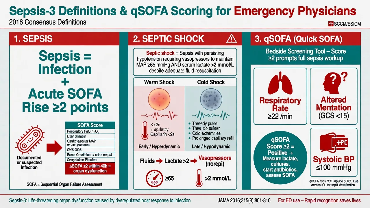
The modern definition of sepsis discards the old SIRS-plus-infection model because the systemic inflammatory response is neither sensitive nor specific — it is generated by every insult from a viral upper-respiratory infection to pancreatitis, and it is absent in some genuinely septic patients. The Sepsis-3 consensus redefines sepsis as life-threatening organ dysfunction caused by a dysregulated host response to infection, where organ dysfunction is operationalised as an acute increase in the SOFA score of 2 points or more from baseline. Septic shock is a subset of sepsis defined by the combination of circulatory and cellular abnormalities — specifically, the need for vasopressors to maintain a mean arterial pressure of 65 mmHg or above, with a serum lactate above 2 mmol per litre, despite adequate fluid resuscitation. This definition cleanly separates the uncomplicated infection (managed as infection), the septic patient (with organ dysfunction requiring the bundle), and the septic shock patient (with the vasopressor-and-lactate criteria requiring intensive care).[1]
[1]The clinical classification the resuscitation bay uses daily is the haemodynamic one. Compensated shock describes a patient with a normal blood pressure but with perfusion failure — tachycardia, mottled or cool skin, prolonged capillary refill, oliguria, altered mentation — and is far commoner than the lay reader expects. Decompensated shock is the state in which the blood pressure has fallen. The further distinction, useful for vasoactive choice, is between warm shock (high cardiac output, low systemic vascular resistance — the vasodilated, warm, flash-refill patient, the commonest adult pattern, responsive to noradrenaline) and cold shock (low output, high resistance — the cool, mottled, thready-pulse patient, responsive to fluid and an inotrope). The adult septic patient is most often warm and vasoplegic; the cold pattern in adults signals a late, decompensated myocardial state.[4]
Pathophysiology
The septic cascade begins when a pathogen-associated molecular pattern — endotoxin in gram-negative sepsis, teichoic acid in gram-positive — engages the innate immune receptor (the toll-like receptor family). The result is a cytokine surge: tumour necrosis factor, interleukin-1 and interleukin-6 within hours, driving endothelial activation, glycocalyx shedding, capillary leak and nitric-oxide-mediated vasodilation. The haemodynamic consequence is a distributive shock — the profound fall in systemic vascular resistance — compounded by hypovolaemia from third-space losses, myocardial depression (a circulating myocardial depressant substance reduces contractility), and disordered microvascular flow with mitochondrial dysfunction. The end-organ injury — acute kidney injury, acute respiratory distress syndrome, disseminated intravascular coagulation, septic encephalopathy — is the product of hypoperfusion, ongoing inflammation and disordered cellular oxygen use rather than a single ischaemic insult. [1]
The physiology explains why the blood pressure is a late and unreliable monitor. Cardiac output is the product of stroke volume and heart rate; the septic patient compensates with tachycardia and a heightened sympathetic drive, and the systemic vascular resistance can fall substantially before the pressure registers a fall. The patient who looks well but has a raised lactate and a widened pulse pressure is already well along the cascade. This is why the contemporary approach measures perfusion — the lactate, the mentation, the urine output, the capillary refill — and not merely the blood pressure, and why the qSOFA prompt exists. [1]
Epidemiology and risk factors
Sepsis remains among the leading causes of in-hospital death worldwide and a dominant consumer of intensive-care resources, with the incidence rising in the elderly, the immunocompromised and the patient with multiple comorbidities. The common foci of infection presenting to the emergency department are the lung (pneumonia), the urinary tract (pyelonephritis and obstructive urosepsis), the abdomen (biliary sepsis, perforated viscus, appendicitis, ischaemic bowel), the skin and soft tissue (cellulitis, necrotising fasciitis, infected ulcer), and device-related infection (the indwelling line, the urinary catheter, the prosthetic joint). Risk factors that raise the pre-test probability and lower the threshold to treat broadly are age over 65, immunosuppression (chemotherapy, steroids, transplant, HIV, neutropenia), diabetes mellitus, chronic kidney or liver disease, indwelling lines and devices, asplenia, alcohol misuse, recent surgery or instrumentation, and antibiotic use within the preceding 90 days (which selects for resistant organisms). The mortality of septic shock reaches 30 to 40 per cent and concentrates in the patient who presents late, the immunocompromised, and the patient in whom the antibiotic is delayed or inappropriate.[1][4]
Recognition at the bedside — the qSOFA score
The bedside recognition of sepsis does not wait for the full SOFA, which requires laboratory data the emergency department does not yet have. The Sepsis-3 task force derived a simple three-item prompt — the qSOFA (the quick SOFA) — from a large retrospective dataset of infected ward and emergency patients, validated to identify those at high risk of a poor outcome. The qSOFA is not the definition of sepsis (the SOFA rise is); it is a prompt that should trigger concern, a fuller workup, and escalation of care when the score reaches 2 or more out of 3. The three components are chosen because they are measured in seconds, at the bedside, from the vital signs alone.[2][3]
The qSOFA score — each component scores 1, a total of 2 or more is positive
A qSOFA of 2 or more in a patient with suspected infection predicts in-hospital mortality with a discrimination that exceeds the old SIRS criteria in the non-intensive-care population, and the Rudd 2018 JAMA analysis confirmed the association across low-, middle- and high-income settings, identifying unrecognised organ dysfunction in patients who looked deceptively well.[3] The limits are real: the qSOFA is less sensitive than SIRS for the early identification of the septic patient (it picks up sicker patients), so a normal qSOFA does not exclude sepsis, and the Surviving Sepsis Campaign continues to recommend a low threshold to investigate and treat any patient with suspected infection and physiological derangement regardless of the score.
Clinical presentation
The septic patient presents along a spectrum from the febrile and alert patient with a source of infection to the cold, mottled and hypotensive patient in established shock. The classic features are fever or hypothermia (a temperature above 38.3 or below 36 degrees Celsius), tachycardia, tachypnoea, an altered mental state, warm or mottled skin, a prolonged capillary refill, oliguria, and, in the late or meningococcal pattern, a non-blanching petechial rash. The elderly and the immunocompromised present atypically — a fall, confusion, a functional decline, hypothermia rather than fever, a soft abdomen — and the threshold to screen for sepsis in these groups is deliberately low. The history targets the source (the cough, the dysuria, the abdominal pain, the cellulitic limb, the line), the comorbidities and the immune status, the recent antibiotics and instrumentation, and the time course. A key point for the candidate: the patient with the warm dry skin, the bounding pulse and the wide pulse pressure is in warm vasodilatory shock and may look deceptively well; the perfusion deficit and the lactate, not the appearance, drive the resuscitation. [1]
Differential diagnosis
Sepsis is the leading diagnosis in any unwell, febrile or shocked patient, but the bedside challenge is to distinguish the septic shock from the other shock states and from the non-infectious causes of a SIRS picture, because the empiric therapy diverges. The Fellowship candidate must run the differential in parallel with the resuscitation, not after it. [1]
Septic shock
- A fever or hypothermia, a source of infection, a warm vasodilated periphery (early) or a cold mottled one (late), a widened pulse pressure
- A raised lactate, a positive qSOFA, the leucocytosis or leucopenia, the source identified on examination and imaging
- The Hour-1 bundle: lactate, cultures, antibiotics, 30 mL per kilogram crystalloid, vasopressors for a MAP below 65, source control
- The archetype of distributive shock; mortality rises with every hour the antibiotic is delayed
Cardiogenic shock
- A cool, mottled periphery, a narrowed pulse pressure, a gallop rhythm, pulmonary oedema, raised jugular venous pressure; a cardiac history or an acute coronary syndrome
- A raised lactate, a troponin, an ECG showing ischaemia, a bedside echo showing a poorly contracting ventricle; the central venous oxygen saturation is low
- Cautious fluids (250 mL aliquots), early inotropes and vasopressors, reperfusion for the infarct, mechanical support in the refractory case
- A fluid bolus in the cardiogenic patient can precipitate pulmonary oedema — examine for the gallop and the crackles before fluids
Hypovolaemic shock
- A history of haemorrhage, vomiting, diarrhoea or burns; a cool periphery, a narrowed pulse pressure, dry mucous membranes, low jugular venous pressure
- A raised lactate, a raised urea-to-creatinine ratio, anaemia in the haemorrhagic cause; the source evident on history and examination
- Blood for the haemorrhagic, balanced crystalloid for the others, control of the source of loss; permissive hypotension in the active bleed
- The periphery is cold and vasoconstricted, distinguishing it from the warm vasoplegia of early septic shock
Obstructive shock
- A massive pulmonary embolism (the hypoxia, the raised JVP, the right-heart strain) or a tension pneumothorax (the unilateral signs, the tracheal deviation) or a tamponade (the muffled sounds, the pulsus paradoxus)
- A bedside ultrasound is diagnostic — the dilated right ventricle, the collapsed lung, the pericardial effusion; the lactate is raised
- Thrombolysis or thrombectomy for the embolism, needle decompression for the pneumothorax, pericardiocentesis for the tamponade
- Distinguished by the obstructive physiology and the ultrasound; fluids alone will not reverse it
Anaphylaxis
- A sudden onset after an allergen, urticaria, angioedema, stridor, wheeze and hypotension; no fever
- A history of allergy or a recent food, drug or sting; the rash is the discriminator
- Intramuscular adrenaline 500 micrograms (0.5 mL of 1 in 1000), fluid, repeat adrenaline, an antihistamine and a steroid
- A shocked patient with a rash is not always sepsis — the timing and the urticaria distinguish the two
SIRS (non-infectious)
- Pancreatitis, adrenal crisis, trauma, burns, the poisons (salicylate, aspirin) and the post-ictal state all generate fever, tachycardia and a raised lactate without infection
- The history and the focused testing — the lipase for pancreatitis, the cortisol for the adrenal crisis, the drug levels — identify the cause
- Treat the cause: aggressive fluid and analgesia for pancreatitis, hydrocortisone for the adrenal crisis, decontamination and antidote for the poison
- SIRS is a syndrome, not a diagnosis; never close on SIRS alone — find the insult driving it
The other distributive shocks — the neurogenic (after a spinal cord injury, with a warm dry periphery and a bradycardia), the anaphylactic (above), and the thyroid storm or the adrenal crisis (the endocrine shocks that overlap with sepsis and that respond to hydrocortisone or a beta-blocker) — complete the differential. The candidate's discipline is to keep the shock differential open during the first hour: examine for the cardiac and the obstructive causes with the bedside ultrasound, check a lipase and a glucose, and treat sepsis empirically while the workup proceeds, because a missed sepsis kills faster than an unnecessary antibiotic. [1]
Bedside assessment and investigations
The investigations run in parallel with — never before — the first fluid bolus and the antibiotic. The venous or arterial blood gas gives the immediate lactate (the single most important prognostic test), the pH and the base deficit; a lactate above 2 mmol per litre signals tissue hypoperfusion and a lactate above 4 mmol per litre defines severe sepsis physiology mandating the full bundle. The full blood count (the leucocytosis, the leucopenia, the bandemia, the thrombocytopenia), the CRP and procalcitonin (more specific for a bacterial infection), the urea, electrolytes and creatinine, the liver function tests, the coagulation (the disseminated intravascular coagulation of late sepsis), and the glucose (hypoglycaemia is dangerous and overlooked) are sent at the first draw. The blood cultures — two sets from separate sites — are drawn before the first antibiotic whenever they can be obtained without delaying the dose, because the yield falls by roughly a third once the antibiotic is given, but the antibiotic is never delayed for the cultures. A urinalysis and urine culture, a chest radiograph for the respiratory source, and a lactate recheck at two to four hours complete the first-round workup. The source imaging — the chest CT for a complex pneumonia, the ultrasound or CT for the biliary or abdominal source, the ultrasound for the urinary tract — is performed once the patient is stabilised, not before the resuscitation. [1]
The Hour-1 bundle and resuscitation
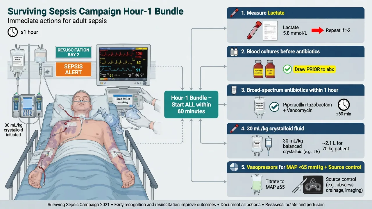
The Surviving Sepsis Campaign Hour-1 bundle is the contemporary replacement for the old three-hour and six-hour bundles, collapsed into a single resuscitation hour because the evidence showed that the earlier the elements are delivered, the better the outcome. The bundle is five elements, delivered in parallel, not in sequence, with the reassessment after every intervention. The candidate must be able to recite them and execute them.[4]
The Hour-1 bundle — the five parallel elements
LABVS
Measure the lactate immediately and recheck it at 2 to 4 hours; a lactate above 4 mmol per litre or a non-clearing lactate drives the aggressive resuscitation
Broad-spectrum antibiotics within the first hour, after the cultures are drawn if possible but never delayed by them
Two sets from separate sites before the antibiotic if it does not delay the dose
Start a vasopressor for a mean arterial pressure below 65 or above 65 target, after or during the fluid; noradrenaline is first-line
Identify and treat the source — drain the abscess, remove the infected line, relieve the obstruction — within the first 6 to 12 hours
(Note the bundle also explicitly includes the 30 mL per kilogram crystalloid for the hypotensive or lactate-over-4 patient, given within the first hour alongside the elements above.) [1]
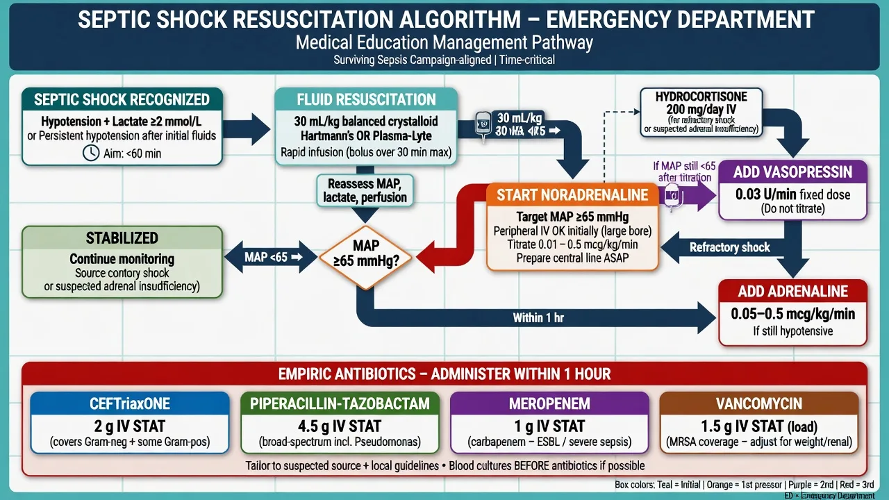
Fluid resuscitation uses a balanced crystalloid (Hartmann's solution or Plasma-Lyte, preferred over 0.9 per cent saline for the lower chloride and the lower risk of hyperchloraemic acidosis), at 30 mL per kilogram intravenously within the first three hours for the patient with sepsis-induced hypoperfusion (the hypotension or the lactate above 4 mmol per litre). The fluid is given as boluses with a reassessment after each — the perfusion, the heart rate, the blood pressure, the mentation, the urine output, and the signs of overload (the rising work of breathing, the crackles, the hepatomegaly). The contemporary Surviving Sepsis Campaign guidance softened the fluid mandate, advising a dynamic reassessment rather than a blind large-volume push, and the candidate should be able to articulate that the fluid is titrated, not automatic: a patient who clears the lactate and reaches the target pressure on a litre does not need three litres. [1]
[1]Vasopressors and the mean arterial pressure
The resuscitation target is a mean arterial pressure of 65 mmHg or above, sufficient to perfuse the brain, the kidneys and the heart. When the fluid does not reach the target — or the patient is in frank shock from the outset — a vasopressor is started early. The first-line agent is noradrenaline (norepinephrine), an alpha agonist that restores the vascular tone lost in the vasoplegic shock. It is run as an intravenous infusion titrated to the mean arterial pressure, classically through a central line, but the current guidance permits a peripheral infusion through a proximal, well-functioning cannula while the central access is secured, because the harm of delaying the vasopressor exceeds the small risk of extravasation. Vasopressin (0.03 units per minute, a fixed dose) is added as a second agent to allow a lower noradrenaline dose and to recruit the V1 receptor pathway. Adrenaline (epinephrine) is added or substituted for the refractory shock and adds an inotropic effect. The hydrocortisone 200 mg intravenously per day (or 50 mg every 6 hours) is considered for the shock that is refractory to adequate fluid and two vasopressors — the rationale is a relative adrenal insufficiency in critical illness, not an absolute deficiency. [1]
Empiric antibiotics — the doses, the source, the host
The empiric antibiotic is the single most time-critical therapy in sepsis: every hour of delay in septic shock adds measurable mortality, and an inappropriate first antibiotic doubles it. The choice is governed by three questions — the suspected source, the host (the immune status and the comorbidities), and the local resistance pattern. The Fellowship candidate must know the standard adult doses and their rationale.[4]
[1]The doses above are the first dose and are given within the first hour. Metronidazole 500 mg IV provides the anaerobic cover for the abdominal and the biliary source when a cephalosporin is the gram-negative backbone. Vancomycin 1.5 g IV (or 25 to 30 mg per kilogram loading) covers the methicillin-resistant Staphylococcus aureus and the resistant pneumococcus. The de-escalation principle governs the days that follow: once the organism and the sensitivities return, the broad regimen is narrowed to the targeted agent, and a short course (typically 7 days) is the default for the responsive patient. The antibiotic is never withheld for the cultures, the imaging or the surgical review — it is the first-hour intervention. [1]
Lactate-guided resuscitation
The lactate is the marker of tissue hypoperfusion and the guide to the depth and the duration of the resuscitation. A lactate above 2 mmol per litre is abnormal; a lactate above 4 mmol per litre defines severe sepsis physiology and mandates the full bundle; a rising lactate after fluids predicts mortality, and a clearing lactate (a fall of 10 per cent or more within two hours, or a normalisation within four to six hours) is the target. The lactate-guided resuscitation rechecks the lactate every two to four hours and titrates the fluid and the vasopressor to its clearance, alongside the mean arterial pressure of 65 or above, the urine output of 0.5 mL per kilogram per hour or above, and a normalising mentation. The candidate should resist the reflex to chase a single elevated lactate with ever more fluid — a non-clearing lactate with adequate perfusion targets may signal the mitochondrial dysfunction of late sepsis (the cells can no longer use oxygen) rather than ongoing hypovolaemia, and the answer is escalation to intensive care, not a seventh litre.
Lactate clearance versus central venous oxygen saturation — the EGDT debate [1]
The single most examined resuscitation-target controversy in the Fellowship viva is the rise and the fall of early goal-directed therapy (EGDT) and the question it forces: when the first hour is over and the patient still has a raised lactate, what is the resuscitation target — the lactate clearance, or the central venous oxygen saturation (ScvO₂)? The answer is now settled, and the candidate who can narrate the arc of the evidence earns the marks. [1]
In 2001, Rivers and colleagues published a single-centre randomised trial of a protocolised six-hour resuscitation targeting a ScvO₂ of 70 per cent or above (using a central venous oximetry catheter), achieved by sequential additions of fluid, packed red cells to a haematocrit of 30 per cent, and dobutamine — alongside a MAP of 65 to 90, a CVP of 8 to 12, and a urine output of 0.5 mL/kg/h. In-hospital mortality fell from 46.5 to 30.5 per cent. This trial single-handedly embedded the ScvO₂ target, the early central line, the protocolised transfusion and the dobutamine into sepsis practice worldwide for over a decade. [1]
Then came three large multicentre randomised trials — ProCESS (the USA, 2014), ARISE (Australia and New Zealand, 2014), and ProMISe (the United Kingdom, 2015) — each comparing formal protocolised EGDT against contemporary usual care in over 4,000 patients combined. All three found no mortality difference. The contemporary usual-care arm had already absorbed the lessons of Rivers — the early antibiotic, the early fluid, the early vasopressor — and the additional ScvO₂ catheter, the protocolised transfusion and the dobutamine added harm (more central lines, more transfusion, more inotrope) without benefit. The ScvO₂-guided EGDT protocol was retired, and the contemporary Surviving Sepsis Campaign Hour-1 bundle replaced it with a lactate-guided approach. [1]
The physiological reconciliation matters for the viva. The ScvO₂ falls in septic shock for two reasons — a low cardiac output (the heart cannot deliver the oxygen) and a high oxygen extraction (the tissues are starving). The Rivers patients, in an era of delayed presentation and late antibiotics, were genuinely hypoxic and under-resuscitated, and the dobutamine and the transfusion rescued a subset. The contemporary patient, resuscitated within the hour with antibiotics and fluid, rarely has a low ScvO₂ — the persistent lactate is more often the mitochondrial dysfunction of late sepsis (cytopathic dysoxia, the cells cannot use the oxygen) than an oxygen-delivery deficit, and pushing more fluid, more blood and more dobutamine at a normal ScvO₂ does not help and may harm. Hence the modern preference for the lactate clearance as the pragmatic, cheap, point-of-care target. [1]
Lactate clearance (modern standard)
- The pragmatic point-of-care target; recheck the lactate at 2 to 4 hours and aim for a fall of 10 per cent or more per hour, or a normalisation within 4 to 6 hours
- No central line required — a venous or arterial gas at the bedside suffices; cheap, rapid, repeatable, and applicable in every setting
- Validated by the Jones 2010 JAMA randomised trial (lactate clearance non-inferior to ScvO₂-guided therapy) and now embedded in the Surviving Sepsis Campaign Hour-1 bundle
- The limitation: a non-clearing lactate with adequate perfusion targets may signal mitochondrial dysfunction (cytopathic dysoxia), not hypovolaemia — escalate, do not flood with fluid
ScvO₂-guided EGDT (retired)
- The Rivers 2001 protocol: a central venous oximetry catheter, target ScvO₂ of 70 per cent or above, achieved by fluid, transfusion to a haematocrit of 30 per cent, and dobutamine
- Reduced mortality in the original single-centre trial (46.5 to 30.5 per cent) and dominated practice for a decade
- Refuted by the ProCESS, ARISE and ProMISe trials (2014 to 2015) — no mortality benefit over contemporary usual care in any of the three
- Added harm: more central lines, more transfusion, more inotrope. Retired from the Surviving Sepsis Campaign bundle in favour of the lactate-guided approach
Capillary refill time (the ANDROMEDA-SHOCK adjunct)
- A peripheral-perfusion target as an alternative to the lactate in the vasopressor-requiring patient; the capillary refill at the fingertip, targeted to under 3 seconds
- The ANDROMEDA-SHOCK trial (2019) showed a peripheral-perfusion-guided strategy was associated with lower mortality and less organ dysfunction than a lactate-guided strategy (a 77 per cent probability of benefit on Bayesian reanalysis)
- Cheap, bedside, repeatable, and a useful adjunct when the lactate is discordant with the clinical appearance — combine the macro (the lactate) with the micro (the capillary refill)
The landmark trials — the evidence that built and rebuilt the sepsis bundle
The Fellowship candidate must be able to name, summarise and place in context the trials that shaped the contemporary sepsis pathway. The arc runs from the Rivers EGDT, through the triple refutation, to the modern fluid-and-vasopressor trials. [1]
Rivers et al — Early Goal-Directed Therapy (NEJM 2001)
New England Journal of Medicine
PMID 11794169
Key finding
A single-centre randomised trial of 263 emergency-department patients with severe sepsis or septic shock, comparing a six-hour protocolised resuscitation to a ScvO₂ of 70 per cent or above (with fluid, transfusion to a haematocrit of 30 per cent, and dobutamine) against standard care. In-hospital mortality fell from 46.5 per cent to 30.5 per cent (p=0.009).
Practice change
Established that the early, aggressive, protocolised resuscitation saves lives — and embedded the ScvO₂ target, the early central line, the protocolised transfusion and the dobutamine into a decade of sepsis practice.
ProCESS — Protocolised Care for Early Septic Shock (NEJM 2014)
New England Journal of Medicine
PMID 24635773
Key finding
A multicentre randomised trial of 1,341 patients with septic shock across 31 US emergency departments, comparing formal protocolised EGDT (the ScvO₂ protocol) against protocolised standard therapy and against usual care. No difference in 60-day mortality (21.0 per cent EGDT, 18.2 per cent protocolised standard, 18.9 per cent usual care) or in any secondary outcome.
Practice change
The first of the triple refutation — formal EGDT conferred no mortality benefit over contemporary usual care in the well-resourced setting.
ARISE — Australasian Resuscitation in Sepsis Evaluation (NEJM 2014)
New England Journal of Medicine
PMID 25272316
Key finding
A multicentre randomised trial of 1,600 patients with septic shock across 51 Australian and New Zealand centres, comparing EGDT against usual care. No difference in 90-day mortality (18.6 per cent EGDT vs 18.8 per cent usual care) or at any time point.
Practice change
The ANZ contribution to the refutation — the ScvO₂-guided protocol added no benefit in the system that already resuscitated early and well.
ProMISe — Protocolised Management in Sepsis (NEJM 2015)
New England Journal of Medicine
PMID 25776532
Key finding
A multicentre randomised trial of 1,260 patients with septic shock across 56 UK emergency departments, comparing EGDT against usual care. No difference in 90-day mortality (29.5 per cent EGDT vs 29.2 per cent usual care); the EGDT group received more fluid, more vasopressors and more transfusion.
Practice change
The third refutation, completing the case for retiring the formal EGDT protocol and replacing it with the contemporary lactate-guided Hour-1 bundle.
Jones et al — lactate clearance vs ScvO₂ (JAMA 2010)
JAMA
PMID 20233819
Key finding
A randomised non-inferiority trial of 300 patients with sepsis-induced hypoperfusion, comparing resuscitation to a lactate clearance of 10 per cent or more against the ScvO₂-guided EGDT protocol. In-hospital mortality was equivalent (17 per cent lactate clearance vs 23 per cent ScvO₂), establishing non-inferiority.
Practice change
The pragmatic trial that legitimised the lactate clearance as a cheap, point-of-care alternative to the central venous oximetry catheter — the foundation of the contemporary lactate-guided approach.
SEPSISPAM — the blood-pressure target (NEJM 2014)
New England Journal of Medicine
PMID 24882793
Key finding
A multicentre randomised trial of 776 patients with septic shock on vasopressors, comparing a high MAP target (80 to 85 mmHg) against a low target (65 to 70 mmHg). No difference in 28-day or 90-day mortality overall; the higher target caused more atrial fibrillation, though a pre-specified subgroup with chronic hypertension appeared to benefit.
Practice change
The MAP target of 65 mmHg is the standard; a higher target is reserved for the chronically hypertensive patient who may need it to perfuse.
ADRENAL — adjunctive corticosteroids (NEJM 2018)
New England Journal of Medicine
PMID 30305074
Key finding
A multicentre randomised trial of 3,658 patients with septic shock on vasopressors, comparing hydrocortisone 200 mg per day against placebo. No difference in 90-day mortality (27.9 per cent hydrocortisone vs 28.7 per cent placebo), but faster shock reversal and more rapid vasopressor cessation with hydrocortisone.
Practice change
Hydrocortisone is reserved for the septic shock refractory to adequate fluid and vasopressors, not routine use — the ANZ trial capped the enthusiasm from the earlier CORTICUS-era debates.
SMART — balanced crystalloids versus saline (NEJM 2018)
New England Journal of Medicine
PMID 29910163
Key finding
A pragmatic cluster-randomised crossover trial of 15,802 critically ill adults, comparing balanced crystalloids against 0.9 per cent saline. The composite of death, new dialysis or persistent renal dysfunction was lower with the balanced crystalloid (14.3 per cent vs 15.4 per cent, p=0.04).
Practice change
The balanced crystalloid (Hartmann's, Plasma-Lyte) is preferred over 0.9 per cent saline for the sepsis fluid bolus — the hyperchloraemic acidosis of the saline worsens the renal and the haemodynamic outcome.
CLOVERS — liberal versus restrictive fluids in septic shock (NEJM 2023)
New England Journal of Medicine
PMID 36630640
Key finding
A randomised trial of 1,563 patients with septic shock, comparing a liberal (early large-volume) against a restrictive (early vasopressor, less fluid) fluid strategy. No difference in 90-day mortality (30.7 per cent liberal vs 30.8 per cent restrictive).
Practice change
Both strategies are acceptable — the fluid is titrated to the dynamic reassessment, and the modern practice sits between the two, neither flooding nor under-resuscitating. The era of the mandatory 30 mL/kg as a fixed target is giving way to the reassessment-guided bolus.
CLASSIC — restricted versus standard fluid therapy (JAMA 2022)
JAMA
PMID 35212749
Key finding
A multicentre randomised trial of 1,554 patients with septic shock, comparing a restricted against a standard IV fluid strategy. No difference in 90-day mortality (31.5 per cent restricted vs 32.3 per cent standard) or in ischaemic events.
Practice change
A more restrictive fluid strategy is safe — the patient who reaches the targets on a litre does not need three, and the patient who clears the lactate does not need a litre more. The reassessment, not the formula, governs the volume.
Source identification and source control
The source of the sepsis is sought actively from the first hour, because the antibiotic alone will not cure an undrained abscess, an obstructed and infected urinary tract, or an infected and indwelling line. The source identification combines the focused history and examination (the cough, the dysuria, the abdominal pain, the cellulitic limb, the line) with the targeted imaging — the chest radiograph and CT for the lung, the ultrasound for the biliary tree and the urinary tract, the CT for the abdominal source. The source control is the physical intervention that removes or relieves the source: the drainage of an abscess (radiologically or surgically), the removal of an infected central line, the relief of a urinary tract obstruction (the nephrostomy or the stent), the debridement of the necrotic soft tissue, the cholecystostomy or the ERCP for the obstructed biliary tree. The Surviving Sepsis Campaign recommends the source control within 6 to 12 hours of recognition, and the candidate must be able to name the source-control options for each common focus.[4]
Source control in the ED — the practical interventions by the focus
The skin and soft-tissue source — drain, debride, explore
A fluctuant abscess is incised and drained at the bedside; a necrotising soft-tissue infection (the pain out of proportion, the induration, the crepitus, the rapid spread, the systemic toxicity) is a surgical emergency — broad-spectrum antibiotics (meropenem or piperacillin-tazobactam plus clindamycin for the toxin suppression), the urgent surgical debridement, and the ICU admission. The clue: the patient looks sicker than the skin suggests, and the laboratory risk index (LRINEC) is a 6 or above.
The line and device source — remove the infected hardware
A central venous catheter, a tunnelled line, a urinary catheter, a prosthetic device, in the febrile or the septic patient, is presumed infected until proven otherwise. Remove it (send the tip for culture) once the alternative access is secure and after the blood cultures are drawn through a separate peripheral site. Never delay the antibiotic for the line removal, but do not leave a clearly infected line in.
The urinary tract source — relieve the obstruction
The obstructed, infected kidney (the pyonephrosis, the obstructing stone, the sloughed papilla in the diabetic) is a urological emergency. The source control is the decompression — a percutaneous nephrostomy or a ureteric stent — within hours. An obstructed infected urinary tract in septic shock cannot be cured by the antibiotic alone.
The biliary source — drain the obstructed tree
The ascending cholangitis (the Charcot triad of the fever, the jaundice, the right-upper-quadrant pain; the Reynolds pentad adds the hypotension and the confusion) needs the biliary decompression — the ERCP with the sphincterotomy and the stone extraction, the percutaneous transhepatic cholangiogram and drain, or the surgical exploration. The antibiotic buys time; the duct must be cleared.
The intra-abdominal source — operate on the perforation and the ischaemia
A perforated viscus (the perforated appendix, the diverticulum, the peptic ulcer), the ischaemic or the gangrenous bowel, the post-operative leak — these need the surgical source control, not the radiological. The prompt surgical review and the operating theatre within the 6-to-12-hour window, alongside the resuscitation.
The pleural source — drain the empyema
A parapneumonic effusion or an empyema (the large collection, the loculations, the low pH under 7.2, the positive culture or the pus) is drained by an intercostal chest drain or a surgical decortication. The chest radiograph and the bedside ultrasound identify it; the chest physician or the surgeon drains it.
ED disposition — the intensive care versus the ward decision
The disposition decision is made at the end of the first hour, on the trajectory and the resource needs of the patient, and it is the recurring Fellowship viva question: who goes to the ICU, who goes to the high-dependency unit, and who is safe on the ward? The principle is that the level of care is matched to the level of organ support, not to the diagnosis, and the patient who needs a vasopressor, a ventilator, or an invasive monitor goes where that support exists. [1]
Intensive care (ICU)
- The septic shock on vasopressors, the lactate above 4 mmol per litre that is not clearing, the multi-organ dysfunction (the acute respiratory distress syndrome, the acute kidney injury needing the renal replacement, the encephalopathy needing the airway protection)
- The need for an invasive ventilation, a vasoactive infusion, or a continuous renal replacement therapy
- The patient in whom the resuscitation is ongoing and the trajectory is uncertain or worsening at one hour
- 1:1 nursing, the arterial line, the central venous access, the continuous monitoring, and the early retrieval team involvement
High dependency (HDU)
- The sepsis with a single organ support need (a single vasoactive at a low dose, a non-invasive ventilation, a close haemodynamic monitoring), the borderline lactate that is clearing, the patient on the cusp of the shock
- The need for a closer monitoring than the ward provides — the hourly urine output, the continuous ECG, the frequent lactate, the vasopressor titration
- The patient who is stable but at risk of deterioration, for whom the early recognition of the decline is the safety net
- 1:2 nursing, the close medical cover, the clear escalation pathway to the ICU
The ward
- The sepsis (organ dysfunction) that has responded to the first-hour bundle — the lactate clearing, the MAP held without the vasopressor, the mentation normal, the urine output adequate
- The patient who needs the intravenous antibiotics and the close observation, but no organ support
- The patient with the chronic or the resolving infection, no shock, and a reliable source of the onward care
- The ward with the early-warning score (the NEWS2, the between-the-flags), the clear escalation criteria, and the senior review within 12 hours
The ED disposition decision — the structured sequence
Reassess at the end of the first hour: the mean arterial pressure, the lactate, the mentation, the urine output, the work of breathing, and the trend of each over the resuscitation.
Apply the organ-support filter: does the patient need a vasopressor, a ventilator, a non-invasive ventilation, a continuous renal replacement, or an invasive monitor? If yes, the intensive care or the high-dependency unit.
Apply the trajectory filter: is the lactate clearing, the MAP held, the mentation improved, the urine output rising? If yes on all, the ward or the high-dependency unit with the close monitoring. If no, the intensive care.
Apply the comorbidity filter: the elderly, the immunocompromised, the patient with the chronic kidney or liver disease, the patient on chronic steroids — lower the threshold for the higher level of care.
Engage the retrieval and the intensive-care teams early through the structured ISBAR handover (the Identity, the Situation, the Background, the Assessment, the Recommendation), with the documented timeline of the resuscitation and the explicit handover of the outstanding tasks.
Secure the patient for the transfer: the airway is protected, the access is secure, the vasopressor is running, the monitoring is continuous, and a clinician competent in the resuscitation accompanies the transfer.
Subtypes and scenarios
Warm septic shock is the commonest adult haemodynamic pattern — the vasodilated, warm, flash-refill patient with a wide pulse pressure — and noradrenaline is the first-line vasopressor. Cold septic shock is the late, decompensated pattern with a low cardiac output and a cool, mottled periphery; adrenaline or noradrenaline with an inotrope is the regimen. Meningococcal sepsis is the archetype of the rapidly progressive petechial-purpuric sepsis with the adrenal haemorrhage (the Waterhouse-Friderichsen syndrome) and a role for the empirical hydrocortisone. The neutropenic septic patient is treated on arrival with a broad anti-pseudomonal beta-lactam (piperacillin-tazobactam or meropenem) and an early decision on the vancomycin and the antifungal. The source-specific sepsis — the biliary, the urinary, the pneumonic, the line-related — is targeted by the antibiotic and the source control above. The sepsis in pregnancy uses ceftriaxone or piperacillin-tazobactam with the obstetric team engaged early, and the physiological changes of pregnancy (a higher heart rate and a lower blood pressure) lower the threshold to act. [1]
Complications and pitfalls
The complications of sepsis are the organ failures the resuscitation is designed to prevent: the acute respiratory distress syndrome, the acute kidney injury, the disseminated intravascular coagulation with purpura fulminans and the digital ischaemia, the hepatic dysfunction, and the septic encephalopathy that drives a long-term cognitive morbidity. The recurring pitfalls mirror the protocol's inversions. The first is delaying the antibiotic for the cultures, the imaging or the surgical review — the first dose is due within the hour, and the yield of the cultures falls by a third once it is given. The second is chasing the blood pressure with fluid past the 30 mL per kilogram mark in a vasoplegic patient who needs a vasopressor — the pulmonary oedema and the worsening gas exchange are iatrogenic. The third is closing the diagnosis on SIRS alone, missing the pancreatitis, the adrenal crisis or the poison that drives the picture. The fourth is failing to look for and treat the source — the undrained abscess and the infected line will kill the patient despite the correct antibiotic. The fifth is the under-resuscitation of the apparently stable patient — the normotensive patient with a lactate of 5 is already in severe sepsis and is not discharged home. [1]
Prognosis and disposition
Treated promptly, the septic shock reverses over the first 24 to 48 hours, with the mortality sitting around 10 per cent for uncomplicated sepsis and rising to 30 to 40 per cent for the septic shock, concentrated in the elderly, the immunocompromised and the delayed presentation. Every patient with the septic shock — the vasopressor requirement, the lactate above 4, or the multi-organ dysfunction — is admitted to the intensive care or the high-dependency unit, with the retrieval and the intensive-care teams engaged early through the structured ISBAR handover. The patient with sepsis (organ dysfunction but no shock) is admitted to the high-dependency or the ward with the close monitoring. The disposition from the emergency department is therefore a decision about the level of care and the timing of the transfer: stabilise the first-hour bundle, secure the airway and the access, start the vasopressor, achieve the source control, and hand over with the documented timeline of the resuscitation. [1]
Special populations
The neutropenic or the immunocompromised patient is treated as septic on arrival at any temperature and any appearance — the broad anti-pseudomonal cover (piperacillin-tazobactam or meropenem) within the hour, the early vancomycin for the line infection, and the aggressive search for the source. The elderly patient presents atypically — the fall, the confusion, the hypothermia, the soft abdomen — and the threshold to screen and treat is deliberately low. The pregnant patient has the physiological changes (a higher baseline heart rate and a lower blood pressure) that lower the action threshold and the obstetric team is engaged early. The asplenic patient carries the overwhelming post-splenectomy infection risk (the pneumococcus, the meningococcus, the Capnocytophaga) and receives ceftriaxone on any febrile presentation. The patient on chronic steroids or with the adrenal insufficiency receives the empirical hydrocortisone for the refractory shock. The patient with the indwelling line raises the device-related infection to the top of the differential, and the source control includes the line removal once the access is secure. [1]
High-yield clinical pearls — the Fellowship viva answers
[1] [1] [1] [1] [1] [1] [1] [1] [1] [1] [1]Warm septic shock
- The vasodilated, warm, flash-refill periphery, the bounding pulse, the wide pulse pressure, the commonest adult pattern (the high cardiac output, the low systemic vascular resistance)
- Noradrenaline is the first-line vasopressor — the alpha-1 agonist restores the vascular tone; vasopressin 0.03 units per minute is the second agent
- The patient may look deceptively well — the warm dry skin and the bounding pulse mask the profound vasoplegia; the lactate and the mentation reveal it
- The progression to the cold pattern signals a late, decompensated myocardial depression — the catecholamine surge can no longer hold the output
Cold septic shock
- The cool, mottled, thready-pulse periphery, the narrowed pulse pressure, the late decompensated pattern (the low cardiac output, the high resistance, the myocardial depression)
- Adrenaline is the first-line, or noradrenaline plus an inotrope (dobutamine) — the heart needs the inotropic support as much as the vasculature needs the tone
- Often accompanied by the acute kidney injury, the ARDS, and the multi-organ failure — the mortality is the highest
- A fluid challenge may help the preload-dependent low output, but the cold periphery after adequate fluid is the inotrope signal, not the more-fluid signal
The first 60 minutes at the bedside — the parallel resuscitation
0 to 5 minutes — recognise and mobilise
Triage to the resuscitation bay; the high-flow oxygen, the full monitoring (the SpO₂, the ECG, the non-invasive blood pressure every 3 minutes); the two large-bore cannulae; the call for the senior and the team. Calculate the qSOFA from the vital signs. The clock starts at the recognition.
5 to 15 minutes — the first blood draw and the antibiotic
Draw the venous or arterial gas (the lactate, the pH, the base deficit), the two sets of blood cultures from separate sites, the full blood count, the CRP, the procalcitonin, the coagulation, the biochemistry, the glucose, and the urinalysis. Give the broad-spectrum antibiotic within the hour, immediately after the cultures if they are quick, never delayed by them.
15 to 30 minutes — the fluid and the reassessment
Start the 30 mL per kilogram of the balanced crystalloid (Hartmann or Plasma-Lyte) in 500 mL boluses, reassessing the perfusion, the MAP, the mentation and the work of breathing after each. Start the noradrenaline peripherally for a MAP below 65 that does not respond to the first bolus, while the central access is secured.
30 to 45 minutes — the source hunt and the imaging
Targeted source imaging — the chest radiograph, the bedside ultrasound of the abdomen and the biliary tree, the urinary tract — once the patient is stabilised. Identify the source and begin the source-control planning (the drain, the line removal, the surgical referral). Engage the intensive-care team with the ISBAR handover.
45 to 60 minutes — the trajectory and the disposition
Reassess the MAP, the lactate, the mentation, the urine output and the trend of each over the hour. Make the disposition decision on the organ-support need and the trajectory — the ICU for the vasopressor or the ventilation, the HDU for the single-organ support, the ward for the resolving sepsis. Document the timeline of the resuscitation and hand over explicitly.
Evidence and regional guidelines
The contemporary framework is built on the Sepsis-3 consensus (Singer and colleagues, 2016), which discarded the SIRS-based definition in favour of the organ-dysfunction (SOFA) definition and introduced the qSOFA prompt.[1] The qSOFA derivation (Seymour and colleagues, 2016) established the bedside score as a mortality predictor in the non-intensive-care population.[2] The Rudd 2018 JAMA analysis validated the qSOFA across resource settings and identified the unrecognised organ dysfunction that the prompt is designed to catch.[3] The Surviving Sepsis Campaign International Guidelines 2021 (Evans and colleagues) are the operational standard for the Hour-1 bundle, the fluid, the vasopressor, the antibiotic and the source control used throughout this topic.[4]
ANZ practice note. The Australian and New Zealand intensive-care and emergency pathways follow the Surviving Sepsis Campaign 2021 bundle and adopt the Sepsis-3 definitions for audit and research. The state emergency-department pathways (the New South Wales, the Victorian and the Starship-aligned pathways) operationalise the Hour-1 bundle into a recogniser-to-retrieval flow, with the early notification of the intensive-care team for the septic shock. Meningococcal disease is notifiable in all Australian states and New Zealand, and the public-health notification and the chemoprophylaxis of the close contacts are initiated from the emergency department. [1]
SAQ — Septic shock and the Hour-1 bundle
10 minutes · 10 marks
A 68-year-old man is brought to the resuscitation bay with a 24-hour cough, fever and progressive confusion. He is warm and vasodilated, the respiratory rate is 28 per minute, the heart rate 124, the blood pressure 84 over 48, and the point-of-care lactate is 5.2 mmol per litre. The qSOFA is 3.
SAQ — Lactate-guided resuscitation and lactate clearance
10 minutes · 10 marks
A 55-year-old woman presents with a three-day history of dysuria, flank pain, rigors and confusion. She is tachycardic at 130, hypotensive at 78 over 44, and the initial venous lactate is 4.8 mmol per litre. She has received 1.5 litres of balanced crystalloid and the blood pressure has improved to 92 over 56, but the repeat lactate at four hours is 4.1 mmol per litre.
Exam pearls
- Sepsis equals suspected infection plus an acute SOFA rise of 2 or more; septic shock equals sepsis needing vasopressors to hold a MAP of 65 or above with a lactate over 2 after adequate fluid.
- The qSOFA (the respiratory rate of 22 or above, the altered mentation, the systolic of 100 or below) is a prompt, not the definition; a score of 2 or more triggers the concern and the workup.
- The Hour-1 bundle is five parallel elements — the lactate, the blood cultures, the antibiotics, the 30 mL per kilogram crystalloid, the vasopressors — delivered in parallel, not in sequence.
- The first antibiotic is given within the hour: ceftriaxone 2 g IV for the community source, piperacillin-tazobactam 4.5 g IV for the abdominal or the resistant, meropenem 1 g IV for the neutropenic, vancomycin 1.5 g IV for the MRSA, metronidazole 500 mg IV for the anaerobic.
- Never close the diagnosis on SIRS alone — pancreatitis, adrenal crisis and the poisons generate SIRS and demand their own therapy.
- Source control within 6 to 12 hours — drain the abscess, remove the line, relieve the obstruction — the antibiotic alone will not cure an undrained source. [1]
Model answer — the first 60 minutes of an adult with septic shock
A 68-year-old man presents with a 24-hour cough, a fever, confusion, a respiratory rate of 28, a heart rate of 124, a blood pressure of 84 over 48, and a lactate of 5.2 mmol per litre. The qSOFA is 3 (the tachypnoea, the altered mentation, the hypotension). Apply oxygen, establish the intravenous access, and draw the blood cultures, the gas, the full blood count, the CRP, the procalcitonin, the coagulation, the electrolytes, the glucose and the urine. Give ceftriaxone 2 g IV (with the consideration of piperacillin-tazobactam 4.5 g IV for the broader cover, and the vancomycin 1.5 g IV if the MRSA or the line infection is plausible) within the first hour. Run in 30 mL per kilogram of the balanced crystalloid — roughly 2 litres over the first hour — and reassess the perfusion, the pressure, the mentation and the work of breathing. Start noradrenaline peripherally for a mean arterial pressure below 65 while the central access is secured. Request the chest radiograph and the CT for the source, and engage the intensive-care team with the ISBAR handover. Recheck the lactate at 2 to 4 hours; a non-clearing lactate with the ongoing vasopressor requirement escalates to the intensive-care admission. [1]
Red flags
[1]References
- [1]Singer M, Deutschman CS, Seymour CW, et al. The Third International Consensus Definitions for Sepsis and Septic Shock (Sepsis-3) JAMA, 2016.PMID 26903338
- [2]Seymour CW, Liu VX, Iwashyna TJ, et al. Assessment of Clinical Criteria for Sepsis: For the Third International Consensus Definitions for Sepsis and Septic Shock (Sepsis-3) JAMA, 2016.PMID 26903335
- [3]Rudd KE, Seymour CW, Aluisio AR, et al. Association of the Quick Sequential (Sepsis-Related) Organ Failure Assessment (qSOFA) Score With Excess Hospital Mortality in Adults With Suspected Infection in Low- and Middle-Income Countries JAMA, 2018.PMID 29800114
- [4]Evans L, Rhodes A, Alhazzani W, et al. Surviving sepsis campaign: international guidelines for management of sepsis and septic shock 2021 Intensive Care Med, 2021.PMID 34599691