EM · Applied anatomy and physiology (basic sciences)
Applied anatomy and physiology for emergency medicine
Also known as High-yield EM anatomy · Procedural anatomy · Applied physiology for resuscitation · Anatomical landmarks for ED procedures
Applied anatomy and physiology for emergency medicine — the body-region tour of anatomy that changes emergency management (the airway and the cricothyroid membrane for the front-of-neck access, the vocal cords and the recurrent laryngeal nerve, the femoral and internal jugular vessels for the line access, the brachial plexus and the facial nerve for the blocks and the palsies, the coronary arteries and the conduction system for the ECG, the biliary tree and the appendiceal positions for the surgical abdomen) paired with the four governing physiology principles — the Starling forces, the oxygen-haemoglobin dissociation curve, the baroreceptor reflex and the Frank-Starling mechanism — and the normal-versus-variant anatomy that misleads. ACEM-primary, globally tagged.
On this page & tools
Your progress
Saved locally on this device.
8 MCQs with explanations
Target exams
Red flags
Related topics
- Front-of-neck access and the emergency surgical airway
- Central and arterial line insertion in the emergency department
- Regional nerve blocks in the emergency department
- Acute coronary syndromes (STEMI, NSTEMI and unstable angina)
- Acute appendicitis
- Fluid resuscitation in the emergency department
- Arterial blood gas interpretation — the systematic emergency department approach
Applied anatomy and physiology is the substrate on which every emergency decision rests, and the Fellowship examiner probes it precisely because a clinician who cannot defend the cricothyroid membrane, the coronary territory of an inferior infarct, or the Starling forces behind a fluid bolus is practising from memory rather than understanding. This topic is not a comprehensive anatomy textbook reduced to a page; it is the high-yield map of the structures and the principles that change a specific emergency management decision — where to cut for the front-of-neck access, which coronary artery to suspect from the ECG, why the fluid bolus helps one patient and drowns the next, and which normal-variant anatomy misleads the unwary. The organising framework is a body-region tour of the anatomy that governs ED procedures and presentations, paired with the four physiology principles that govern resuscitation, and closed with the normal-versus-variant anatomy that the examiner uses to separate the candidate who has read the textbook from the one who understands the patient.[1][1]
Why applied anatomy and physiology is examined
Anatomy and physiology are examined in the Fellowship because the emergency physician operates on, accesses, and resuscitates the body in real time without the luxury of a planned list. The drug dose, the procedure, and the disposition are all derived from the underlying structure and function. Three principles govern the framework. First, know the landmark before you need it: the cricothyroid membrane, the femoral artery, the internal jugular vein and the brachial plexus are identified by surface anatomy and confirmed by ultrasound while the patient is stable, because the can't-intubate-can't-oxygenate situation and the crashing child leave no time to look them up. Second, defend every resuscitation step from first principles: a fluid bolus works because it raises ventricular preload and moves the heart up the Frank-Starling curve, and it fails — and causes pulmonary oedema — when the failing heart is on the flat or descending limb of that same curve. Third, expect the variant: the coronary circulation is right-dominant in only roughly 85 per cent of patients, the appendix is retrocaecal in roughly two-thirds, and the right hepatic artery is aberrant in roughly one in six — the atypical presentation is usually the variant anatomy declaring itself.[5][4]
The airway — cricothyroid membrane, vocal cords and the recurrent laryngeal nerve
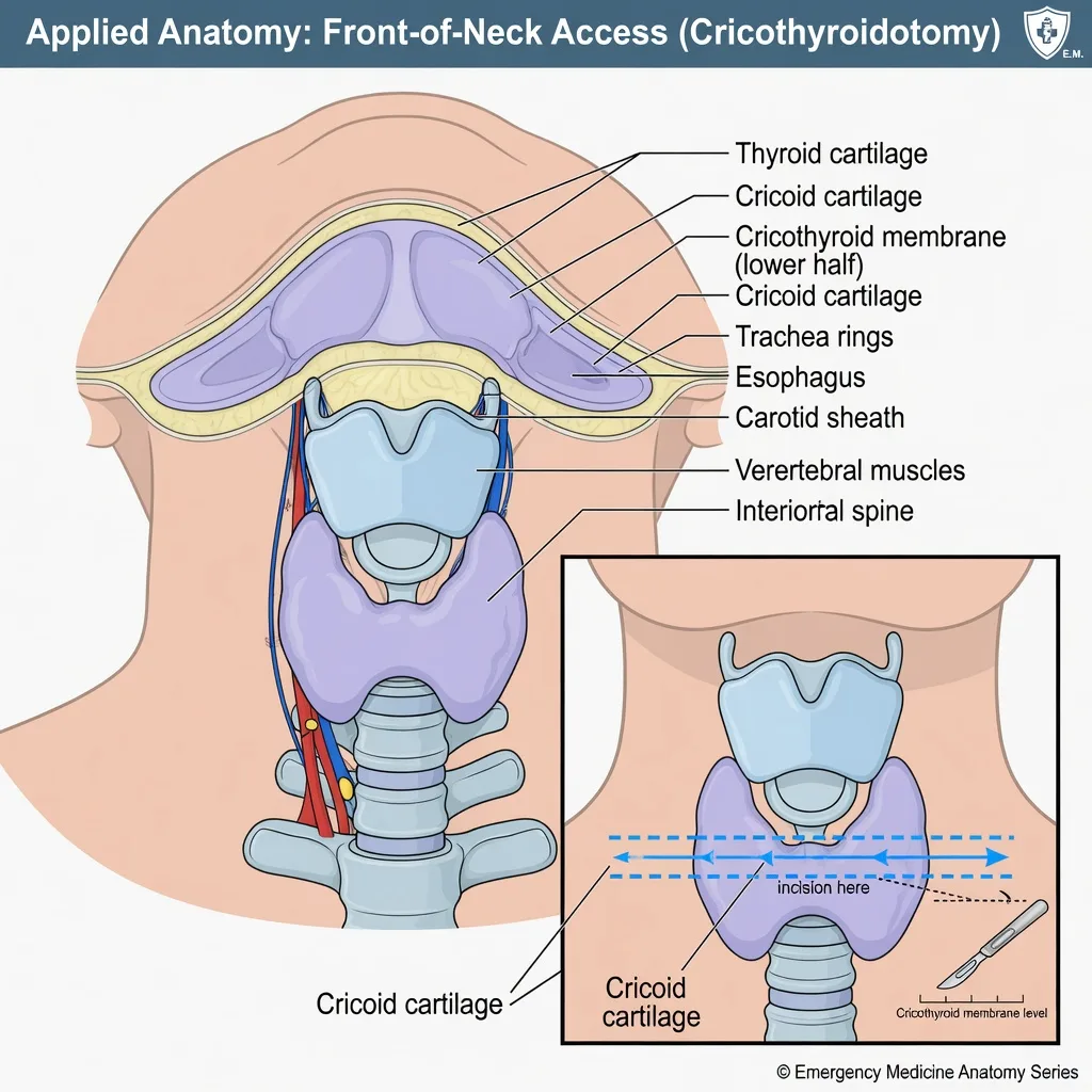
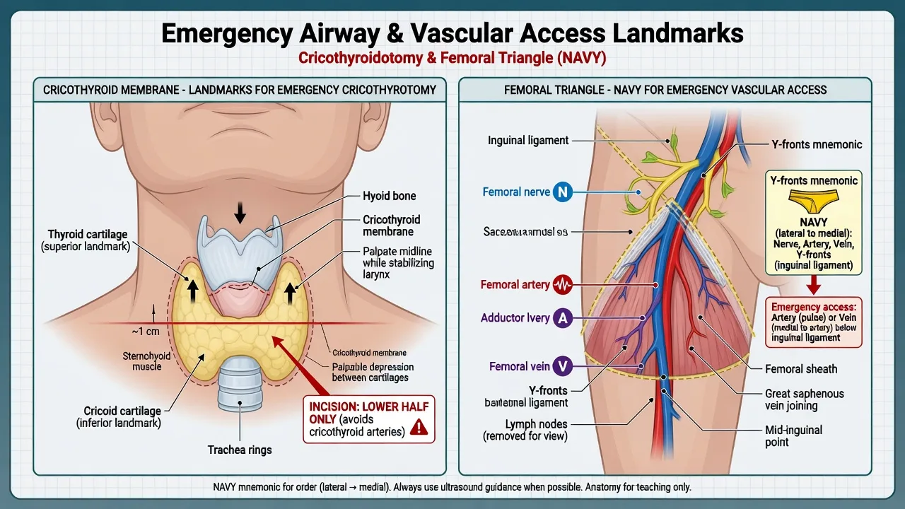
The laryngeal framework is the substrate for both the difficult airway and the emergency surgical airway, and its surface anatomy must be automatic. The thyroid cartilage forms the laryngeal prominence (the Adam's apple) and is the principal anterior landmark; the cricoid cartilage lies immediately below it as a complete signet ring, and the cricothyroid membrane stretches between them in the midline. In the adult the membrane is roughly 8 to 12 millimetres tall and 25 to 30 millimetres wide, and it is the target of the scalpel-bougie-tube cricothyroidotomy performed in the can't-intubate, can't-oxygenate situation.[3] A computed-tomography study of the laryngeal framework confirmed that the membrane is smaller and more variable than the traditional teaching implies, that it is frequently asymmetric, and that in a substantial minority of adults it lies partly or wholly hidden behind the thyroid cartilage promontory — which is why the emergency physician palpates it deliberately and, where possible, marks it with ultrasound before rapid sequence induction.[3]
[1]The vocal cords (the true vocal folds) meet anteriorly at the anterior commissure and form the glottis, the narrowest part of the adult larynx. Their movement is controlled by the intrinsic laryngeal muscles, all of which are supplied by the recurrent laryngeal nerve — a branch of the vagus — except the cricothyroid muscle, which is supplied by the external branch of the superior laryngeal nerve. The recurrent laryngeal nerve's long course (looping under the subclavian on the right and under the aortic arch on the left) makes it vulnerable to tumour, surgery, and thoracic injury. A unilateral injury paralyses the cord in a paramedian position and causes hoarseness; a bilateral injury apposes both cords in the midline and causes stridor and airway obstruction — a post-thyroidectomy patient with stridor has a bilateral recurrent laryngeal nerve injury until proven otherwise. The internal laryngeal nerve (the other branch of the superior laryngeal) carries sensation from above the cords, which is why cricothyroidotomy and awake intubation respect the supraglottic sensitivity.[1]
The vascular system — femoral artery, internal jugular and subclavian veins
Vascular access in the shocked or arrested patient depends on knowing three relationships by heart. The femoral artery lies in the femoral triangle just below the inguinal ligament, within the femoral sheath. The contents of the sheath, from lateral to medial, are recalled as the femoral artery, the femoral vein and the lymphatics; the femoral nerve lies outside the sheath, immediately lateral to the artery — which is why a lateral needle pass can hit the nerve, and why ultrasound guidance is used for arterial and venous cannulation alike. The artery is punctured at the groin crease over the femoral head, where it is compressible against bone, and it is the fallback central and arterial access site when the upper-body vessels are inaccessible. [1]
The internal jugular vein runs deep to the sternocleidomastoid, lateral or anterolateral to the carotid artery, and is cannulated under ultrasound at the apex of the triangle formed by the two heads of sternocleidomastoid. The right internal jugular is preferred because it offers a straight, valveless run to the superior vena cava and right atrium, avoids the thoracic duct (which is on the left), and carries a lower risk of carotid puncture than the landmark technique when ultrasound is used. The subclavian vein runs below the clavicle, anterior to the scalenus anterior muscle (the subclavian artery lies posterior to it, separated by the muscle), and over the first rib; its fixed position keeps it patent in hypovolaemia, but the apex of the lung sits behind it, so pneumothorax is the defining risk and the patient cannot be compressed if coagulopathic.[1]
[1]The nervous system — brachial plexus and the facial nerve
The brachial plexus supplies the upper limb and is the target of the upper-limb regional blocks and the source of the birth and traction injuries that present to the department. It is formed from the ventral rami of C5 to T1, which pass between the anterior and middle scalene muscles (the interscalene groove, the target of the interscalene block), then form trunks, divisions, cords and finally the five terminal branches — the musculocutaneous, axillary, radial, median and ulnar nerves. The interscalene block anaesthetises the shoulder but predictably paralyses the ipsilateral phrenic nerve (C3 to C5 runs on the anterior scalene) and so causes a transient hemidiaphragmatic paresis that is tolerable in most patients but dangerous in the respiratory-compromised; the supraclavicular, infraclavicular and axillary approaches block progressively more distal territory with progressively less phrenic risk. Upper-trunk injury (C5 to C6, Erb's palsy) produces the waiter's-tip posture; lower-trunk injury (C8 to T1, Klumpke's) produces a claw hand and, with a T1 sympathetic injury, a Horner syndrome.[1]
The facial nerve (the seventh cranial nerve) supplies the muscles of facial expression, carries taste from the anterior two-thirds of the tongue, and provides parasympathetic fibres to the lacrimal, submandibular and sublingual glands. After emerging from the stylomastoid foramen it runs forward through the parotid gland — which is why a parotid mass with ipsilateral facial weakness is malignant until proven otherwise, and why the nerve is at risk in parotid surgery and in forceps delivery. The EM-critical distinction is the level of the lesion. A lower motor neurone lesion (Bell's palsy, the parotid malignancy, the stroke in the pontine facial nucleus) paralyses the entire ipsilateral hemiface, forehead included, because the lower facial muscles have only contralateral cortical input. An upper motor neurone lesion (the cortical stroke) spares the forehead, because the upper facial muscles have bilateral cortical input. The patient who cannot wrinkle the forehead on the weak side has a lower motor neurone palsy and needs the workup for the cause; the patient who can wrinkle the forehead has an upper motor neurone lesion and the stroke pathway.[1]
The heart — coronary artery territories, dominance and the conduction system
The coronary anatomy is read off the ECG every shift, and the territory governs both the treatment and the complication. The left main stem divides into the left anterior descending (supplying the anterior wall, the apex, the anterior two-thirds of the septum, and the anterolateral papillary muscle) and the left circumflex (the lateral and posterior walls). The right coronary artery supplies most of the right ventricle, the inferior wall, the posterior third of the septum, and — in the roughly 85 per cent of people in whom it gives off the posterior descending artery — the inferior septum. The artery that supplies the posterior descending artery defines coronary dominance: right-dominant in roughly 85 per cent, left-dominant (the circumflex gives the posterior descending artery) in roughly 8 per cent, and codominant in the remainder. A cadaveric study of the external cardiac vasculature emphasises that the branch pattern is substantially more variable than the schematic implies, with accessory, duplicated and early-branching vessels the rule rather than the exception.[5]
The conduction system is the other cardiac anatomy the examiner expects verbatim. The sinoatrial node sits high in the right atrium near the entry of the superior vena cava and is supplied by the right coronary artery in roughly 60 per cent; the atrioventricular node sits low in the right atrium on the interatrial septum, is the only electrical bridge between atria and ventricles across the otherwise insulating fibrous skeleton, is supplied by the right coronary artery in roughly 90 per cent, and is the reason an inferior infarct causes atrioventricular block. From the atrioventricular node the bundle of His divides into the bundle branches and then the Purkinje fibres. The atrioventricular node is also the substrate of atrioventricular nodal re-entrant tachycardia (dual slow and fast pathways), and an accessory pathway across the fibrous skeleton — as in Wolff-Parkinson-White — bypasses the node and creates the pre-excitation circuit.[1]
The abdomen — biliary tree and the appendiceal positions
The biliary tree begins as the right and left hepatic ducts, which join to form the common hepatic duct; the cystic duct from the gallbladder joins it to form the common bile duct, which runs to the ampulla of Vater and empties through the sphincter of Oddi into the duodenum. The EM-critical anatomy is the Calot triangle (the cystohepatic triangle), bounded inferiorly by the cystic duct, medially by the common hepatic duct, and superiorly by the liver edge. It normally contains the cystic artery (a branch of the right hepatic artery) and the cystic lymph node, and its meticulous dissection is the critical-view-of-safety step in cholecystectomy. The danger is the aberrant or replaced right hepatic artery, which arises from the superior mesenteric artery in roughly 15 to 20 per cent of people and courses through or alongside the triangle — it is the unrecognised structure that bleeds catastrophically and that is injured in biliary and hepatic trauma.[1]
The appendix arises from the posteromedial caecal wall at the convergence of the taeniae coli, a fixed base whose surface marking is McBurney's point (one-third of the way from the anterior superior iliac spine to the umbilicus). The tip is highly variable, and that variability is the whole clinical problem. A computed-tomography study of appendiceal positions found the retrocaecal position to be the commonest (roughly two-thirds), followed by pelvic, subcaecal and preileal positions, with the position determining the clinical picture far more than any other single factor.[4] A retrocaecal appendix irritates the psoas and the ureter and produces flank or back pain that mimics pyelonephritis; a pelvic appendix produces diarrhoea, urinary frequency and rectal or pelvic tenderness and mimics pelvic inflammatory disease; a long appendix reaching the right upper quadrant produces pain and signs indistinguishable from biliary disease and has been reported to present with recurrent right-upper-quadrant pain.[6] A normal right-lower-quadrant examination therefore never excludes appendicitis, and the appendix is localised by imaging, not by the textbook.
Core physiology — Starling forces and capillary fluid exchange
Fluid movement across the capillary wall is governed by the balance of hydrostatic and oncotic pressures set out by Ernest Starling and revised in the light of modern microvascular biology. Net fluid flux is driven by the capillary hydrostatic pressure (pushing fluid out), the plasma oncotic pressure (pulling fluid in, generated almost entirely by albumin), the interstitial hydrostatic pressure and the interstitial oncotic pressure.[1]
[1]The revised Starling principle is the physiology behind every oedema.[1] A raised capillary hydrostatic pressure drives fluid out — the pulmonary and peripheral oedema of heart failure and the pulmonary oedema of over-resuscitation. A fall in plasma oncotic pressure lets fluid escape — the oedema of hypoalbuminaemia from nephrosis, liver failure, sepsis and burns. A fall in the reflection coefficient (the capillary becomes leaky to protein) collapses the oncotic gradient — the leak of sepsis, anaphylaxis and burns. And lymphatic obstruction prevents the return of the filtrate that does escape — the unilateral limb oedema of malignancy and post-surgical lymphoedema. The emergency physician reads these at the bedside: the septic patient is oedematous not from too much fluid alone but from a leaky capillary that cannot hold it, and the diuresis that follows source control is the capillary sealing.[1]
Core physiology — the oxygen-haemoglobin dissociation curve
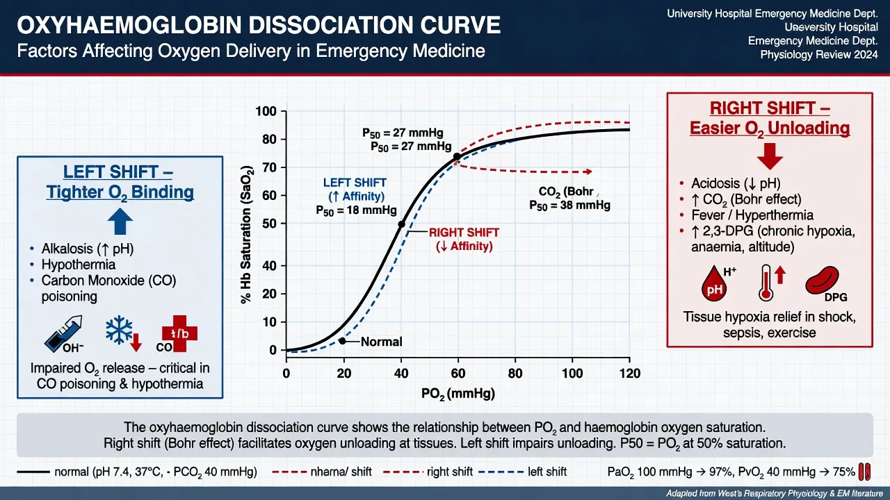
The oxygen-haemoglobin dissociation curve relates the saturation of haemoglobin to the partial pressure of oxygen in blood, and its sigmoid shape is the molecular signature of cooperative binding: each oxygen molecule bound increases the affinity for the next, so haemoglobin loads oxygen avidly in the lung and releases it efficiently at the tissue. The P50 — the partial pressure at which haemoglobin is half-saturated — is about 26 to 27 millimetres of mercury (3.5 kilopascals) at normal pH and temperature. The curve is the physiology of loading and unloading, and the emergency physician manipulates it constantly.[1]
[1]The curve explains two clinical traps. Carbon monoxide poisoning produces a near-normal pulse-oximetry saturation because carboxyhaemoglobin absorbs light like oxyhaemoglobin, yet the oxygen content of the blood is catastrophically low — carbon monoxide both occupies the binding sites and shifts the surviving curve to the left, starving the tissues. Anaemia lowers the oxygen content through the haemoglobin term even when the saturation is perfect: arterial oxygen content is (1.34 times haemoglobin times saturation) plus a negligible dissolved term, so a haemoglobin halved by bleeding halves the content irrespective of the saturation. A normal saturation therefore never excludes hypoxia, and the oxygen content, not the saturation, is what the tissue consumes.[1]
Core physiology — the baroreceptor reflex and the Frank-Starling mechanism
Two linked mechanisms hold the blood pressure and the cardiac output moment to moment, and both are probed in the viva. The baroreceptor reflex is the fast, beat-to-beat control of arterial pressure. High-pressure stretch receptors in the carotid sinus (innervated by the glossopharyngeal nerve) and the aortic arch (innervated by the vagus) fire in proportion to the arterial pressure; a rise in pressure increases their firing, which the medullary cardiovascular centres translate into increased vagal (parasympathetic) outflow and reduced sympathetic outflow, slowing the heart rate, reducing contractility and vasodilating. A fall in pressure does the reverse — reduced firing, withdrawal of vagal tone, sympathetic activation, tachycardia, vasoconstriction and the cold, clammy periphery of compensated shock. The reflex explains the vagal manoeuvres that terminate supraventricular tachycardia (carotid sinus massage and the Valsalva manoeuvre work by transiently raising then releasing the baroreceptor firing), the syncope of carotid sinus hypersensitivity in the older patient, and the orthostatic hypotension of autonomic failure in diabetes and Parkinson disease. The related Bezold-Jarisch reflex — bradycardia, hypotension and vasodilation from ventricular mechanoreceptors — underlies the vasovagal faint, the bradycardia of the inferior infarct, and the sudden collapse of the standing or pregnant patient.[1]
The Frank-Starling mechanism is the intrinsic match of stroke volume to venous return: within the physiological range, an increase in ventricular end-diastolic volume (the preload) stretches the sarcomeres, increases the sensitivity of the contractile proteins to calcium, and increases the force of the next contraction, so stroke volume rises.[2] The heart therefore pumps what it receives, moment to moment, without waiting for a neural signal. The mechanism has a plateau, set by the sarcomere length at which overlap is optimal, and beyond it further stretch reduces force — the descending limb that defines decompensated heart failure.
[1]Differential diagnosis — normal versus variant anatomy
The variant anatomy is not a curiosity; it is the explanation for the atypical presentation, and the Fellowship examiner uses it to test whether the candidate expects it. The recurring theme is that the textbook picture describes the majority, and the dangerous presentation is the minority declaring itself. [1]
Right-dominant circulation (standard)
- The right coronary artery gives the posterior descending artery in roughly 85 per cent of people and supplies the inferior wall, the right ventricle and the atrioventricular node
- An inferior ST-elevation on the ECG is a right coronary artery lesion, nitrates are given with care, and right-sided leads are checked for right ventricular involvement
- The AV node supply is from the RCA, so inferior infarcts cause atrioventricular block — usually transient and nodal, responding to atropine
- The textbook inferior infarct, and the pattern most candidates are taught
Left-dominant circulation (the variant)
- The circumflex artery gives the posterior descending artery in roughly 8 per cent; an inferior infarct is then a circumflex occlusion with posterolateral extension
- The ECG may be subtle or nondiagnostic because the lateral and posterior forces are partially cancelling — the "silent" occlusion
- A left-dominant inferior infarct implies a larger myocardium at risk (inferior plus posterolateral plus AV nodal supply) and a worse prognosis
- The atrioventricular node is supplied by the circumflex, so AV block in this group carries the same prognostic weight as in right-dominant disease
Typical appendix (pelvic, RLQ)
- The classical right-lower-quadrant pain, McBurney point tenderness, migration from the periumbilical region, and the Rovsing and psoas signs
- The picture every candidate recognises, and the easiest diagnosis on the ward
- Ultrasound or CT confirms, and the disposition is straightforward
- The minority of actual appendices — the retrocaecal position is commoner
Retrocaecal appendix (the variant)
- Roughly two-thirds of appendices lie retrocaecally and irritate the psoas and the ureter, producing flank or back pain rather than right-lower-quadrant pain
- Mimics pyelonephritis, with flank tenderness, a positive urinalysis from ureteric irritation, and an absent right-lower-quadrant sign
- A normal right-lower-quadrant examination does not exclude appendicitis — the appendix is localised by imaging
- A long retrocaecal appendix reaching the right upper quadrant presents as recurrent right-upper-quadrant pain indistinguishable from biliary disease
Standard biliary vasculature
- The cystic artery arises from the right hepatic artery within the Calot triangle and is the standard vessel ligated at cholecystectomy
- The right hepatic artery arises from the common hepatic artery in the standard pattern
- Bleeding from the triangle is a cystic artery problem and is controlled at its origin
- The anatomy the surgeon expects and the radiologist reports
Aberrant right hepatic artery (the variant)
- The right hepatic artery is replaced or accessory from the superior mesenteric artery in roughly 15 to 20 per cent and courses through the Calot triangle
- It is mistaken for the cystic artery, ligated or avulsed, and bleeds catastrophically — the unrecognised structure in biliary and hepatic trauma
- A pulsatile structure in the triangle that is larger than expected is the aberrant artery, not the cystic, and is left alone
- The variant that converts a straightforward procedure into a damage-control haemorrhage
Applied procedural landmarks and drug dosing
The anatomy is operationalised at the bedside through the procedures and the drugs that depend on it, and the candidate is expected to give the landmark, the technique and the dose as one answer.[1] For the scalpel-bougie-tube cricothyroidotomy, palpate the cricothyroid membrane in the midline between the thyroid and cricoid cartilages, make a transverse incision through the skin and the lower half of the membrane, confirm tracheal entry with the little finger, pass a bougie, and railroad a size 6.0 cuffed tracheal tube; the membrane is identified by ultrasound in the anticipated difficult airway. For internal jugular and femoral venous access, ultrasound is used in plane or out of plane, the vein is confirmed as compressible and non-pulsatile, and the right internal jugular is the default central line.
The local anaesthetic doses are derived from the innervation mapped above and are the highest-yield drug knowledge in this topic. The maximum safe dose of lidocaine is 3 mg per kilogram plain and 7 mg per kilogram with adrenaline (the adrenaline-induced vasoconstriction slows systemic absorption and raises the ceiling). The maximum dose of bupivacaine is 2 mg per kilogram plain and 2 to 3 mg per kilogram with adrenaline. The total is calculated from the patient's weight and the concentration: 1 per cent lidocaine contains 10 mg per millilitre, so a 70-kilogram adult receives a maximum of 21 millilitres plain or 49 millilitres with adrenaline. Bupivacaine toxicity is particularly dangerous because the drug is highly protein-bound, causes refractory ventricular arrhythmia and cardiac arrest, and is reversed by 20 per cent lipid emulsion at a bolus of 1.5 mL per kilogram followed by an infusion — a dose every emergency physician should be able to give from memory.[1]
Confirming the anatomy — imaging, ultrasound and endoscopy
The anatomy is rarely seen directly in the emergency department; it is inferred through the imaging that confirms the suspected diagnosis. Point-of-care ultrasound confirms the gallbladder (a distended, tender, thick-walled viscus with a stone impacted at the neck, a positive sonographic Murphy sign), the appendix (a non-compressible blind-ending loop over 6 millimetres in diameter in the right lower quadrant), the internal jugular and femoral vessels for access, and the gallbladder and aortic calibre in the shocked patient. Computed tomography with intravenous contrast defines the appendiceal position and the retrocaecal variant, the biliary tree and the aberrant artery, the coronary arteries by angiography, and the source of the obstruction or the perforation. Endoscopic retrograde cholangiopancreatography is both diagnostic and therapeutic for the common bile duct stone and the ascending cholangitis, removing the obstruction at the ampulla. The principle is that the imaging confirms the anatomy the variant has disturbed: the retrocaecal appendix on CT, the aberrant artery on angiography, the dominant circulation on the coronary study.[4][6]
Special populations — paediatric airway, pregnancy and the elderly
The paediatric airway differs from the adult in ways that change every airway manoeuvre. The larynx sits higher in the neck (around C3 to C4 in the infant versus C6 in the adult), the epiglottis is longer, U-shaped and floppy, the tongue is relatively large, and the narrowest point is the cricoid cartilage rather than the vocal cords — which is why uncuffed tubes were traditionally used in the young child and why a slightly oversized tube obstructs at the cricoid ring rather than at the glottis. The large occiput flexes the infant airway forward, so the sniffing position is achieved with a towel under the shoulders, not the head. These differences mean a paediatric difficult airway is anticipated, the equipment is size-matched, and the surgical airway in the young child is a needle cricothyroidotomy rather than a scalpel technique.[1]
In pregnancy, the gravid uterus compresses the inferior vena cava and the aorta from about 20 weeks in the supine position (the aortocaval compression syndrome), reducing venous return, preload, and cardiac output by up to a third, and the emergency response is the left lateral tilt or manual uterine displacement. The elevated diaphragm lowers functional residual capacity and raises the risk of rapid desaturation, while the engorged epidural venous plexus and the laryngeal oedema make the airway friable and difficult — the pregnant airway is a feared airway, and the cricothyroid membrane is harder to find. The elderly patient carries the calcified, tortuous and ectatic vessels that defeat arterial and central lines, the kyphotic chest and the rigid rib cage that alter the surface anatomy, and the smaller, higher-riding or calcified cricothyroid membrane that defeats the surgical airway — the landmarks are reassessed in every older patient, and ultrasound is used liberally.[1]
Common errors and pitfalls
The recurring errors follow the anatomy directly. Incising the upper half of the cricothyroid membrane cuts the cricothyroid artery and turns the rescue airway into a bleeding field — the incision is transverse and low. Mistaking the femoral artery for the vein, or the internal jugular for the carotid, is avoided by ultrasound, but in the shocked patient the vein is small and the artery is mistaken for it; colour Doppler or pulsed-wave confirmation is used when the picture is unclear. Assuming every inferior infarct is a right coronary artery lesion misses the left-dominant circulation, the silent circumflex occlusion, and the worse prognosis. Discharging a right-lower-quadrant-painless patient with a retrocaecal appendix is the classic appendiceal miss; the appendix is imaged, not assumed. Ligating the aberrant right hepatic artery as if it were the cystic is the biliary catastrophe. Reading the bell's-palsy face as a stroke (or the stroke face as a Bell's) is the facial-nerve error — the forehead is the discriminator. Over-resuscitating past the Frank-Starling plateau converts a treatable shock into pulmonary oedema; the response to fluid is tested, not assumed. And treating a normal oxygen saturation as proof of adequate oxygen delivery misses the carbon-monoxide-poisoned and the profoundly anaemic patient, in both of whom the content, not the saturation, is the problem.[1][1]
Evidence and regional guidelines
The evidence base for applied anatomy and physiology is the primary anatomical and physiological literature anchored to the standard emergency textbooks. Levick's review of microvascular fluid exchange is the definitive statement of the revised Starling principle and the physiology of oedema.[1] Han and colleagues' work on cardiac efficiency and Starling's law frames the Frank-Starling mechanism in modern molecular terms.[2] Sagiv and colleagues' computed-tomography study of the laryngeal framework is the contemporary reference for the dimensions and the variability of the cricothyroid membrane that governs the emergency surgical airway.[3] Singh and colleagues' computed-tomography study of the appendiceal positions quantifies the retrocaecal, pelvic and subcaecal variants that underlie the atypical presentation.[4] Lane and colleagues' cadaveric study of the external cardiac vasculature documents the coronary branch variability that the dominance concept only approximates.[5] Ferrie and colleagues' case of a retrocaecal appendix presenting with right-upper-quadrant pain is the clinical illustration of the appendiceal variant.[6] Roberts and Hedges' Clinical Procedures in Emergency Medicine is the standard procedural reference for the landmark-based technique and the local anaesthetic doses.[1] Guyton and Hall's Textbook of Medical Physiology is the standard physiology reference for the oxygen-haemoglobin dissociation curve, the baroreceptor reflex and the conduction system.[1]
ANZ practice note. The Australasian College for Emergency Medicine expects the Fellowship candidate to defend the surface anatomy for the front-of-neck access and the central and arterial lines from first principles, and the ACEM Policy on the Emergency Surgical Airway and the ANZCA airway guidance inform the technique. Ultrasound guidance for vascular access and for the difficult airway is the Australasian standard, and the difficult-airway societies (the Australian and New Zealand Airway Emergency Group) promote pre-procedural ultrasound marking of the cricothyroid membrane. The local anaesthetic doses and the lipid-emulsion protocol follow the ANZCA and the Australian Resuscitation Council guidance. [1]
Exam pearls
- The cricothyroid membrane is palpated and ultrasound-marked before it is needed — it is smaller, higher-riding and more variable than the textbook implies, and the incision is transverse and low to avoid the cricothyroid artery.
- An inferior ST-elevation is a right coronary artery lesion only in the right-dominant majority — suspect the left-dominant circumflex occlusion in the patient with a subtle or nondiagnostic ECG and inferior signs.
- The atrioventricular node is supplied by the right coronary artery in roughly 90 per cent, which is why the inferior infarct causes atrioventricular block — and why the right ventricle, also from the RCA, must be checked with right-sided leads before any nitrate is given.
- A normal right-lower-quadrant examination never excludes appendicitis — the retrocaecal appendix is the commonest position and produces flank, back or right-upper-quadrant pain.
- The aberrant right hepatic artery from the superior mesenteric artery runs through the Calot triangle in roughly one in six patients — it is the unrecognised structure that bleeds catastrophically in biliary and hepatic procedures.
- The forehead is the discriminator on the weak face — a lower motor neurone (Bell's) palsy paralyses the forehead, an upper motor neurone (stroke) palsy spares it.
- Arterial oxygen content is (1.34 times haemoglobin times saturation) plus a negligible dissolved term — a normal saturation never excludes hypoxia in carbon monoxide poisoning or in anaemia, because the content, not the saturation, is what the tissue consumes.
- The fluid bolus helps the patient on the ascending limb of the Frank-Starling curve and drowns the patient on the descending limb — test the response with a passive leg raise, do not resuscitate by rote. [1]
SAQ — The predicted-difficult airway progressing to can't-intubate-can't-oxygenate: front-of-neck access anatomy
10 minutes · 10 marks
A 58-year-old man with ankylosing spondylitis, a fixed cervical flexion deformity and severe rheumatoid arthritis of the temporomandibular joints is brought to the emergency department after a fall. He is in type 1 respiratory failure from pneumonia, with SpO2 86 per cent on 15 L oxygen, RR 32 and exhaustion. After a thorough airway assessment you predict a difficult airway, position him with cushions and induce anaesthesia with careful pre-oxygenation. Both direct and video laryngoscopy yield a Cormack-Lehane grade 4 view, a bougie and a second-generation supraglottic airway both fail, and oxygen saturations fall to 58 per cent despite optimal two-person bag-valve-mask ventilation with oral and nasal airways. You proceed to a front-of-neck access.
SAQ — Penetrating precordial trauma with tamponade: cardiac anatomy in the injured mediastinum
10 minutes · 10 marks
A 26-year-old man is brought to the trauma bay 12 minutes after a single stab wound to the precordium, the entry 1 cm left of the sternal edge in the fourth intercostal space. He is agitated and cold, GCS 14, BP 76/52 with a narrowed pulse pressure of 24 mmHg, HR 134, JVP distended to the angle of the jaw at 45 degrees, muffled heart sounds, and equal air entry bilaterally. The extended FAST shows a large pericardial effusion with diastolic collapse of the right ventricle. He loses cardiac output on arrival to the bay.
Red flags
[1]References
- [1]Levick JR. Microvascular fluid exchange and the revised Starling principle Cardiovasc Res, 2010.PMID 20200043
- [2]Han JC, et al. Cardiac efficiency and Starling's Law of the Heart J Physiol, 2022.PMID 35998082
- [3]Sagiv D, et al. Novel Anatomic Characteristics of the Laryngeal Framework: A Computed Tomography Evaluation Otolaryngol Head Neck Surg, 2016.PMID 26861235
- [4]Singh N, et al. Computed tomography evaluation of variations in positions and measurements of appendix in patients with non-appendicular symptoms: time to revise the diagnostic criteria for appendicitis Pol J Radiol, 2023.PMID 37808175
- [5]Lane CP, et al. Anatomical variation in the external vasculature of the human heart: A cadaveric investigation of dominance in coronary artery branching and cardiac venous drainage Ann Anat, 2026.PMID 41161614
- [6]Ferrie A, et al. A Retrocaecal Appendix Presenting With Recurrent Right Upper Quadrant Pain (RUQ): A Rare Presentation of a Rather Common Surgical Pathology Cureus, 2025.PMID 41189815