EM · ABG interpretation (approach)
Arterial blood gas interpretation — the systematic emergency department approach
Also known as ABG interpretation · Blood gas analysis · Acid-base interpretation · Arterial blood gas
The systematic approach to the arterial blood gas for the emergency medicine trainee — the six values (pH, PaO2, PaCO2, HCO3, base excess, lactate), the respiratory-versus-metabolic rule (pH and PaCO2 move opposite in respiratory disorders, together in metabolic), the compensation formulas (Winter's, the respiratory rules), the anion gap Na minus Cl plus HCO3, the delta-delta for mixed disorders, and the venous-versus-arterial comparison. Includes the gas-driven drug doses — sodium bicarbonate 8.4 percent, insulin 0.1 units per kg per hour, fomepizole 15 mg per kg, naloxone 400 micrograms. ACEM-primary, globally tagged.
On this page & tools
Your progress
Saved locally on this device.
Target exams
Red flags
The arterial blood gas is the highest-yield point-of-care test in the emergency department. A single sample read in under two minutes quantifies oxygenation, ventilation, the acid-base status and the lactate, and it can trigger intubation, non-invasive ventilation, an antidote, or an insulin infusion before any other result returns. The Fellowship examiner is not testing whether a candidate can recognise one favourite gas; the test is whether a candidate can apply a reproducible systematic method to any blood gas, in any patient, under pressure, and not miss a mixed disorder or an incompletely compensated one. Pattern recognition fails on the unusual gas; a system does not. This topic is the diagnostic-skill framework, not a disease. [1]
When to use the framework and the principles of a systematic read
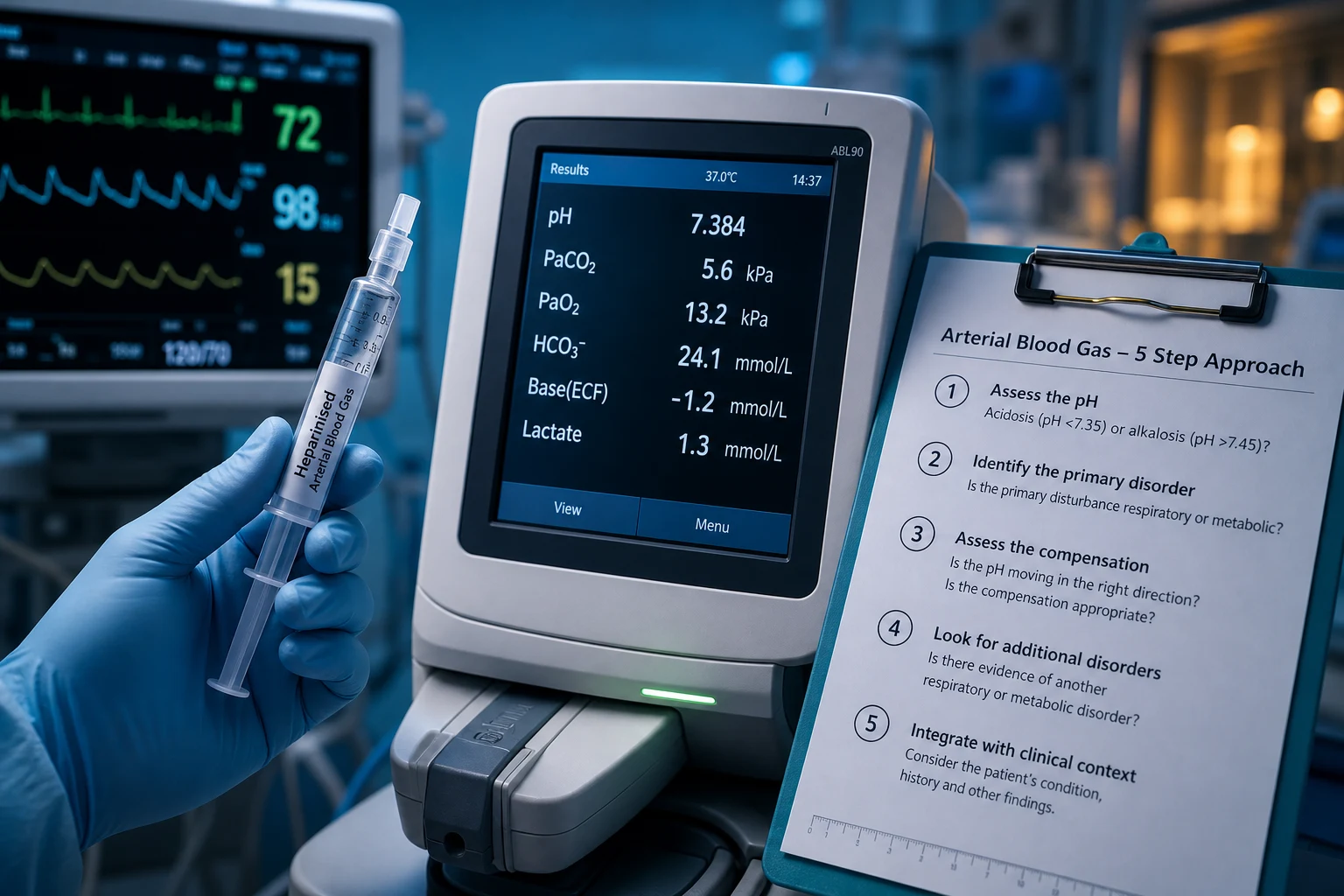
An arterial (or venous) blood gas is obtained on any patient in respiratory distress or respiratory failure, any shocked or septic patient, any patient with diabetic ketoacidosis or a suspected toxic ingestion, any patient with a reduced conscious level, and any patient in or near peri-arrest. The gas is interpreted by the treating clinician at the bedside, not deferred to the laboratory, and the result is acted on in parallel with resuscitation — an unstable patient with a pH under 7.1 is treated before every value is parsed. Three principles govern the read.[3] First, run the same ordered sequence every time so that no compartment is skipped under cognitive load. Second, treat the patient, not the number: a lactate of 6 mmol per litre is meaningless without the cause, and a "normal" gas does not exclude a sick patient. Third, always finish the sequence — the commonest error is to stop at the first abnormality and miss the second, third, or fourth disorder hiding in the same gas.[10]
The sample, the units, and the normal values
The radial artery is the first-choice site because it is superficial, compressible, and has collateral flow from the ulnar artery; a modified Allen's test confirms the collateral circulation before sampling, and ultrasound guidance improves success and reduces complications. The brachial and femoral arteries are alternatives in the shocked or oedematous patient, and the femoral is the pragmatic default in cardiac arrest. A heparinised syringe is used, the air bubble is expelled to avoid a falsely elevated PaO2, and the sample is analysed within 15 minutes or transported on ice. Local anaesthesia — lignocaine 1 percent infiltrated at the puncture site — reduces pain and does not impair sampling. In Australia and New Zealand blood gases are reported in kilopascals (kPa); the conversion to millimetres of mercury (mmHg) is to multiply by 7.5, so PaCO2 of 5.3 kPa equals 40 mmHg. [1]
The normal arterial blood gas values
The physiology that underpins acid-base
Acid-base homeostasis is governed by the Henderson–Hasselbalch relationship: pH is set by the ratio of bicarbonate to dissolved carbon dioxide, not by either value alone. Carbon dioxide is a volatile acid generated by metabolism and excreted in the breath by the lung, so its accumulation or loss is the respiratory limb of acid-base. Bicarbonate is the renal buffer, regenerated over hours to days, and its depletion or excess is the metabolic limb. A change in PaCO2 therefore produces a respiratory disorder, and a change in HCO3 (or the base excess, which mirrors it) produces a metabolic disorder. The two systems compensate for each other: a primary metabolic acidosis drives hyperventilation to lower PaCO2, and a primary respiratory acidosis drives renal bicarbonate retention. The compensation is predictable and quantifiable, which is why every interpretation ends with a formula.[3]
Step 1 — pH: acidosis, alkalosis, or normal
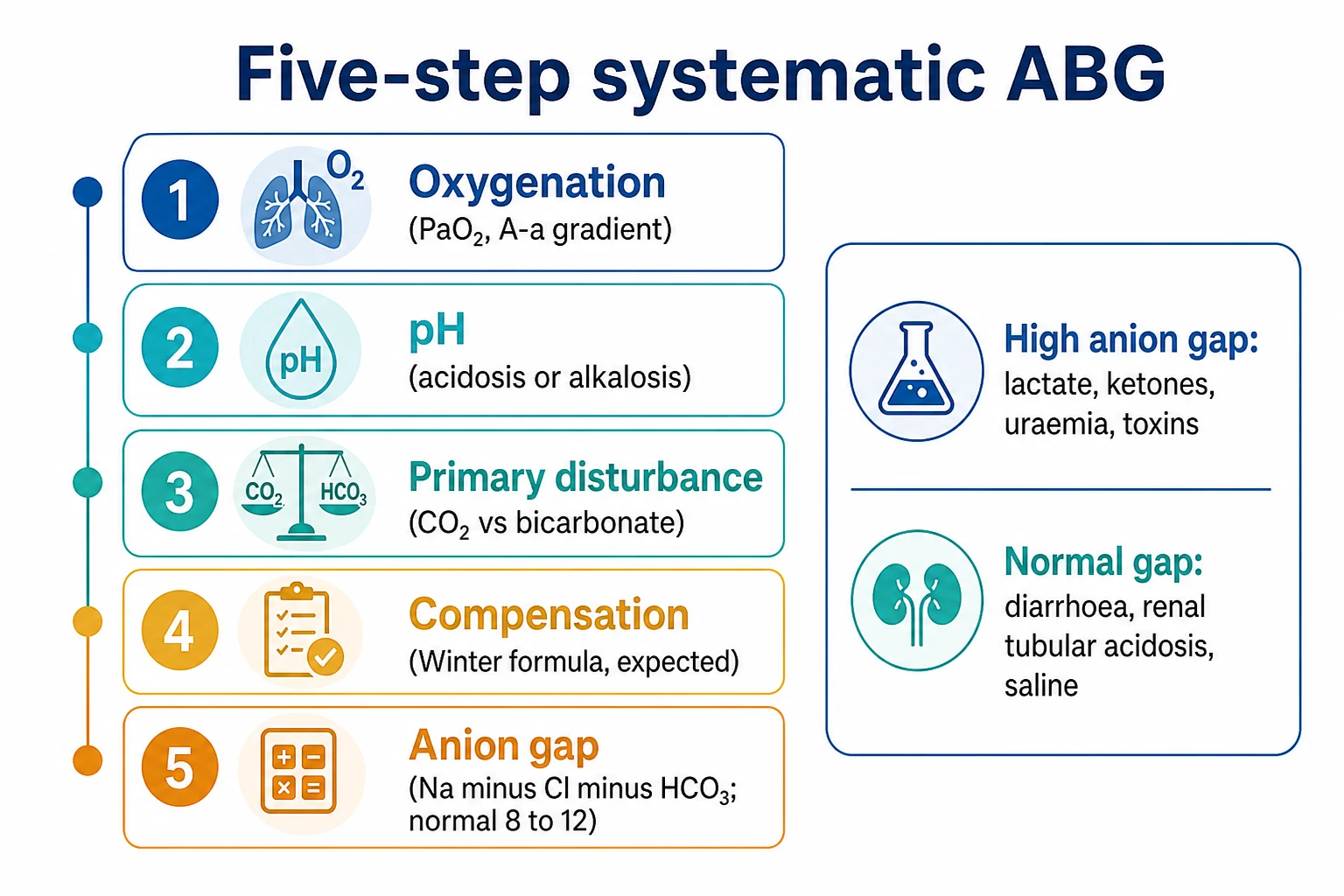
The first value read is the pH. A pH under 7.35 is an acidaemia, indicating that the net process in the blood is acid accumulation; a pH over 7.45 is an alkalaemia. A normal pH does not exclude an acid-base disorder — a compensated disorder, or two opposing disorders, can hold the pH inside the normal range, and the diagnosis is then read from the PaCO2 and the HCO3. The pH is also the first prognostic marker: a pH under 7.1 with haemodynamic instability is a medical emergency and triggers the gas-driven drug doses set out below.[2]
Step 2 — The primary disorder: respiratory or metabolic
The second step decides whether the primary disorder is respiratory or metabolic, using a single rule. If the pH and the PaCO2 move in opposite directions, the primary disorder is respiratory; if they move in the same direction, the primary disorder is metabolic.[3] A low pH (acidosis) with a high PaCO2 is a respiratory acidosis from alveolar hypoventilation; a low pH with a low PaCO2 is a metabolic acidosis, and the low PaCO2 is the compensatory hyperventilation. A high pH (alkalosis) with a low PaCO2 is a respiratory alkalosis from hyperventilation; a high pH with a high PaCO2 is a metabolic alkalosis with compensatory hypoventilation. The HCO3 (or base excess) confirms the metabolic limb: a low HCO3 with a low pH is metabolic acidosis; a high HCO3 with a high pH is metabolic alkalosis. Once the primary disorder is named, the next step tests whether the compensation is appropriate.
[1]Step 3 — Compensation: the predicted PaCO2 or HCO3
Compensation is judged by comparing the measured value to the value predicted by a formula, not by eyeballing it. A measured value within the predicted range is appropriate compensation; a value outside it indicates a second, mixed disorder.[2] The formulas are committed to memory because they are scored in the written paper and the viva.
Step 4 — The anion gap: raised or normal
The fourth step is the anion gap, calculated from the electrolytes on the same sample. Anion gap = Na − (Cl + HCO3). The normal range is 8 to 12 mmol per litre.[4] A raised anion gap means an unmeasured anion is present — lactate, ketones, a toxin or its metabolite, sulphate, oxalate, or phosphate — and it sorts the metabolic acidoses into the two diagnostic families: high-anion-gap metabolic acidosis (HAGMA) and normal-anion-gap (hyperchloraemic) metabolic acidosis (NAGMA). The mnemonic GOLD MARK (Glycols, Oxoproline, L-lactate, D-lactate, Methanol, Aspirin, Renal failure, Ketoacidosis) is the modern list; the older MUDPILES (Methanol, Uraemia, DKA, Paraldehyde, Iron/INH, Lactic acidosis, Ethylene glycol, Salicylates) is equally acceptable in the exam.[4] The normal-anion-gap acidoses are the chloride-excess acidoses: diarrhoea, large-volume saline resuscitation, renal tubular acidosis, and ureteric diversion.
High-anion-gap metabolic acidosis — GOLD MARK
GOLD MARK
Ethylene glycol, propylene glycol; calcium oxalate crystals in the urine, raised osmolar gap
5-oxoproline toxicity from chronic paracetamol, especially in malnutrition
Sepsis, shock, mesenteric ischaemia, metformin; the commonest cause in the ED
Short-bowel syndrome, bacterial overgrowth
Formate toxicity; raised osmolar gap, blurry vision, basal ganglia infarct
Salicylate; early respiratory alkalosis, then mixed high-anion-gap acidosis
Uraemia; sulphate and phosphate accumulate
Diabetic, alcoholic, starvation; ketones positive
Step 5 — The delta-delta: the search for mixed disorders
The fifth step is the delta-delta, the tool that unmasks the second metabolic disorder hiding inside the first. It asks whether the fall in bicarbonate is fully accounted for by the rise in the anion gap. The delta ratio = (anion gap − 12) ÷ (24 − HCO3).[10] A ratio of 1 to 2 is a pure high-anion-gap metabolic acidosis: every mmol of unmeasured anion has consumed a mmol of bicarbonate. A ratio under 1 means the bicarbonate has fallen further than the anion gap has risen, so a normal-anion-gap acidosis is layered on top — the classic example is septic shock (lactic acidosis) combined with renal failure or diarrhoea (hyperchloraemic acidosis). A ratio over 2 means the bicarbonate has fallen less than expected, so a metabolic alkalosis is coexisting — the patient with vomiting and uraemia, or DKA with volume contraction. The corrected bicarbonate (corrected HCO3 = HCO3 + (anion gap − 12)) gives the same answer by a second route: a corrected HCO3 over 26 means a coexisting metabolic alkalosis, under 22 a coexisting metabolic acidosis.[10]
[1]Step 6 — The osmolar gap and the toxidromes
The sixth step is invoked when a toxic alcohol or another small solute is suspected. The osmolar gap = measured osmolality − calculated osmolality, where calculated osmolality = 2 × Na + glucose + urea (in mmol per litre). A normal osmolar gap is under 10. A raised anion-gap metabolic acidosis combined with a raised osmolar gap is a toxic alcohol (methanol or ethylene glycol) until proven otherwise, and fomepizole is started before the level returns.[2] Salicylate poisoning produces a characteristic mixed disorder — an early respiratory alkalosis (direct stimulation of the medulla) combined with a high-anion-gap metabolic acidosis (uncoupling of oxidative phosphorylation and accumulation of salicylate, lactate, and ketones) — and the diagnosis is confirmed at the bedside by a salicylate level.
Step 7 — Lactate, oxygenation, and the parallel reads
The seventh step reads the lactate and the PaO2 alongside the acid-base. A lactate at or above 2 mmol per litre is hyperlactataemia; at or above 4 mmol per litre it is severe and, in the septic patient, it signals a high mortality and triggers the sepsis bundle.[5] Lactate accumulates whenever oxygen delivery fails to meet demand (type A, from shock, hypoxia, seizures, mesenteric ischaemia, or exercise) or when aerobic metabolism is deranged (type B, from metformin, malignancy, or toxins); the type A causes dominate the emergency department.[5] The PaO2 is read against the inspired oxygen: a PaO2 under 8 kPa (60 mmHg) on room air is type 1 respiratory failure, and a PaCO2 over 6.5 kPa (50 mmHg) is type 2 respiratory failure. The A-a gradient — calculated as PAO2 − PaO2, where PAO2 = FiO2 × (760 − 47) − PaCO2 divided by 0.8 — distinguishes hypoxaemia from a low inspired oxygen (normal gradient) from hypoxaemia from a lung problem (raised gradient).
Venous versus arterial — when the venous gas is enough
The venous blood gas is faster, safer, and less painful than the arterial, and for most ED questions it is sufficient. The venous pH is on average 0.02 to 0.04 lower than the arterial; the venous PaCO2 is 0.5 to 0.8 kPa (4 to 8 mmHg) higher; the venous HCO3 and base excess are within 2 of the arterial; and the venous lactate and electrolytes are clinically equivalent.[8] The venous gas is therefore reliable for screening, for monitoring the DKA or septic patient, and for tracking the lactate trend. The venous gas is not reliable for the PaO2 (which is meaningless in a venous sample) or for the precise PaCO2 in respiratory failure — whenever hypercapnia is suspected and non-invasive ventilation is being considered, an arterial sample is taken to confirm.[8]
Arterial blood gas
- The reference standard — accurate PaO2, PaCO2, pH, HCO3, base excess, and lactate
- Painful; small risk of haematoma, thrombosis, distal ischaemia, and infection
- Required for: suspected hypercapnia before NIV, exact PaO2 in respiratory failure, CO poisoning (carboxyhaemoglobin)
- Also required when the venous gas is discordant with the clinical picture
Venous blood gas
- Drawn from any cannula — quick, safe, and repeatable
- pH within 0.03, HCO3 within 2, base excess within 2, lactate equivalent to arterial
- Use for screening, for septic and DKA monitoring, and for the lactate trend
- Unreliable for PaO2 and for the precise PaCO2 in respiratory failure
Capillary blood gas
- Used in neonates and small children — heel or earlobe sample
- Correlates well with arterial pH and HCO3, less well with PaO2
- Useful for serial monitoring in paediatric DKA and bronchiolitis
- Warm the heel first to arterialise the sample
Differential diagnosis — the gas pattern by disorder
The blood gas does not give a diagnosis; it gives a pattern, and the pattern points to a differential. The four commonest abnormal patterns each carry a short list of can't-miss causes. [1]
High-anion-gap metabolic acidosis
- Lactic acidosis — sepsis, shock, mesenteric ischaemia, metformin, seizures
- Ketoacidosis — diabetic (hyperglycaemia, ketones), alcoholic, starvation
- Renal failure — uraemia with retained sulphate and phosphate
- Toxins — methanol, ethylene glycol, salicylate, paraldehyde, 5-oxoproline (paracetamol)
Normal-anion-gap (hyperchloraemic) acidosis
- Diarrhoea — bicarbonate loss through the gut
- Large-volume normal saline resuscitation — hyperchloraemia suppresses bicarbonate
- Renal tubular acidosis — failure to excrete acid or reclaim bicarbonate
- Ureteric diversion (sigmoid loop) — reabsorption of chloride and ammonium
Respiratory acidosis (high PaCO2, low pH)
- Central depression — opioids, sedatives, brainstem stroke
- Neuromuscular weakness — Guillain–Barré, myasthenia, neuromuscular blockers
- Chest-wall failure — kyphoscoliosis, flail chest, obesity hypoventilation
- Airway obstruction and COPD exacerbation — the commonest in the ED
Respiratory alkalosis (low PaCO2, high pH)
- Salicylate poisoning — early and direct medullary stimulation
- Sepsis, pneumonia, pulmonary embolism, asthma — hypoxia- or pain-driven hyperventilation
- Pregnancy (progesterone-driven) — a normal finding, PaCO2 3.7 to 4.2 kPa
- Anxiety and panic — a diagnosis of exclusion after the organic causes are sought
The mixed disorder is the recurring trap and earns the most marks when named unprompted. Salicylate poisoning is the classic mixed respiratory alkalosis with high-anion-gap metabolic acidosis. Sepsis combines a high-anion-gap acidosis (lactate) with a respiratory alkalosis (hyperventilation), and when complicated by renal failure or diarrhoea it adds a normal-anion-gap acidosis — a triple disorder read from the delta ratio.[10] Vomiting adds a metabolic alkalosis to any primary metabolic acidosis, seen in DKA with volume contraction and in the patient with a small-bowel obstruction.
Management driven by the gas result
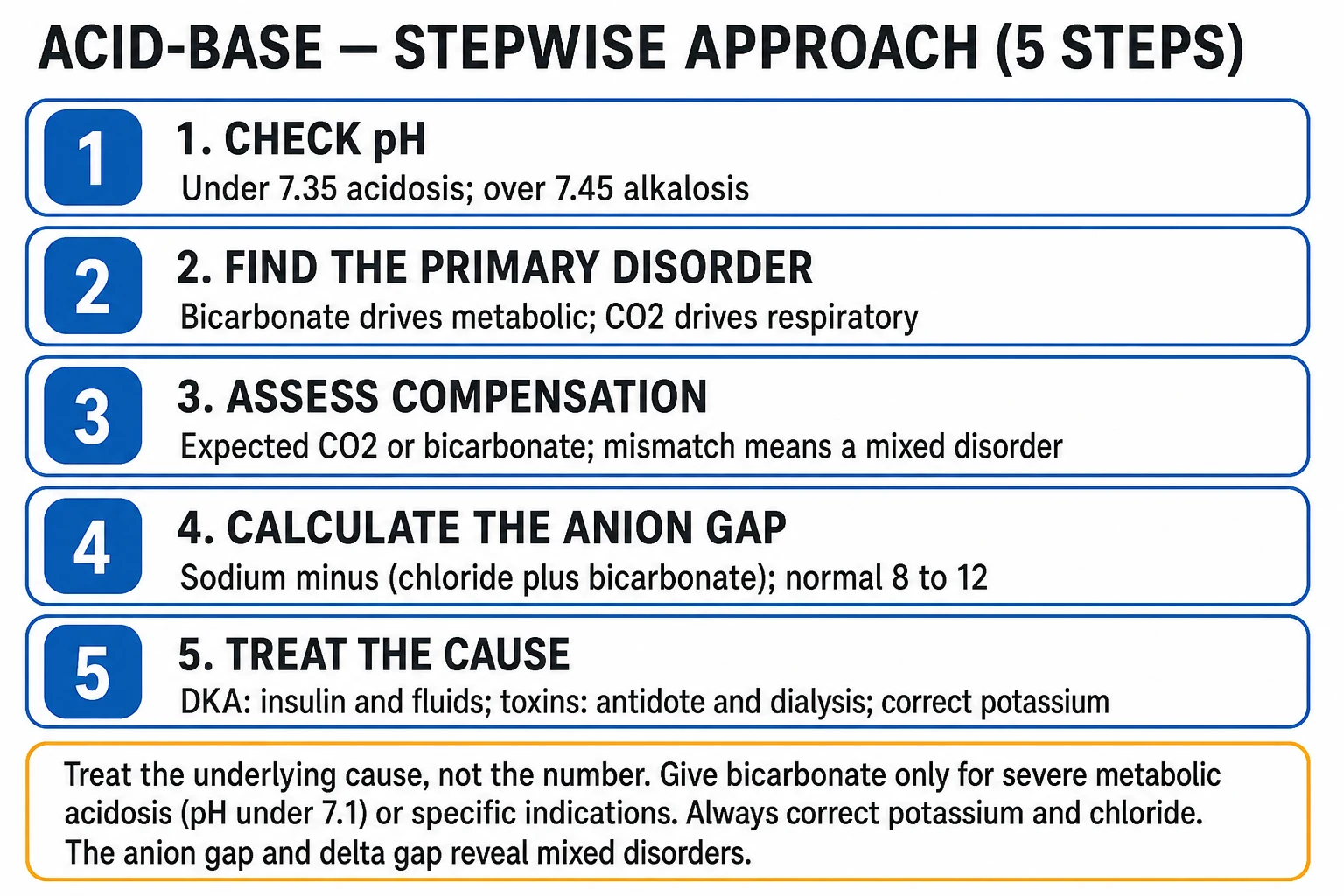
The gas does not stop at diagnosis; several results immediately trigger a drug. The doses are committed to memory because the unstable patient cannot wait for a lookup. The overriding principle is to treat the cause, not the number — sodium bicarbonate corrects the pH but does not fix the sepsis or the methanol, and over-correction carries its own harm.[2]
The gas-driven emergency drug doses
Special populations
The COPD patient is a chronic carbon-dioxide retainer with a baseline high PaCO2 and a compensatory high HCO3; the target saturation is 88 to 92 percent to avoid suppressing the hypoxic drive and precipitating hypercapnic respiratory failure, and a gas showing a pH under 7.35 with a high PaCO2 indicates an acute-on-chronic respiratory acidosis treated with controlled oxygen and non-invasive ventilation. The chronic kidney disease patient runs a chronic high-anion-gap metabolic acidosis with a low baseline HCO3, and a small further fall signals acute deterioration. Pregnancy produces a mild respiratory alkalosis as a normal finding — progesterone-driven hyperventilation lowers PaCO2 to 3.7 to 4.2 kPa, with a compensatory low HCO3 — and a PaCO2 of 5.0 kPa in the third trimester is, in effect, hypercapnia. The paediatric gas accepts slightly lower PaO2 values, the neonate runs a lower baseline pH, and the capillary (heel-prick) gas is the practical monitoring tool. The post-cardiac-arrest patient is intentionally kept at a PaCO2 in the normal range — both hypocapnia (cerebral vasoconstriction) and hypercapnia (raised intracranial pressure) are avoided. [1]
Common errors and pitfalls
The recurring errors are well described. Treating a venous gas as arterial and acting on the PaO2 — the venous PaO2 is meaningless. Stopping at the first disorder and missing the mixed one — the delta ratio is run on every raised anion gap. Declaring "compensated metabolic acidosis" without applying Winter's formula — the formula decides whether the compensation is appropriate, and a discordant value is a second disorder. Giving bicarbonate for a moderate metabolic acidosis — it does not improve outcome outside the BICAR-ICU subgroup and can worsen intracellular acidosis and hypocalcaemia.[7] Failing to measure the lactate — lactate is the single most prognostic value on the gas and is missed if the panel is not requested. Sampling from a heparin-contaminated syringe, which falsely lowers sodium and bicarbonate; failing to expel the air bubble, which falsely raises PaO2; or delaying analysis, which lowers pH and raises PaCO2 and lactate.[3] Calling a primary respiratory disorder "compensated metabolic" by misreading the direction rule. And forgetting the temperature — in hypothermia the gas is interpreted uncorrected, the value the patient's tissues actually see.
ANZ practice note. Blood gases in Australia and New Zealand are reported in kilopascals for the partial pressures (multiply by 7.5 for mmHg) and in mmol per litre for the bicarbonate, base excess, and lactate. The systematic six-step read — pH, primary disorder, compensation, anion gap, delta-delta, lactate and oxygenation — is the expected viva method, and the compensation formulas are scored verbatim. Sodium bicarbonate is reserved for severe metabolic acidaemia with instability; insulin and fomepizole are the gas-driven antidotes for DKA and toxic alcohol respectively. The venous gas is accepted for screening, septic and DKA monitoring, and the lactate trend; an arterial sample is taken to confirm hypercapnia before non-invasive ventilation and to measure PaO2 in respiratory failure. [1]
Evidence and regional guidelines
The compensation formulas trace to Albert, Dell, and Winters' original derivation in 1967.[1] The two-part review by Adrogué and Madias in the New England Journal of Medicine (1998) remains the standard reference for the management of life-threatening acid-base disorders.[2] The physiological approach to assessment — the pH–PaCO2 direction rule, the predicted-compensation formulas, and the anion-gap and delta-ratio framework — is laid out in Berend and colleagues' New England Journal of Medicine review.[3] The interpretation and limitations of the anion gap are detailed by Kraut and Madias.[4] The modern approach to lactic acidosis — including the type A versus type B distinction and the prognostic weight of the lactate — is from Kraut and Madias (2015).[5] The DKA protocol and its insulin 0.1 units per kg per hour infusion are from the Kitabchi consensus.[6] The role of sodium bicarbonate in severe metabolic acidaemia is informed by the BICAR-ICU randomised trial, which restricted benefit to the subgroup with a pH under 7.2.[7] The venous-versus-arterial equivalence for pH, bicarbonate, base excess, and lactate is supported by Middleton's agreement study.[8] Lactate-guided resuscitation in the critically ill was tested by Jansen and colleagues.[9] The framework for recognising mixed disorders through the anion gap and the delta ratio was set out by Narins.[10]
Exam pearls
- Run the six-step sequence on every gas: pH, primary disorder, compensation, anion gap, delta-delta, lactate and oxygenation. The method is the mark, not the one-liner.
- The direction rule: pH and PaCO2 opposite = respiratory; pH and PaCO2 together = metabolic. This single test sorts any gas in seconds.
- Quote the formula before declaring compensation. Winter's: PaCO2 = 1.5 × HCO3 + 8 (±2). Respiratory acidosis: HCO3 rises 1 per 10 mmHg acutely, 4 chronically.
- Anion gap = Na − (Cl + HCO3); normal 8 to 12. Raised = GOLD MARK; normal = diarrhoea, saline, renal tubular acidosis.
- Delta ratio = (AG − 12) ÷ (24 − HCO3). One to two pure HAGMA; under one adds NAGMA; over two adds metabolic alkalosis.
- Venous pH, HCO3, base excess and lactate ≈ arterial. Use the venous gas for screening and trends; take the arterial gas for PaO2 and for hypercapnia before NIV.
- Treat the cause, not the number. Bicarbonate 8.4 percent 1 to 2 mmol per kg is reserved for pH under 7.1 with instability; insulin 0.1 units per kg per hour for DKA; fomepizole 15 mg per kg for the toxic alcohol.
- In hypothermia, interpret the gas uncorrected. The temperature-corrected value is a mathematical fiction; treat the patient in front of you. [1]
Exam practice
SAQ — Acute respiratory acidosis with lactic acidosis in COPD sepsis
10 minutes · 10 marks
A 72-year-old man with severe COPD (FEV1 35 percent predicted, on home oxygen 2 L per minute, long-term tiotropium and salbutamol) presents with three days of purulent sputum, worsening breathlessness, and increasing drowsiness. He is a 50 pack-year smoker. On arrival: GCS 13 (E3V4M6), temperature 38.6 degrees C, HR 128, BP 88/56 (MAP 60), RR 30 with accessory muscle use, SpO2 84 per cent on room air. Chest auscultation reveals widespread wheeze and right basal crackles. ABG on 2 L per minute nasal cannula: pH 7.10, PaCO2 10.6 kPa (80 mmHg), PaO2 6.8 kPa (51 mmHg), HCO3 18 mmol/L, base excess minus 9, lactate 5.2 mmol/L, sodium 138, chloride 98, potassium 5.4.
SAQ — Delta gap in DKA with vomiting: unmasking the hidden metabolic alkalosis
10 minutes · 10 marks
A 22-year-old woman with type 1 diabetes presents with two days of nausea, profuse vomiting (10 to 12 episodes), diffuse abdominal pain, and polyuria. She has not taken her insulin for 48 hours due to the vomiting. She is drowsy but rousable (GCS 14), markedly dehydrated with dry mucosae and sunken eyes, deep sighing Kussmaul respirations at 30 per minute, HR 126, BP 98/62 with a postural drop to 78/44, and a fruity breath odour. Capillary glucose reads high on the meter. Venous blood gas: pH 7.22, HCO3 17 mmol/L, base excess minus 10, glucose 29 mmol/L, beta-hydroxybutyrate 5.8 mmol/L, lactate 2.0 mmol/L, sodium 132, chloride 86, potassium 5.1. Urinalysis: 4+ glucose, large ketones.
Red flags
[1]References
- [1]Albert MS, Dell RB, Winters RW. Quantitative displacement of acid-base equilibrium in metabolic acidosis Ann Intern Med, 1967.PMID 6016545
- [2]Adrogué HJ, Madias NE. Management of life-threatening acid-base disorders. First of two parts N Engl J Med, 1998.PMID 9414329
- [3]Berend K, de Vries APJ, Gans ROB. Physiological approach to assessment of acid-base disturbances N Engl J Med, 2014.PMID 25295502
- [4]Kraut JA, Madias NE. Serum anion gap: its uses and limitations in clinical medicine Clin J Am Soc Nephrol, 2007.PMID 17699401
- [5]Kraut JA, Madias NE. Lactic acidosis N Engl J Med, 2015.PMID 25760366
- [6]Kitabchi AE, Umpierrez GE, Miles JM, Fisher JN. Hyperglycemic crises in adult patients with diabetes Diabetes Care, 2009.PMID 19564476
- [7]Jaber S, Paugam C, Futier E, et al. Sodium bicarbonate therapy for patients with severe metabolic acidaemia in the intensive care unit (BICAR-ICU): a multicentre, open-label, randomised controlled, phase 3 trial Lancet, 2018.PMID 29910040
- [8]Middleton P, Kelly AM, Brown J, Robertson M. Agreement between arterial and central venous values for pH, bicarbonate, base excess, and lactate Emerg Med J, 2006.PMID 16858095
- [9]Jansen TC, van Bommel J, Schoonderbeek FJ, et al. Early lactate-guided therapy in intensive care unit patients: a multicenter, open-label, randomized controlled trial Am J Respir Crit Care Med, 2010.PMID 20463176
- [10]Narins RG, Jones ER, Stom MC, et al. Simple and mixed acid-base disorders: a practical approach Medicine (Baltimore), 1980.PMID 6774200