EM · Facial nerve and cranial nerve palsies
Bell palsy and cranial nerve palsies
Also known as Bell palsy · Idiopathic facial paralysis · Lower motor neurone facial palsy · Facial nerve palsy · Ramsay Hunt syndrome · Cranial nerve palsy
Bell palsy — acute, idiopathic, unilateral lower motor neurone facial (CN VII) paralysis. The load-bearing distinction is lower motor neurone (Bell palsy, forehead involved) versus upper motor neurone (stroke, forehead spared). Management is oral corticosteroid started within 72 hours (prednisolone 50 mg daily for 5 days then taper), antivirals reserved for Ramsay Hunt syndrome (herpes zoster oticus), and mandatory corneal protection. Roughly 70 per cent recover fully. Differential includes stroke, Ramsay Hunt, Lyme, otitis media and bilateral causes (Guillain–Barre). The broader cranial nerve lens adds the dangerous CN III pupil-involving palsy (posterior communicating artery aneurysm). ACEM-primary, globally tagged.
On this page & tools
Your progress
Saved locally on this device.
8 MCQs with explanations
Target exams
Red flags
Bell palsy is an acute, idiopathic, unilateral lower motor neurone paralysis of the facial nerve (CN VII), and is the commonest cause of acute unilateral facial weakness. The Fellowship decision the candidate must make at the bedside is single and load-bearing: is this a lower motor neurone (peripheral, usually Bell) palsy or an upper motor neurone (central, stroke) lesion? — because that distinction decides whether the patient is discharged on a steroid taper with eye care or sent to the stroke pathway. The second decision is eye protection, because the unprotected open eye ulcerates. Roughly 70 per cent of patients recover full facial function.[6]
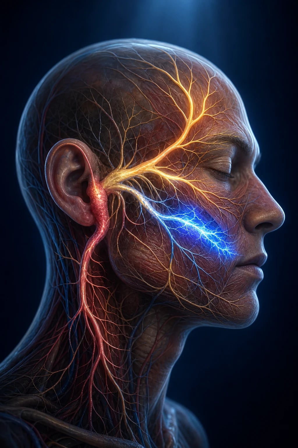
Definition and classification
Bell palsy is defined as acute, unilateral, idiopathic paralysis of the facial nerve of lower motor neurone type, reaching maximal weakness within 72 hours, in the absence of an identifiable cause. It is a diagnosis of exclusion — the label applies only once stroke, infection (otitis media, Ramsay Hunt, Lyme), tumour and systemic causes have been ruled out at the bedside and by targeted testing.[7]
Classification runs along two axes. By lesion level: a lower motor neurone (LMN) lesion — Bell palsy, Ramsay Hunt, Lyme — denervates all ipsilateral facial muscles including frontalis; an upper motor neurone (UMN) lesion — cortical stroke — spares the forehead because frontalis receives bilateral cortical innervation while the lower face receives only contralateral input. By aetiology: idiopathic (Bell), viral reactivation (herpes simplex virus type 1 is the leading proposed trigger; varicella zoster virus causes Ramsay Hunt), bacterial (Lyme, otitis media), and structural (cholesteatoma, parotid or cerebellopontine angle tumour).[1][6]
Pathophysiology — the fallopian canal and the five facial nerve functions
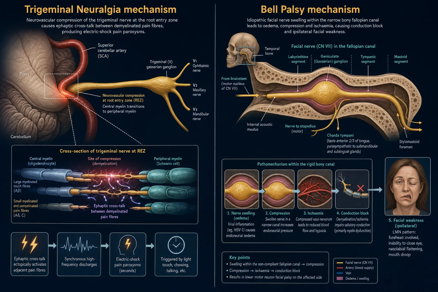
The facial nerve is a mixed nerve carrying five functional components, and a Bell palsy can impair any combination of them: motor to the muscles of facial expression (forehead, eye closure, smile); parasympathetic to the lacrimal and submandibular/sublingual glands (lacrimation, salivation); special sensory taste from the anterior two-thirds of the tongue; and a small somatic sensory branch to the external ear. The clinical signature of a complete LMN palsy is therefore drooping of one entire hemiface, an eye that will not close (with eyeball rolling upwards on attempted closure — the Bell phenomenon), hyperacusis from stapedius paralysis, reduced taste, and reduced lacrimation.[6]
The mechanism is oedema of the nerve within the narrow bony fallopian canal, most critically at the narrowest segment near the geniculate ganglion. Swelling (proposed viral, ischaemic, or inflammatory) compresses the nerve against unyielding bone, producing conduction block and, in severe cases, axonal degeneration — which is why early anti-oedema therapy with corticosteroids improves recovery and why delay beyond 72 hours forfeits the benefit.[1][3]
Clinical presentation
The onset is rapid, over hours to a maximum of 72 hours, and the patient (or a mirror, or a photo) usually notices the asymmetry first. The face droops on one side: the forehead is smooth and will not wrinkle on attempted frowning, the palpebral fissure is widened and the eye cannot be closed, the nasolabial fold is flattened, and the corner of the mouth droops, causing drooling and a slurred quality to labial consonants. Associated features reflect the loss of the other facial nerve functions: hyperacussis (sounds seem loud and harsh on the affected side because the stapedius cannot dampen the ossicles), altered or reduced taste over the anterior two-thirds of the tongue, and reduced tearing. Pain around the ear or jaw, reduced hearing, tinnitus or vertigo may accompany the palsy but prominent ear pain with vesicles or hearing loss should redirect the diagnosis to Ramsay Hunt.[6][7]
The red flags that argue against simple Bell palsy are a forehead-sparing pattern (UMN stroke), vesicles in the ear or mouth or on the tympanic membrane (Ramsay Hunt), otitis media or mastoiditis on otoscopy, bilateral weakness (GBS, Lyme, sarcoid), slowly progressive weakness beyond 72 hours (tumour), recurrent palsy (rule out tumour), involvement of other cranial nerves (basilar meningitis, cerebellopontine angle tumour), and any limb weakness, dysarthria, or visual field defect (stroke).[7]
Differential diagnosis — LMN versus UMN, and the dangerous mimics
The differential is the heart of the Fellowship question, because every unilateral facial weakness must first be classified as LMN or UMN, and then the LMN group subdivided into the dangerous secondary causes. The single discriminating sign is the forehead. [1]
Bell palsy (LMN, idiopathic)
- Forehead INVOLVED — cannot wrinkle the brow or close the eye on the affected side
- Bell phenomenon on attempted eye closure; hyperacussis, altered taste, reduced lacrimation
- Rapid onset, maximal within 72 h; no limb signs; normal otoscopy
- Oral prednisolone within 72 h + eye protection; ~70% full recovery
Cortical stroke (UMN)
- Forehead SPARED — lower face only; patient can wrinkle both brows and close both eyes
- Associated limb weakness, dysarthria, hemianopia, NIHSS findings; vascular risk factors
- CN VII is a common stroke mimic and missed-stroke presentation — do a full neuro exam
- Stroke pathway: CT/CTA or MRI, thrombolysis assessment if eligible
Ramsay Hunt (VZV, herpes zoster oticus)
- Painful vesicles in the ear canal, on the pinna, or on the anterior tongue/palate; otalgia out of proportion
- Often with hearing loss, tinnitus, vertigo; can involve multiple cranial nerves
- Worse prognosis for facial recovery and hearing than Bell palsy
- Steroid PLUS high-dose antiviral (aciclovir 800 mg 5x/day or valaciclovir 1 g TDS)
Lyme disease facial palsy
- Tick exposure or endemic area; possible erythema migrans, arthralgia, meningitis
- Often bilateral; commonest cause of bilateral facial palsy in endemic regions
- Serology (two-tier); CSF if neuroborreliosis suspected
- Doxycycline (or ceftriaxone for neuroborreliosis); prognosis generally good
Otitis media / cholesteatoma
- Otoscopy abnormal — bulging, erythematous TM, or cholesteatoma (pearly mass, attic crust)
- Ear discharge, conductive hearing loss; deep otalgia; can erode into the facial nerve
- CT temporal bone if suspicious; ENT urgently
- IV antibiotics for acute otitis media with complications; surgical decompression for cholesteatoma
Guillain–Barre (bilateral)
- Bilateral, usually descending or ascending; areflexia, progressive limb weakness
- May follow a gastrointestinal or respiratory illness; respiratory muscle involvement
- LP shows albuminocytologic dissociation (raised protein, normal cells)
- Admit for monitoring; IVIG or plasma exchange; spirometry for FVC
The broader cranial nerve lens adds the dangerous ocular motor palsies the ED candidate must not miss. A pupil-involving CN III palsy — ptosis, the eye deviated "down and out," and a dilated unreactive pupil — is a posterior communicating artery aneurysm compressing the nerve until proven otherwise, and demands emergency CT angiography. (The pupil is spared in a microvascular/diabetic third, because the parasympathetic pupilloconstrictor fibres run in the nerve's periphery and survive an ischaemic core lesion.) A CN VI palsy (failure of abduction, horizontal diplopia worse on lateral gaze) can be a false-localising sign of raised intracranial pressure. Any isolated ocular motor palsy with pain, a dilated pupil, trauma, or a stepwise progression is imaged.[7]
Bedside assessment
Examine all the cranial nerves, not just the face. On the facial nerve itself, grade severity by asking the patient to raise the eyebrows (frontalis), gently close the eyes (orbicularis oculi — observe for Bell phenomenon and incomplete closure), and smile, puff out the cheeks, and show the teeth (orbicularis oris and buccinator). The standard severity scale is House–Brackmann (below). Perform otoscopy on the affected ear — looking for vesicles (Ramsay Hunt), a bulging or erythematous tympanic membrane (otitis media), or a pearly mass (cholesteatoma). Examine the tongue and oral mucosa for vesicles. Perform a full neurological examination including the limbs (strength, tone, reflexes), speech, and visual fields to exclude stroke, and look for a rash (erythema migrans) and lymphadenopathy.[6][7]
[1]House–Brackmann facial nerve grading
The House–Brackmann scale (1985) grades facial nerve function from normal to total paralysis and is the universal reporting and prognostic standard.[6]
| Grade | Function | Findings |
|---|---|---|
| I | Normal | Symmetrical face in all regions; normal forehead and eye closure |
| II | Mild dysfunction | Slight weakness on close inspection; slight synkinesis; forehead 2/4 to 3/4; eye closes with minimal effort; mouth slightly weak |
| III | Moderate dysfunction | Obvious but not disfiguring asymmetry; mild synkinesis/contracture; forehead 1/2 to 2/4; eye closes with effort; mouth slightly weak with maximal effort |
| IV | Moderate–severe | Obvious disfiguring asymmetry; no synkinesis/contracture; forehead 1/4; eye cannot close; mouth asymmetric with maximal effort |
| V | Severe | Barely perceptible movement; forehead none; eye cannot close; mouth barely moves |
| VI | Total paralysis | No movement at any level; no tone |
Investigations — a clinical diagnosis, with targeted testing
Bell palsy is a clinical diagnosis. Routine bloods (full blood count, electrolytes, glucose, inflammatory markers) and an HbA1c screen for diabetes are reasonable baseline tests, because diabetes is both a risk factor and a cause of alternative cranial nerve palsies. Add Lyme serology in an endemic area or with bilateral disease, an HIV test when risk factors are present, and a pregnancy test in women of childbearing potential (pregnancy multiplies the risk of Bell palsy roughly three-fold). [7]
Imaging is not required for a typical first Bell palsy. Order CT or, preferably, MRI of the brain and temporal bone when the palsy is atypical — slow progression beyond 72 hours, recurrent, bilateral, involving other cranial nerves, with an abnormal ear, after head trauma, or with any stroke feature — to exclude stroke, tumour, cholesteatoma, or demyelination. Electroneurography (ENoG) and electromyography grade axonal degeneration and prognosticate, but these are outpatient investigations for the 10 to 15 per cent with no recovery by three weeks, not ED tests. A lumbar puncture is reserved when Guillain–Barre, Lyme neuroborreliosis, or basilar meningitis is suspected.[6][7]
Immediate management — steroids, antivirals, and the cornea
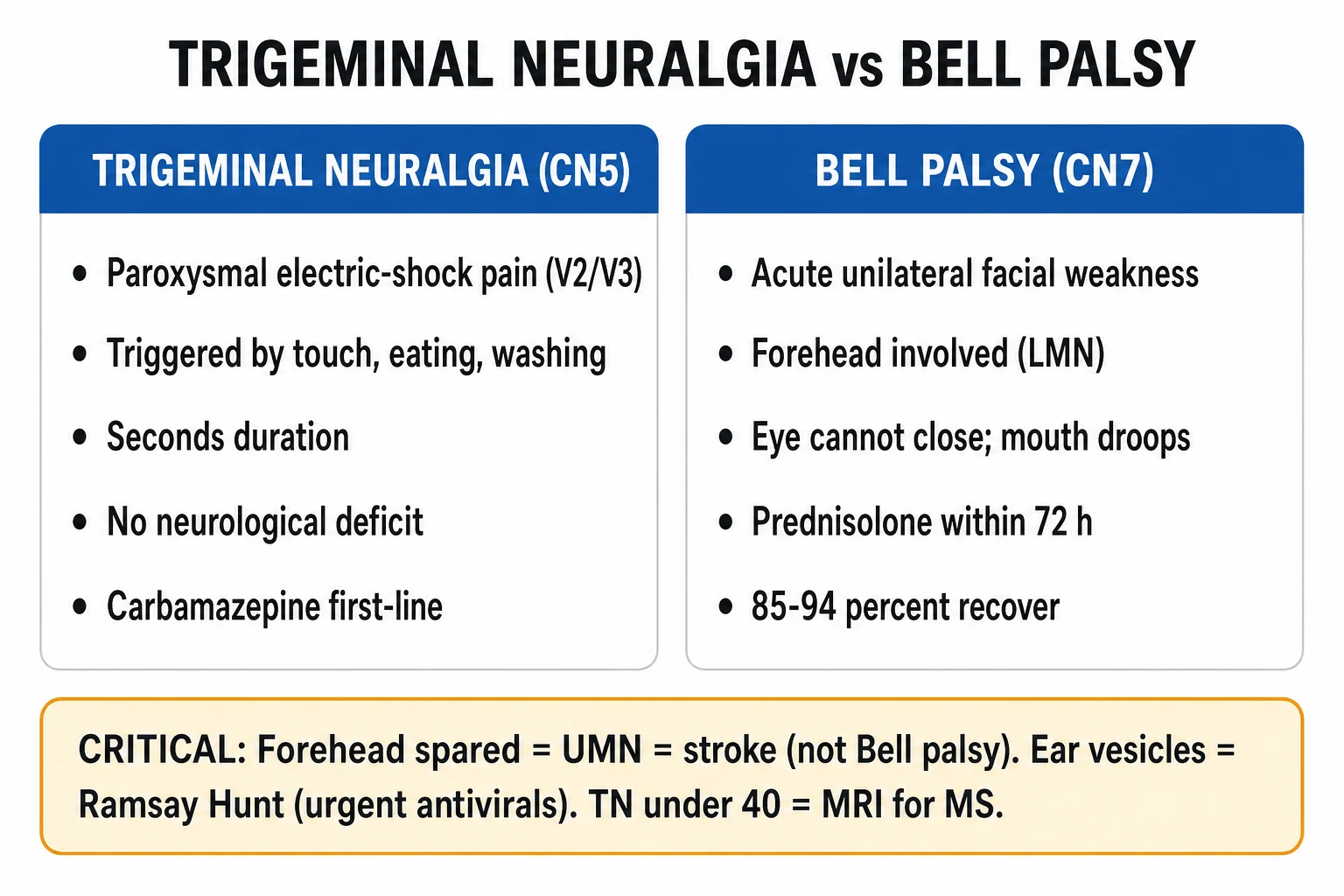
Three interventions define ED management: oral corticosteroid within the time window, antiviral therapy in the appropriate context, and immediate corneal protection. Intravenous access and resuscitation are rarely required unless vomiting or an alternative diagnosis dominates.[1][7]
Corticosteroid. Give oral prednis(ol)one to every patient seen within 72 hours of onset, at a dose of 50 mg once daily for 5 days, then tapered over the following 5 days (for example 50 mg daily on days 1–5, then 25 mg, 20 mg, 15 mg, 10 mg, 5 mg). The landmark Scottish trial (Sullivan 2007) used 60 mg daily for 5 days then tapered, and the AAO-HNS guideline recommends a total of approximately 1 mg/kg per day (typically 50–60 mg daily) for 5 to 7 days with a taper. The Cochrane meta-analysis confirms corticosteroids increase the rate of complete recovery at nine months (number needed to treat approximately 6 to 11).[1][3][7]
Antiviral therapy. Antivirals alone confer no benefit in idiopathic Bell palsy, and the addition of an antiviral to corticosteroid in Bell palsy shows no convincing added benefit over corticosteroid alone (Sullivan 2007 aciclovir; Engström 2008 valaciclovir; Cochrane 2009). Antivirals are therefore not routine for Bell palsy. They are reserved for Ramsay Hunt syndrome, where varicella zoster is the cause: combine high-dose antiviral — aciclovir 800 mg orally five times daily for 7 to 10 days, or valaciclovir 1 g three times daily — with corticosteroid. (The aciclovir 400 mg five-times-daily regimen used in the Bell palsy trials is sub-therapeutic for herpes zoster oticus; use the higher zoster dose.) Severe Ramsay Hunt or immunocompromise warrants intravenous aciclovir and ENT admission.[1][2][4][5][7]
Eye protection. Corneal exposure is the principal acute complication of an unprotectable eye and can progress to exposure keratitis, ulceration, and sight loss. Prescribe preservative-free lubricating eye drops hourly by day, lubricating ointment at night, and mechanical closure of the eyelid at night with surgical tape or a moisture chamber; advise the patient not to drive if binocular vision is impaired. If the eye cannot be closed at all (House–Brackmann IV–VI), refer urgently to ophthalmology — a temporary tarsorrhaphy or botulinum-induced ptosis may be required.[7]
Model answer — drug regimen for a Bell palsy seen within 24 hours
Complications and pitfalls
The complications, in descending danger, are the missed stroke (a UMN lesion mislabelled as Bell palsy — the forehead test is the safeguard), corneal exposure keratitis and ulceration from the unprotected eye, residual facial weakness and synkinesis (aberrant regeneration causing eye closure on smiling, or "crocodile tears"), missed Ramsay Hunt (with consequent worse facial and hearing recovery), and missed bilateral or systemic disease (GBS, Lyme, sarcoid). The recurring pitfalls at the bedside are: failing to examine the forehead and so misclassifying a stroke; omitting otoscopy and missing Ramsay Hunt or otitis media; prescribing an antiviral without a steroid in routine Bell palsy; starting steroids too late (beyond 72 hours) or in inadequate dose; and forgetting eye care entirely.[1][5][7]
Prognosis and disposition
Prognosis is favourable for the majority. In Peitersen's 2500-patient natural-history series, 71 per cent recovered completely without treatment and 84 per cent attained a good functional outcome; recovery began within three weeks in most and was complete by six months.[6] Corticosteroid within 72 hours raises the complete-recovery rate further.[1][3] Poor prognostic markers are complete paralysis at onset (House–Brackmann V–VI), age over 60, severe pain, diabetes, hypertension, and Ramsay Hunt syndrome (only about half recover good facial function).[5][6]
Disposition is outpatient for a typical Bell palsy: steroid course, eye care, and a written safety-net for new neurological symptoms, with GP or ENT review at 3 to 4 weeks. Admit for Ramsay Hunt with severe pain or systemic features (for intravenous antiviral), any bilateral or atypical palsy requiring work-up (GBS, Lyme, meningitis), and any patient in whom a dangerous mimic cannot be confidently excluded. Refer to ophthalmology if the eye is unprotected, and to ENT/neurology for the 10 to 15 per cent without recovery by three weeks.[7]
Special populations
Pregnancy multiplies the risk of Bell palsy roughly three-fold, particularly in the third trimester and early puerperium; oral prednisolone is used, with ophthalmology oversight of the eye, and the prognosis is similar to non-pregnant adults. Diabetes is both a risk factor and a prognostic marker — screen for it, and remember that an isolated CN III palsy in a diabetic is usually a pupil-sparing microvascular palsy that recovers over months (still image if painful or the pupil is involved). Children have an excellent prognosis; exclude otitis media as the cause. Recurrent Bell palsy (roughly 7 per cent) and familial cases warrant imaging to exclude a structural lesion. Immunocompromised patients are at higher risk of Ramsay Hunt and disseminated zoster — use intravenous antivirals.[6][7]
The systematic cranial nerve examination in the ED (CN I–XII)
Every facial-weakness patient deserves a full cranial nerve screen, both to confirm the lesion is isolated (Bell palsy) and to surface the dangerous companion signs of a cerebellopontine angle tumour, basilar meningitis, or brainstem stroke. Examine cranially to caudally, in order, so nothing is omitted.[6][7]
| CN | Nerve | Test in the ED | Lesion significance |
|---|---|---|---|
| I | Olfactory | Test each nostril separately with a non-irritant odour (coffee, vanilla) after confirming patency; avoid ammonia (it stimulates the trigeminal nerve) | Anosmia: head trauma (cribriform plate fracture), frontal tumour, Parkinson disease; rarely ED-critical but new anosmia after head injury is a red flag |
| II | Optic | Visual acuity (Snellen), fields (confrontation), pupils (direct/consensual), fundoscopy | Sudden monocular blindness: optic neuritis, ophthalmic/ICA occlusion, giant cell arteritis, raised ICP (papilloedema) |
| III, IV, VI | Oculomotor, trochlear, abducens | Eyelid (ptosis), eye position at rest, all six directions of gaze, pupils (size, symmetry, reactivity) | Painful CN III with pupil involved = posterior communicating artery aneurysm; CN VI palsy = raised ICP false-localiser |
| V | Trigeminal | Facial sensation (all three divisions), corneal reflex (V afferent, VII efferent), jaw clench (masseter), jaw jerk | Sensory loss: stroke, MS; trigeminal neuralgia; corneal anaesthesia risks exposure keratopathy |
| VII | Facial | Forehead (frontalis), eye closure (orbicularis oculi — Bell phenomenon), smile / puff cheeks / show teeth (orbicularis oris) | Forehead INVOLVED = LMN (Bell); forehead SPARED = UMN (stroke) |
| VIII | Vestibulocochlear | Whispered-voice hearing (each ear), Rinne and Weber tuning fork; nystagmus, head-impulse (HINTS if vertigo) | Sudden sensorineural hearing loss; vertigo (central vs peripheral); cerebellopontine angle tumour |
| IX, X | Glossopharyngeal, vagus | Palate elevation (uvula deviates AWAY from the lesion), gag reflex (IX afferent, X efferent), voice quality, cough | Palatal weakness: bulbar stroke, GBS; absent gag = aspiration risk; the palate check precedes feeding |
| XI | Accessory | Shoulder shrug (trapezius), turn head against resistance (sternocleidomastoid) | Isolated palsy: neck trauma or surgery; bilateral weakness in neuromuscular disease |
| XII | Hypoglossal | Tongue protrusion — deviates TOWARDS the weak side; look for fasciculations and atrophy | Bulbar stroke (LMN or UMN); motor neurone disease; base-of-skull tumour |
The reflex trio — pupillary, corneal, and gag
These three reflexes interrogate pairs of cranial nerves at the bedside and are disproportionately high-yield in the Fellowship viva because they localise the lesion precisely.[9]
Pupillary reflex (CN II + III)
- Afferent = optic nerve (II); efferent = oculomotor nerve (III, parasympathetic Edinger–Westphal nucleus)
- Shine light in ONE eye — BOTH pupils should constrict (direct in the lit eye, consensual in the other)
- Afferent defect (e.g. optic neuritis): the swinging-light test reveals a relative afferent pupillary defect (RAPD) — the affected pupil dilates when the light swings to it
- Efferent defect (CN III compression): the affected pupil is dilated and unreactive; the consensual reflex STILL constricts the opposite normal pupil, localising the lesion to the III nerve
Corneal reflex (CN V + VII)
- Afferent = ophthalmic division of the trigeminal nerve (V1); efferent = facial nerve (VII) to orbicularis oculi
- Touch the cornea (not the sclera) with a wisp of cotton from the lateral aspect — both eyes should blink
- Loss of the blink with an intact V (test the other side) localises the lesion to the facial nerve and flags a cornea at risk of exposure keratopathy
- A depressed reflex in a sedated or comatose patient tracks brainstem depth and is part of the brainstem death examination
Gag reflex (CN IX + X)
- Afferent = glossopharyngeal (IX) from the posterior pharynx; efferent = vagus (X) — palate elevation and pharyngeal contraction
- Stimulate the posterior pharynx (not the soft palate) with a tongue depressor; observe symmetric palate lift and gag
- An absent gag alone is a poor predictor of aspiration in a conscious patient — never use it in isolation to decide feeding safety
- Bilateral loss in a comatose patient indicates lower brainstem depression and is part of the brainstem death examination
The other cranial nerve palsies — the complete ED reference
Beyond the facial nerve, the cranial nerve palsies that present to the emergency department fall into a small number of patterns the candidate must recognise instantly, because each carries a specific dangerous diagnosis.[9]
CN III (oculomotor)
- Ptosis; eye deviated "down and out" (unopposed IV and VI); diplopia in all directions except lateral gaze
- PUPIL INVOLVED (dilated, unreactive) = compressive — posterior communicating artery aneurysm until proven otherwise; emergency CT angiography
- PUPIL SPARED = microvascular (diabetes, hypertension); the parasympathetic fibres run in the periphery and survive an ischaemic core lesion; recovers over ~3 months
- Image (CTA / MRA) any painful, pupil-involving, or rapidly progressive III palsy; observe a painless pupil-sparing microvascular palsy and re-image if no recovery at 3 months
CN IV (trochlear)
- Vertical diplopia, worse on DOWN-gaze and when reading or descending stairs; head tilt AWAY from the affected side (compensatory)
- The superior oblique intorts the adducted eye and depresses the abducted eye; the patient tilts the head to the opposite shoulder to minimise diplopia
- Commonest ED cause = CLOSED HEAD TRAUMA — the trochlear nerves have the longest intracranial course and are contused against the free edge of the tentorium
- Also congenital, microvascular (diabetes), and tumour; most traumatic palsies resolve spontaneously over months
CN VI (abducens)
- Failure of ABDUCTION; horizontal diplopia worse on lateral gaze to the affected side; the affected eye sits adducted at rest
- A FALSE-LOCALISING SIGN of raised intracranial pressure — the long, steeply angled intracranial course is stretched over the petrous tip as the brain shifts
- ALWAYS exclude raised ICP: headache, papilloedema, vomiting, depressed GCS; consider a space-occupying lesion or idiopathic intracranial hypertension
- A microvascular (diabetic) VI palsy also occurs but is a diagnosis of exclusion — neuroimaging is mandatory for any new isolated VI palsy
CN VII (facial)
- The subject of this topic — LMN (Bell, forehead involved) vs UMN (stroke, forehead spared) is the discriminator
- Look for vesicles (Ramsay Hunt), otitis media, bilateral disease (GBS, Lyme, sarcoid), and atypical features mandating imaging
- Prednisolone within 72 h + eye protection; antiviral only for Ramsay Hunt
- ~70% full recovery; worse if complete paralysis, age over 60, diabetes, Ramsay Hunt
CN VIII (vestibulocochlear)
- Hearing loss — Rinne: air > bone is normal or conductive; bone > air is sensorineural. Weber lateralises TOWARDS a conductive loss and AWAY from a sensorineural loss
- Vertigo — the HINTS examination separates central (stroke: normal head impulse, direction-changing nystagmus, skew) from peripheral (vestibular neuritis, BPPV)
- Sudden sensorineural hearing loss is a time-critical ENT emergency (oral steroids within 2 weeks); a cerebellopontine angle tumour adds VI/VII signs
- Auditory and vestibular limbs are tested separately — never assume a single "CN VIII" lesion
CN IX/X (glossopharyngeal/vagus)
- Dysphagia, dysphonia, nasal regurgitation, palatal droop; uvula deviates AWAY from the lesion; absent gag
- Bulbar palsy (LMN) = wasted fasciculating tongue, absent gag — stroke, GBS, motor neurone disease; pseudobulbar (UMN) = spastic tongue, brisk jaw jerk, emotional lability
- Airway protection is the ED priority — a drooping palate and absent cough reflex aspirate; keep nil by mouth until a formal swallow assessment
- A lateral medullary (Wallenberg) stroke classically strikes the nucleus ambiguus (IX/X) with ipsilateral Horner, ataxia, and contralateral body sensory loss
CN XII (hypoglossal)
- Tongue weakness on protrusion — deviates TOWARDS the weak side (LMN); fasciculations and atrophy in a chronic LMN lesion
- A bulbar stroke (medial medullary) is the acute cause; motor neurone disease and base-of-skull tumour are the chronic causes
- A unilateral XII palsy is rarely an emergency in itself, but it signals a brainstem or skull-base lesion that mandates imaging
- Bilateral XII palsies give a "bag of worms" fasciculating tongue that cannot protrude, with aspiration and profound dysarthria
Corticosteroid dosing across the landmark Bell palsy trials
The dose and duration of oral corticosteroid vary slightly between the major trials and guidelines, but all converge on roughly 1 mg/kg per day (50–60 mg) started within 72 hours, with a taper.[1][2][3][7]
Sullivan 2007 (Scottish trial)
- Prednisolone 60 mg daily for 5 days, then 25/20/15/10/5 mg (a 5-day taper)
- Started within 72 h of onset; the aciclovir arm showed no added benefit
- Complete recovery at 9 months 83% (steroid) vs 63.6% (placebo) for severe palsies
Engström 2008 (Swedish trial)
- Prednisolone 60 mg daily for 5 days, then taper by 10 mg/day to zero
- The valaciclovir arm (1000 mg TDS ×3 days) showed no added benefit over steroid alone
- Recovery at 12 months similar; supported corticosteroid benefit over antiviral
AAO-HNS guideline 2013
- A total of ~1 mg/kg/day prednis(ol)one for 5–7 days with a taper
- Recommends steroids within 72 h; does NOT recommend routine antivirals
- Eye care is mandatory; severity graded with House–Brackmann
Madhok 2016 (Cochrane)
- Meta-analysis: corticosteroids increase complete recovery (NNT ~6–11)
- Confirms no benefit of antivirals alone; a possible small added benefit of combination is not statistically robust
- Establishes steroid within 72 h as the standard of care
Trial cards — the evidence base for Bell palsy and cranial nerve palsies
Sullivan 2007 (NEJM) — early prednisolone or aciclovir in Bell palsy
Design
Double-blind, placebo-controlled, factorial RCT — 551 patients presenting within 72 h of onset
Intervention
Prednisolone 60 mg ×5d then taper; aciclovir 400 mg 5×/d ×5d; both; or double placebo (2×2 factorial)
Result
Prednisolone improved complete recovery at 9 months (83.0% vs 63.6% in severe palsies). Aciclovir alone or added to steroid showed NO benefit
Bottom line
Oral corticosteroid within 72 h is the standard of care; antivirals are not routine in idiopathic Bell palsy
Engström 2008 (Lancet Neurology) — prednisolone and valaciclovir in Bell palsy
Design
Randomised, double-blind, placebo-controlled, multicentre trial — 829 patients
Intervention
Prednisolone 60 mg ×5d then taper; valaciclovir 1000 mg TDS ×3d; combination; or double placebo (2×2 factorial)
Result
Prednisolone improved recovery at 12 months. Valaciclovir showed no added benefit over corticosteroid alone
Bottom line
Independent confirmation of corticosteroid benefit; no support for routine antivirals in Bell palsy
Madhok 2016 (Cochrane) — corticosteroids for Bell palsy
Design
Cochrane systematic review and meta-analysis of corticosteroid RCTs
Result
Corticosteroids significantly increased the rate of complete recovery at 6 months (NNT ~11) and 9 months (NNT ~6)
Antivirals
No benefit of antivirals alone; combination therapy showed a possible but non-robust small benefit
Bottom line
Establishes corticosteroid within 72 h as the evidence-based standard
Lee 2002 (Survey of Ophthalmology) — evaluating the isolated third nerve palsy
Source
Authoritative review of the imaging strategy for an isolated CN III palsy
Key teaching
A pupil-involving complete CN III palsy demands imaging for an aneurysm (CTA first-line, MRA or catheter angiography as needed); a pupil-sparing palsy in a vasculopath may be observed, but pain, progression, or pupillary involvement mandates imaging
Bottom line
Frames the 'image the painful pupil-involving third, observe the pupil-sparing microvascular third' rule
House & Brackmann 1985 — the facial nerve grading system
Source
The original description of the universal six-grade facial nerve recovery scale
Grades
I = normal; II = mild; III = moderate; IV = moderate–severe; V = severe; VI = total paralysis
Use
Reports severity at presentation and recovery at follow-up; prognostic — Grades V–VI at onset predict incomplete recovery
Bottom line
Grade every facial palsy at the bedside; the grade is the shared language between ED, ENT, and ophthalmology
ED disposition pathway — Bell palsy and the dangerous mimics
Disposition of the unilateral facial-weakness patient — the ED algorithm
Step 1 — Test the forehead
If the forehead is SPARED (it wrinkles on the weak side), this is an UMN (cortical stroke) lesion — activate the stroke pathway: CT/CTA or MRI, thrombolysis assessment if eligible, admit.
Step 2 — Examine the ear and oropharynx
Otoscopy for vesicles, a bulging tympanic membrane, cholesteatoma; inspect the tongue and palate for vesicles. Vesicles + otalgia + hearing loss = Ramsay Hunt — add high-dose antiviral (aciclovir 800 mg 5×/d or valaciclovir 1 g TDS) to the steroid, and admit if severe.
Step 3 — Confirm the palsy is isolated
A full CN I–XII exam, limb power, reflexes, speech, and visual fields. A bilateral palsy enters the GBS / Lyme / sarcoid pathway. Any other CN involvement, limb signs, or atypical course mandates imaging and admission.
Step 4 — Steroid within 72 h
Prednis(ol)one 50–60 mg PO daily ×5 days then taper over 5 days, for any LMN facial palsy seen within 72 h of onset. Beyond 72 h the benefit is unproven; discuss with ENT.
Step 5 — Protect the eye
Grade with House–Brackmann; if the eye will not close (Grade IV–VI) prescribe preservative-free lubricant drops hourly by day, ointment and eyelid taping or a moisture chamber at night, and refer urgently to ophthalmology (tarsorrhaphy or botulinum ptosis if unprotected).
Step 6 — Screen the mimics
Glucose/HbA1c (diabetes), Lyme serology if endemic or bilateral, HIV if risk factors, a pregnancy test in women of childbearing age. Image if recurrent, progressive, traumatic, or accompanied by other CN signs.
Step 7 — Safety-net and follow-up
Discharge with written advice to return for new limb weakness, visual change, vesicles, or fever. GP/ENT review at 3–4 weeks; neurology/ENT referral for the 10–15% with no recovery by three weeks.
Workup of a pupil-involving CN III palsy — the aneurysm pathway
Recognise the syndrome
Ptosis, the eye deviated "down and out", and a DILATED unreactive pupil — often with thunderclap headache if an aneurysm is expanding or has ruptured. This is a posterior communicating artery aneurysm until proven otherwise.
Immediate imaging — CT angiography
CT brain (for subarachnoid haemorrhage) PLUS CT angiography of the intracranial circulation. MRA is an alternative if CTA is contraindicated. Do not delay imaging to "observe" a pupil-involving palsy.
If subarachnoid haemorrhage — neurosurgery and the SAH pathway
A pupil-involving III palsy with SAH is a ruptured PCOM aneurysm — activate the SAH pathway: neurosurgery, blood-pressure control, nimodipine, and definitive securing by coiling or clipping.
If CTA negative but suspicion high — catheter angiography
CTA is ~95% sensitive but not perfect. A high-suspicion painful pupil-involving palsy with a negative CTA still warrants catheter digital subtraction angiography before the diagnosis is abandoned.
Distinguish the microvascular (pupil-sparing) III
A pupil-sparing III palsy in an older vasculopath (diabetes, hypertension) that is painless or minimally painful is usually microvascular — observe, control vascular risk factors, and re-image only if the pupil becomes involved or there is no recovery by 3 months.
High-yield Fellowship viva pearls
[1] [1] [1] [1] [1] [1] [1] [1] [1] [1] [1] [1]ANZ practice note. Australian and New Zealand emergency practice follows the AAO-HNS (2013) and Cochrane-based standard: oral prednis(ol)one within 72 hours of onset, eye protection with preservative-free lubricants and night taping, and antivirals reserved for Ramsay Hunt. Lyme disease is rare in Australia and not the default for bilateral facial palsy — Guillain–Barre and sarcoid are pursued first; Lyme is considered in returned travellers from endemic regions (North America, Europe). Ophthalmology and ENT are accessible for the unprotected eye and severe Ramsay Hunt. GPs are the usual 3–4 week follow-up, with neurology/ENT referral if recovery is incomplete. [1]
SAQs
SAQ — Bell palsy within the corticosteroid window
10 minutes · 10 marks
A 34-year-old man presents 20 hours after the onset of a right-sided facial droop with drooling from the right corner of the mouth and difficulty closing the right eye. He describes harsh sounds on the right side and altered taste over the anterior tongue. He has type 1 diabetes. Examination shows right forehead weakness, a right eye that will not close with a positive Bell phenomenon, a flattened right nasolabial fold, and House–Brackmann grade IV. Otoscopy is normal with no vesicles. Limb power, speech and visual fields are normal. Finger-prick glucose is 7.2 mmol/L.
SAQ — Stroke mimic masquerading as facial weakness
10 minutes · 10 marks
A 72-year-old woman is brought in by her husband with two hours of right-sided facial droop and slurred speech that began at the dinner table. She has atrial fibrillation on apixaban 5 mg twice daily (last dose 4 hours ago), hypertension and type 2 diabetes. On examination the forehead wrinkles symmetrically on attempted frown, but the right lower face is weak with a flattened nasolabial fold and drooling. Limb power is full but there is mild right-arm pronator drift. NIHSS is 4 (mild dysarthria, right facial palsy, mild right-arm drift). Finger-prick glucose is 6.8 mmol/L.
Red flags
[1]References
- [1]Sullivan FM, Swan IR, Donnan PT, et al. Early treatment with prednisolone or acyclovir in Bell's palsy N Engl J Med, 2007.PMID 17942873
- [2]Engström M, Berg T, Stjernquist-Desatnik A, et al. Prednisolone and valaciclovir in Bell's palsy: a randomised, double-blind, placebo-controlled, multicentre trial Lancet Neurol, 2008.PMID 18849193
- [3]Madhok VB, Gagyor I, Daly FD, et al. Corticosteroids for Bell's palsy (idiopathic facial paralysis) Cochrane Database Syst Rev, 2016.PMID 27428352
- [4]Lockhart P, Daly F, Pitkethly M, Comerford N, Sullivan F. Antiviral treatment for Bell's palsy (idiopathic facial paralysis) Cochrane Database Syst Rev, 2009.PMID 19821283
- [5]Uscategui T, Doree C, Chamberlain IJ, Burton MJ. Antiviral therapy for Ramsay Hunt syndrome (herpes zoster oticus with facial palsy) in adults Cochrane Database Syst Rev, 2008.PMID 18843734
- [6]Peitersen E. Bell's palsy: the spontaneous course of 2,500 peripheral facial nerve palsies of different etiologies Acta Otolaryngol Suppl, 2002.PMID 12482166
- [7]Baugh RF, Basura GJ, Ishii LE, et al. Clinical practice guideline: Bell's palsy Otolaryngol Head Neck Surg, 2013.PMID 24189771
- [8]House JW, Brackmann DE. Facial nerve grading system Otolaryngol Head Neck Surg, 1985.PMID 3921901
- [9]Lee AG, Hayman LA, Brazis PW. The evaluation of isolated third nerve palsy revisited: an update on the evolving role of magnetic resonance, computed tomography, and catheter angiography Surv Ophthalmol, 2002.PMID 11918895