EM · Transient ischaemic attack
Transient ischaemic attack
Also known as Transient ischemic attack · TIA · Mini-stroke · Cerebral transient ischaemic attack
Transient ischaemic attack — a transient episode of focal neurological dysfunction from brain, spinal cord or retinal ischaemia WITHOUT infarction (the tissue-based definition); an infarct on diffusion-weighted MRI is a stroke, not a TIA. The ABCD2 score (Age, Blood pressure, Clinical features, Duration, Diabetes) stratifies early stroke risk; the danger is the first 48 hours. Exclude the mimics (migraine aura, postictal Todd paresis, hypoglycaemia), then urgent carotid imaging within 24 hours, aspirin 300 mg stat, short-course dual antiplatelet therapy, a high-intensity statin, and carotid endarterectomy within two weeks for symptomatic severe stenosis. A warning event, not a reassurance. ACEM-primary, globally tagged.
On this page & tools
Your progress
Saved locally on this device.
3 MCQs with explanations
Target exams
Red flags
A transient ischaemic attack is a brief episode of focal neurological dysfunction caused by ischaemia in a defined arterial territory that resolves and leaves no permanent deficit. Its importance is not the episode itself — which is usually over by the time the patient arrives — but what it predicts: an unstable vascular lesion that is about to produce a completed stroke. The risk of stroke after a TIA is highest in the first 48 hours, and urgent assessment combined with antiplatelet therapy, blood-pressure control and carotid intervention reduces the 90-day stroke risk from about 10.6 per cent to 2 per cent, an 80 per cent relative reduction shown by the EXPRESS study.[3] The modern tissue-based definition recasts TIA as a transient deficit without infarction: a lesion on diffusion-weighted MRI is, by definition, a stroke and not a TIA.[1] For the Fellowship candidate the task is to separate the true ischaemic event from its mimics, to risk-stratify with the ABCD2 score, to obtain urgent carotid imaging in those who would benefit from endarterectomy, and to start secondary prevention at once — because the window in which prevention works is measured in days.
Definition and classification
The time-based definition, from the 1970s and still widely used in practice, calls any focal neurological deficit of vascular origin that resolves within 24 hours a TIA. The tissue-based definition, set out in the AHA/ASA 2009 scientific statement, replaces the clock with the scan: TIA is a transient episode of neurological dysfunction caused by focal brain, spinal cord or retinal ischaemia without acute infarction.[1] The shift matters because duration is a poor discriminator — about a third of deficits resolving within an hour show infarction on diffusion MRI, and roughly 30 to 50 per cent of clinically-defined TIAs are diffusion-positive. Under the tissue-based definition these are strokes, and they carry a higher recurrence risk and demand stroke-grade investigation. A transient neurological attack (TNA) is the umbrella term for any transient deficit regardless of cause, of which ischaemic TIA is one (and the one that matters).
The clinical classification mirrors that of stroke by vascular mechanism: large-artery atherothromboembolism (typically from internal carotid stenosis, the lesion that endarterectomy cures), cardioembolism (atrial fibrillation dominant), small-vessel lacunar disease, and other determined cause — dissection, patent foramen ovale, thrombophilia, vasculitis. Identifying the mechanism is the whole point of the workup, because each mechanism has a different and specific secondary-prevention therapy. [1]
Mechanisms and their tailored secondary prevention
The whole point of the TIA workup is to find the mechanism, because each mechanism has a different and specific prevention. The table pairs the mechanism with its investigation and its tailored therapy — the structure to recite in the Fellowship viva. [1]
Large-artery atherothrombotic
- Internal carotid stenosis — the lesion endarterectomy cures; artery-to-artery embolism from an ulcerated atherosclerotic plaque
- Investigate: carotid duplex within 24 h, CTA or MRA to grade the stenosis by the NASCET method
- Prevention: antiplatelet (aspirin 300 mg + clopidogrel 300 mg then dual 75 mg for 21 days, then monotherapy), high-intensity statin, BP control, endarterectomy within 2 weeks if symptomatic stenosis is 50 per cent or more
- Hallmarks: amaurosis fugax, a carotid bruit, contralateral cortical or lacunar symptoms, ipsilateral monocular visual loss
Cardioembolic
- Atrial fibrillation dominant; also a recent MI, a prosthetic valve, endocarditis, a dilated cardiomyopathy or a left-ventricular thrombus
- Investigate: 12-lead ECG in EVERY patient; ambulatory or prolonged monitoring for paroxysmal AF; transthoracic echo, with transoesophageal echo if a cardioembolic source is suspected
- Prevention: anticoagulation (a DOAC preferred over warfarin for non-valvular AF) — NOT an antiplatelet, which is inferior for cardioembolic prevention
- Can start early after a TIA because there is no infarct to transform — the mirror image of a completed cardioembolic stroke
Small-vessel (lacunar)
- Lipohyalinosis or microatheroma of the deep penetrators; a pure motor or pure sensory deficit with no cortical signs
- Investigate: MRI (a small subcortical lesion on DWI if infarcted); the mechanism is often inferred after the carotid and cardioembolic workup are both negative
- Prevention: antiplatelet and aggressive vascular risk-factor control — BP and glucose above all; endarterectomy adds nothing for lacunar disease
- Hypertension and diabetes are the dominant drivers — BP control is the single most important prevention
Cryptogenic (undetermined)
- No carotid stenosis, no AF on the initial monitoring, and no lacunar syndrome — the residual after a complete workup
- Investigate: prolonged cardiac monitoring (an implantable loop recorder detects paroxysmal AF in around 20 per cent), a bubble echo or TCD for a PFO, and a thrombophilia screen in the young
- Prevention: antiplatelet as the default; anticoagulation if a source (AF, thrombophilia) surfaces on extended monitoring; PFO closure for selected young patients after a cryptogenic stroke
- A cryptogenic label is a prompt for further investigation, not a stopping point
Epidemiology and risk factors
TIA carries an incidence of roughly 50 per 100 000 per year and, like stroke, rises steeply with age. Its single epidemiological fact is the early stroke risk: untreated, about 5 per cent at two days and 10 to 15 per cent at 90 days, with the steepest hazard in the first 48 hours.[3] This is why TIA is reclassified as a medical emergency requiring same-day assessment. The risk factors are those of ischaemic stroke: age, hypertension (the largest modifiable contributor), atrial fibrillation, diabetes, smoking, dyslipidaemia, obesity, physical inactivity and prior TIA or stroke. Two are decisive at the bedside because they change the acute plan — atrial fibrillation, the source of the cardioembolic clot that demands anticoagulation, and symptomatic carotid stenosis, the lesion cured by endarterectomy.
Pathophysiology
TIA arises when arterial flow to a territory is briefly interrupted and then restores before permanent infarction. The mechanism is usually embolic: a fragment of platelet-fibrin or atherosclerotic debris from an ulcerated carotid plaque (artery-to-artery embolism) or a clot from the left atrium in atrial fibrillation lodges transiently in a distal vessel, then lyses, fragments or migrates. Less often it is haemodynamic: a critical stenosis produces marginal perfusion that fails transiently with a blood-pressure drop. Small-vessel lipohyalinosis of the deep penetrators produces lacunar TIAs. Spontaneous reperfusion is the reason the deficit resolves; the failure of that reperfusion is the reason a TIA may become a stroke hours later. Diffusion-positive TIAs represent microinfarction — tissue death below the threshold of a persistent clinical deficit — and mark a more unstable lesion. [1]

Clinical presentation
The deficit is sudden, focal, maximal at onset, and maps to a vascular territory exactly as a stroke does — the only difference is that it resolves. Anterior-circulation TIAs produce contralateral face and arm weakness (the arm worse than the leg), aphasia for dominant-hemisphere lesions, neglect for non-dominant, and a homonymous hemianopia. Amaurosis fugax — retinal (ocular) TIA — is the classic carotid-territory sign: a painless monocular visual loss described as a curtain or shutter descending over one eye for seconds to minutes, with full recovery, and it points directly to ipsilateral internal carotid stenosis. Posterior-circulation TIAs are the subtle ones: diplopia, vertigo, dysarthria, dysphagia, ataxia, bilateral or crossed weakness, and drop attacks. Lacunar TIAs give a pure motor or pure sensory deficit without cortical signs. Crescendo TIA — two or more events in succession — is a high-risk presentation mandating admission. [1]
Risk stratification — the ABCD2 score
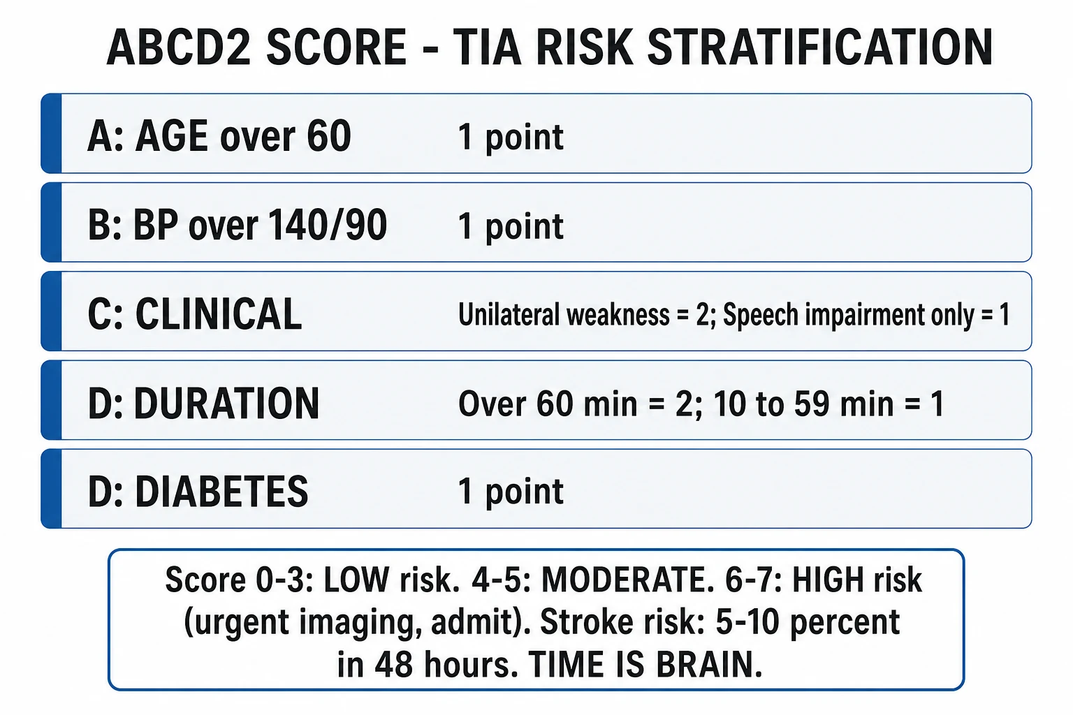
The validated bedside tool for early stroke risk is the ABCD2 score, derived by Johnston and colleagues from the California and Oxford TIA cohorts.[2] It scores five features available at the bedside and stratifies the risk of stroke over the next two, seven and 90 days.
| Component | Criterion | Points |
|---|---|---|
| Age | 60 years or older | 1 |
| Blood pressure | Systolic 140 mmHg or higher, or diastolic 90 mmHg or higher | 1 |
| Clinical features | Unilateral weakness | 2 |
| Speech impairment without weakness | 1 | |
| Duration | 60 minutes or longer | 2 |
| 10 to 59 minutes | 1 | |
| Diabetes | Present (on diet, oral or insulin) | 1 |
| Total | 0 to 7 |
ABCD2 — the five predictors of early stroke risk
ABCD2
One point — age 60 years or older
One point — systolic 140 mmHg or higher, or diastolic 90 mmHg or higher at first assessment
Two points for unilateral weakness, one point for speech impairment without weakness, none otherwise
Two points if 60 minutes or longer, one point if 10 to 59 minutes, none if under 10 minutes
One point — known diabetes on diet, oral therapy or insulin
ABCD2 risk strata and the two-day stroke risk (Johnston 2007)
Differential diagnosis
The mimic rate in emergency department series is high — around half of suspected TIAs are something else. The bedside job is to separate the true ischaemic event from its imitators by the tempo, the presence of positive phenomena, the seizure history, and above all the glucose. The single most dangerous mimic to miss is hypoglycaemia. [1]
Ischaemic TIA
- Sudden onset, maximal at once, maps to a vascular territory; resolves fully
- Negative symptoms (loss of function — weakness, sensory loss, visual loss); no march
- Glucose normal; deficit resolved; vascular risk factors present
- Urgent carotid imaging and secondary prevention; high-risk features warrant admission
Migraine aura
- Positive symptoms — scintillating scotoma, tingling — that MARCH over 20 to 30 min across a body part or visual field
- Slow spread is the key discriminator; headache often follows; prior similar episodes; younger patient
- Glucose normal; imaging normal; diagnosis of exclusion after vascular causes excluded
- Do not over-diagnose TIA on a classic marching aura; no antiplatelet needed
Postictal Todd paresis
- Focal weakness after a seizure; resolves over minutes to hours
- History of epilepsy or a witnessed seizure (tongue-biting, incontinence, postictal confusion)
- Glucose normal; a seizure at onset is a relative thrombolysis exclusion — distinguish from ischaemic stroke
- EEG may show epileptiform activity; treat the underlying seizure disorder
Hypoglycaemia
- Reproduces a focal deficit with complete fidelity; may cause confusion or coma
- Diabetic on insulin or sulfonylurea; often preceded by autonomic symptoms
- Finger-prick glucose (typically below 2.5 mmol/L) — reversed by IV glucose in minutes
- Check glucose FIRST in every transient deficit; a missed hypoglycaemia can brain-damage or kill
Functional (non-organic)
- Deficit does not fit a vascular territory; Hoover sign, give-way weakness, inconsistent examination
- Onset after stress; fixed rather than episodic; past somatisation
- Imaging normal; diagnosis requires positive functional signs AND exclusion of organic disease
- Never assume functional before vascular disease is excluded
Syncope / anoxic convulsion
- Brief loss of consciousness with pallor and rapid recovery; may be followed by brief limb jerking (anoxic convulsion, not epilepsy)
- Prodrome, situational triggers, postictal fatigue; no focal deficit
- Glucose normal; ECG to exclude arrhythmia; no fixed territorial deficit
- Cardiac workup driven by history — an ECG is mandatory in any collapse
The discriminating moves at the bedside: check the glucose; ask explicitly about a seizure and the minutes before it; establish the tempo (TIA is maximal at onset and resolves, migraine marches, Todd paresis follows a seizure); and look for positive phenomena (scintillations, tingling spreading — migraine) versus negative phenomena (loss of function — ischaemia). [1]
Investigations
Investigation runs in parallel with risk stratification and is aimed at the mechanism, because each mechanism has its own prevention. The first test is the finger-prick glucose, before anything else. Bloods include a full blood count, coagulation screen, urea and electrolytes, glucose, a fasting lipid profile, and an erythrocyte sedimentation rate (raised in giant-cell arteritis, a mimic of amaurosis fugax in the older patient). A 12-lead ECG screens for atrial fibrillation and an acute coronary syndrome; where paroxysmal atrial fibrillation is suspected, ambulatory or prolonged cardiac monitoring increases the yield. [1]
Neuroimaging is increasingly MRI-based. Diffusion-weighted MRI is the most sensitive test for acute ischaemia and is the discriminator that defines the tissue-based diagnosis: a diffusion-positive lesion is an infarct, and the event is a stroke, not a TIA.[1] Non-contrast CT is less sensitive for early ischaemia but excludes haemorrhage and a mass lesion, and remains the first-line scan when MRI is unavailable or when any deficit is still present (the stroke pathway). The pivotal vascular test is carotid imaging within 24 hours in any patient who would be a candidate for endarterectomy — carotid duplex ultrasound first, with CT or MR angiography to confirm and grade stenosis. Echocardiography (transthoracic, with transoesophageal imaging when a cardioembolic source is suspected) and prolonged cardiac monitoring complete the search for a source in the cryptogenic or young patient.
Immediate management and disposition
If any focal deficit is still present on assessment the patient is on the acute stroke pathway — non-contrast CT, thrombolysis decision, blood-pressure targets — because a deficit that persists is a stroke until imaging proves otherwise. A deficit that has fully resolved is managed as a suspected TIA. Resuscitation is otherwise supportive: oxygen only if saturation falls below 94 per cent, nil by mouth pending a swallow screen if any residual symptom, and correction of hypoglycaemia with intravenous glucose. [1]
Disposition turns on risk. Admit — or fast-track directly to the stroke team — any patient with high-risk features: a crescendo course, atrial fibrillation, suspected severe carotid stenosis, a definite TIA while on an anticoagulant (treatment failure), a diffusion-positive lesion, or recurrence despite antiplatelet therapy. Patients without these features are referred for specialist assessment and carotid imaging within 24 hours via a rapid-access TIA service. The evidence for urgency is EXPRESS: a system that delivered same-day assessment and treatment lowered the 90-day stroke risk from 10.6 per cent to 2 per cent.[3] Give aspirin 300 mg immediately (after haemorrhage is excluded) to every patient in whom a non-cardioembolic TIA is the working diagnosis.
Definitive management and secondary prevention
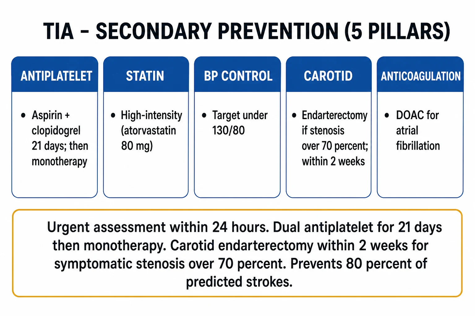
Secondary prevention is begun at once and is mechanism-specific. For non-cardioembolic TIA the current standard is short-course dual antiplatelet therapy: aspirin 300 mg immediately then combined with clopidogrel 300 mg loading dose and 75 mg daily, continued for 21 days, after which antiplatelet monotherapy (clopidogrel 75 mg daily) continues long-term. The 21-day dual regimen rests on CHANCE, which showed clopidogrel plus aspirin reduced the 90-day stroke risk from 11.7 per cent to 8.2 per cent without excess bleeding,[4] and POINT, which confirmed the ischaemic benefit but showed that bleeding accrues when dual therapy is extended beyond 21 days.[5] A high-intensity statin — atorvastatin 80 mg daily — is started regardless of baseline cholesterol on the strength of SPARCL, which reduced recurrent stroke over five years in patients with recent stroke or TIA.[6] Blood pressure is controlled to standard secondary-prevention targets once the acute event has settled.
The single most impactful intervention for the right patient is carotid endarterectomy for symptomatic severe stenosis. The pooled analysis of the endarterectomy trials showed a large, durable benefit for stenosis of 70 per cent or greater (NNT about 3 to prevent one ipsilateral stroke or death over two to five years), a smaller benefit for 50 to 69 per cent stenosis, and no benefit below 50 per cent.[7] The benefit is time-dependent: it is greatest when surgery is performed within two weeks of the presenting event and falls steeply with delay, so carotid imaging within 24 hours is what makes the operation possible in its window of benefit. For cardioembolic TIA from atrial fibrillation, the secondary-prevention therapy is anticoagulation (a direct oral anticoagulant preferred over warfarin for non-valvular atrial fibrillation) rather than an antiplatelet, which is inferior for cardioembolic prevention.
Anticoagulation timing after a cardioembolic TIA
Special populations
The young patient (under 50) with a TIA demands a search for a mechanism not seen in older patients — carotid or vertebral artery dissection (classically after neck trauma or manipulation), patent foramen ovale with paradoxical embolism, a thrombophilia (antiphospholipid syndrome, factor V Leiden), vasculitis, or drug use — because the mechanism changes prevention (endovascular or surgical closure for PFO, anticoagulation for thrombophilia, antiplatelet for dissection). The anticoagulated patient who has a definite TIA has failed that treatment: confirm adherence and the time of the last dose, check renal function and anti-Xa levels where available, reconsider the mechanism (a new carotid stenosis, a missed cause), and escalate with the stroke team rather than simply layering on an antiplatelet (which adds bleeding without clear benefit). Elderly patients carry the highest absolute risk and present more often with atypical or posterior-circulation events; the same urgency applies. TIA in pregnancy is rare and is worked up and treated in collaboration with obstetrics and stroke teams; the imaging and antiplatelet choices are adapted to the trimester. [1]
Evidence and regional guidelines
The evidence base is strong and practice-defining. The tissue-based definition is the AHA/ASA 2009 scientific statement.[1] The ABCD2 score was validated by Johnston in 2007.[2] The case for urgent treatment is EXPRESS,[3] and for short-course dual antiplatelet therapy it is CHANCE[4] and POINT.[5] High-dose statin secondary prevention is SPARCL,[6] and the carotid endarterectomy benefit by stenosis grade is the pooled analysis of the endarterectomy trials.[7] Regionally, ANZ practice follows the Stroke Foundation of Australia Clinical Guidelines for Stroke Management (a living guideline) — short-course aspirin plus clopidogrel for high-risk TIA or minor stroke, carotid imaging within 24 to 48 hours, and endarterectomy within two weeks for symptomatic severe stenosis. The United Kingdom follows NICE guideline NG128 (2019), which sets same-day specialist assessment and carotid imaging for every suspected TIA and no longer uses ABCD2 to triage access. The United States follows the AHA/ASA secondary prevention guideline, and Europe the ESO guidelines. The agents, the 21-day dual window, and the two-week endarterectomy window are converging globally.
ANZ practice note. Australian and New Zealand practice follows the Stroke Foundation Clinical Guidelines for Stroke Management. A suspected TIA is referred for specialist assessment and carotid imaging on the same day through the local stroke service or rapid-access TIA clinic; high-risk patients (crescendo, atrial fibrillation, severe stenosis, recurrence on antiplatelet) are admitted. High-risk non-cardioembolic TIA and minor stroke receive aspirin plus clopidogrel for 21 days then clopidogrel monotherapy. Symptomatic severe carotid stenosis is referred for endarterectomy within two weeks via the regional vascular or stroke service, and carotid imaging is obtained within 24 hours of presentation to make that referral possible. [1]
SAQ — A high-risk TIA in atrial fibrillation
10 minutes · 10 marks
A 68-year-old man presents to the emergency department one hour after a 25-minute episode of right-sided weakness and slurred speech that has fully resolved. He is in atrial fibrillation. The blood pressure is 156 over 92, the bedside glucose is 6.8 mmol per litre, and a carotid bruit is audible on the left.
SAQ — Crescendo amaurosis fugax and the severe carotid stenosis
10 minutes · 10 marks
A 72-year-old woman presents with three episodes of the left-eye visual loss over 24 hours, each described as a curtain descending for two to three minutes then clearing. She has a loud left carotid bruit and is on the aspirin 75 mg daily for the hypertension.
Exam pearls
- Tissue-based definition — a diffusion-positive lesion is a stroke, not a TIA; the clock has been replaced by the scan.
- ABCD2 — Age, Blood pressure, Clinical features, Duration, Diabetes; learn the point allocations cold and the three risk strata.
- Carotid endarterectomy within two weeks for symptomatic severe stenosis is the single highest-impact intervention — carotid imaging within 24 hours is what makes it possible.
- Short-course dual antiplatelet — aspirin plus clopidogrel for 21 days (CHANCE + POINT), then monotherapy; the 21-day limit exists because POINT's bleeding signal accrued after that point.
- Aspirin 300 mg stat is the acute load for every non-cardioembolic TIA after haemorrhage is excluded.
- Glucose first — hypoglycaemia mimics TIA exactly and is reversed in minutes.
- Tempo distinguishes the mimics — TIA is maximal at once and negative; migraine marches and is positive; Todd paresis follows a seizure.
- A TIA on an anticoagulant is treatment failure — reassess mechanism, check adherence and the last dose, do not simply add an antiplatelet.
- Anticoagulation (not antiplatelet) is the secondary prevention for cardioembolic TIA from atrial fibrillation, and can start early because there is no infarct to transform. [1]
Red flags
[1]References
- [1]Easton JD, Saver JL, Albers GW, et al. Definition and evaluation of transient ischemic attack: a scientific statement for healthcare professionals from the American Heart Association/American Stroke Association Stroke Council; Council on Cardiovascular Surgery and Anesthesia; Council on Cardiovascular Radiology and Intervention; Council on Cardiovascular Nursing; and the Interdisciplinary Council on Peripheral Vascular Disease. The American Academy of Neurology affirms the value of this statement as an educational tool for neurologists Stroke, 2009.PMID 19423857
- [2]Johnston SC, Rothwell PM, Nguyen-Huynh MN, et al. Validation and refinement of scores to predict very early stroke risk after transient ischaemic attack Lancet, 2007.PMID 17258668
- [3]Rothwell PM, Giles MF, Chandratheva A, et al. Effect of urgent treatment of transient ischaemic attack and minor stroke on early recurrent stroke (EXPRESS study): a prospective population-based sequential comparison Lancet, 2007.PMID 17928046
- [4]Wang Y, Wang Y, Zhao X, et al. Clopidogrel with aspirin in acute minor stroke or transient ischemic attack N Engl J Med, 2013.PMID 23803136
- [5]Johnston SC, Easton JD, Farrant M, et al. Clopidogrel and Aspirin in Acute Ischemic Stroke and High-Risk TIA N Engl J Med, 2018.PMID 29766750
- [6]Amarenco P, Bogousslavsky J, Callahan A 3rd, et al. High-dose atorvastatin after stroke or transient ischemic attack N Engl J Med, 2006.PMID 16899775
- [7]Rothwell PM, Eliasziw M, Gutnikov SA, et al. Analysis of pooled data from the randomised controlled trials of endarterectomy for symptomatic carotid stenosis Lancet, 2003.PMID 12531577
- [8]Barnett HJ, Taylor DW, Eliasziw M, et al. Benefit of carotid endarterectomy in patients with symptomatic moderate or severe stenosis. North American Symptomatic Carotid Endarterectomy Trial Collaborators N Engl J Med, 1998.PMID 9811916
- [9]European Carotid Surgery Trialists' Collaborative Group. Randomised trial of endarterectomy for recently symptomatic carotid stenosis: final results of the MRC European Carotid Surgery Trial (ECST) Lancet, 1998.PMID 9593407