EM · Procedural & diagnostic ED skills
Fracture and dislocation reduction in the ED
Also known as Joint reduction · Closed reduction of dislocations · Shoulder relocation · Nursemaid elbow reduction · Radial head subluxation reduction · Hip relocation · Pulled elbow
Closed reduction of the dislocations the emergency physician reduces in the ED — anterior and posterior shoulder, pulled elbow (nursemaid elbow), finger interphalangeal joints, and traumatic hip dislocation. The technique set is joint-specific: traction-countertraction, Stimson, scapular manipulation, Cunningham and FARES for the anterior shoulder; hyperpronation for the pulled elbow; a digital block then longitudinal traction for the finger; Allis, Stimson and the Captain Morgan lever for the hip. Analgesia and sedation follow the procedural sedation standard — ketamine 1 mg/kg IV, fentanyl 1 mcg/kg IV, propofol 1 mg/kg IV — with a pre-procedure fast, full monitoring and a trained team. The non-negotiables are the pre- and post-reduction neurovascular examination and a post-reduction radiograph on every patient. The differential that disqualifies an ED closed attempt is the fracture-dislocation and the pathological fracture. ACEM Fellowship, globally tagged.
On this page & tools
Your progress
Saved locally on this device.
8 MCQs with explanations
Target exams
Red flags
Related topics
- Procedural sedation in the emergency department
- Local anaesthesia and topical agents
- Paediatric trauma — the modified approach
- Consent, capacity and the medico-legal framework in the emergency department
- Major trauma resuscitation — the team-based systematic approach
- Acute limb ischaemia (the emergency of the 6 Ps)
Closed reduction of joint dislocations is core emergency medicine practice — the joints the emergency physician is expected to reduce without orthopaedic support are the shoulder, the elbow (pulled elbow in the child), the finger, and, with urgency and often a general anaesthetic, the hip.[1][8] The Fellowship candidate must hold three things simultaneously for each joint: the technique (named, step-by-step, recited as if to an examiner at a viva), the analgesia and sedation (the drug, the dose, the route, the timing and the rationale, recited to the procedural sedation standard), and the safety net (the pre- and post-reduction neurovascular examination and the post-reduction radiograph, on every patient, without exception).[10] The recurring Fellowship trap is to know the manoeuvre but to forget the safety net — the shoulder reduced without a documented post-reduction axillary-nerve check and radiograph is an incomplete answer and a real-world liability. The examiner mental model is built joint-by-joint, because the techniques and the complications differ, but the discipline — consent, drugs, technique, re-image, neurovascular, disposition — is the same for every reduction.
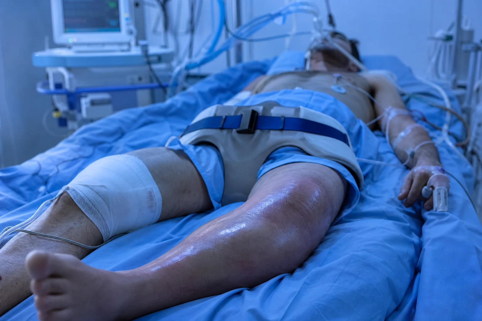
Definition and indications
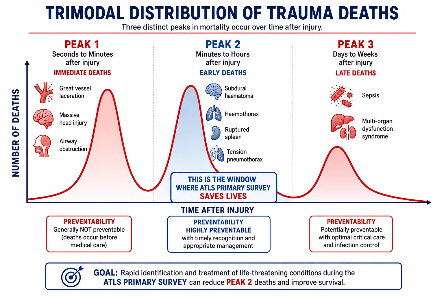
A dislocation is the complete loss of articular congruity between two opposing joint surfaces; a subluxation is a partial loss that spontaneously reduces or remains incompletely displaced. Closed reduction is the manipulation that restores the normal joint relationship through the skin, without an incision; it is distinguished from open reduction (in theatre) and from the percutaneous pinning or internal fixation that a fracture-dislocation requires.[1][8]
The indication for ED closed reduction is a radiographically confirmed dislocation without an associated fracture that mandates operative management, in a patient in whom procedural sedation is safe or not required. The joints the emergency physician reduces routinely are the glenohumeral (shoulder) joint — the commonest major-joint dislocation presenting to the ED, of which the anterior variant is roughly 95 per cent — the radial head subluxation (pulled elbow, nursemaid elbow) of the toddler, the finger interphalangeal and metacarpophalangeal joints, and the hip, which is the most time-critical dislocation the ED encounters because of the avascular-necrosis risk to the femoral head.[1][5][8] The elbow, wrist and knee dislocations are reduced in the ED but commonly in concert with orthopaedics, because the rate of associated fracture and neurovascular injury is high — the knee dislocation in particular is a limb-threatening injury with a high rate of popliteal artery injury, and it is managed as a vascular emergency, not a routine reduction.
The timing of reduction matters as much as the indication. A hip dislocation is reduced as soon as the diagnosis is made — the target is within 6 hours from injury, because the risk of avascular necrosis of the femoral head climbs steeply beyond that window; a shoulder or finger dislocation is reduced promptly to relieve pain and to relieve the stretch on the neurovascular structures, but without the same clock. A vascular-compromised dislocation (the cold, pulseless limb) is reduced immediately, before radiography if the films would delay the reduction, and is handed to the vascular and orthopaedic surgeons in parallel.[8]
Contraindications
There is no absolute contraindication to reducing a dislocation that is compromising the limb — a vascular-compromised joint is reduced on the spot, and the radiograph follows. The contraindications are the factors that change the setting of the reduction from the ED to the operating theatre, or the technique from a single closed attempt to a planned orthopaedic procedure.[1][8]
The fracture-dislocation is the principal contraindication to a blind ED closed reduction. A displaced greater tuberosity fracture, a surgical neck fracture of the humerus, a Hill-Sachs or reverse Hill-Sachs impression fracture of significance, a Barton or paediatric physeal fracture through the joint, and a fracture of the femoral head or an associated acetabular wall fracture all change the reduction from a manipulative act to an operative one — the fragments will not reduce cleanly by traction, and a forceful attempt displaces the fracture further or injures the neurovascular bundle. The emergency physician reviews the radiograph for a fracture before each reduction, and if a fracture-dislocation is recognised the reduction moves to theatre with orthopaedics.[1][8]
The open dislocation (a wound communicating with the joint), the dislocation with an associated vascular injury that does not recover after reduction, and the dislocation in the pathological bone (a metastasis or a simple bone cyst through which the joint has dislocated) are reduced or stabilised in theatre. The uncooperative or unconsentable patient who cannot hold still for a closed reduction under procedural sedation is converted to a general anaesthetic, and the multiply-attempted dislocation that has not reduced after two or three good attempts under adequate sedation is escalated rather than forced — repeated forceful attempts convert a simple dislocation into a fracture-dislocation or a neurovascular injury, and the discipline of stopping is itself an examinable decision.[10]
Relevant anatomy and the landmarks that govern each technique
The anatomy that matters is joint-specific, because each reduction technique exploits a particular anatomical relationship. The glenohumeral joint is a shallow ball-and-socket — the large humeral head on the small glenoid fossa — which is why the shoulder is the most frequently dislocated major joint; the anterior dislocation displaces the head inferiorly and anteriorly, stripping the capsule and the labrum (the Bankart lesion) and impressing the head against the glenoid rim (the Hill-Sachs lesion). The axillary nerve exits the quadrilateral space and supplies sensation to the skin over the deltoid (the regimental-badge patch) — the nerve stretched or injured in up to a third of anterior dislocations and the one the post-reduction examination must test.[1]
The posterior shoulder dislocation (under 5 per cent of shoulder dislocations) drives the head posteriorly, locking it against the glenoid rim and fixing the arm in internal rotation — the anatomical reason a single AP film looks deceptively normal and the axillary lateral is the essential second view. The pulled elbow (radial head subluxation) is a displacement of the annular ligament, which in the young child (peak age 2 to 3 years) is thin and inserts distally; a longitudinal pull on the arm (being lifted by the hand, a yank on the wrist) allows the ligament to slip over the radial head and into the radiocapitellar joint, where it interposes and locks the forearm pronated.[5][7] The finger joints — the proximal interphalangeal (PIP), the most commonly dislocated, and the metacarpophalangeal — receive their sensory supply from the four digital nerves (two palmar, two dorsal), which the digital block anaesthetises at the base of the finger or the web space.
The hip is a deep, congruent ball-and-socket — the femoral head seated in the acetabulum — that dislocates only with high energy (a dashboard injury in a motor-vehicle crash, a fall from height). The posterior hip dislocation (the commonest) displaces the head posteriorly and superiorly, shortening the leg and internally rotating it, and threatens the sciatic nerve (the peroneal division first — test dorsiflexion) and the blood supply to the femoral head, which enters via the retinacular vessels and is torn by the persistent dislocation — the anatomical basis of the 6-hour avascular-necrosis clock.[8]
Equipment, monitoring and procedural sedation drugs
The reduction of a major joint in the adult is performed under procedural sedation and analgesia (PSA), and the equipment is therefore the PSA set plus the reduction adjuncts. The ACEP clinical policy on procedural sedation — the framework the Fellowship answer invokes — requires a trained team (the sedationist dedicated to the airway and the drugs, the operator performing the reduction, and a nurse monitoring the patient), full monitoring (continuous pulse oximetry, ECG, blood pressure, capnography), supplemental oxygen, suction at the bedside, a reversible agent available where one exists, and the reversal and resuscitation equipment immediately to hand.[10]
The procedural sedation drugs — agent, dose, route, onset and rationale
The non-pharmacological setup matters as much as the drugs. A pre-procedure fasting history is taken (the ACEP policy regards recent oral intake as a consideration, not an absolute contraindication, because the aspiration risk is balanced against the urgency of the reduction), the patient is on a trolley that tilts head-down for the Stimson and scapular techniques, a sheet is available for the traction-countertraction counteranchor, weights of 5 to 10 kilograms are available for the hanging-arm Stimson, and the post-reduction radiograph is ordered before the sedation begins so that the patient is imaged as soon as the joint is reduced.[10][2]
Patient preparation, consent and the pre-reduction assessment
Preparation has four steps that the Fellowship answer states in order: the history and mechanism, the examination with the neurovascular check, the radiograph, and the consent and pre-medication. The mechanism is recorded because it predicts the dislocation pattern (the abduction-external-rotation force of a tackle or a fall produces the anterior shoulder; the dashboard force produces the posterior hip; the longitudinal pull on the child's arm produces the pulled elbow). The pre-reduction examination documents the deformity, the skin (an open wound converts the case to theatre), and — critically — the neurovascular status before any manipulation, because a deficit present before the reduction is a pre-existing injury and a deficit appearing after the reduction is a procedural one, and the two are distinguished only by the pre-reduction record.[1]
The radiograph is taken before the reduction in almost every case, to confirm the dislocation and to exclude a fracture-dislocation that would change the plan. The two exceptions are the classic pulled elbow in the typical age group with the classic mechanism (where the radiograph is normal and is reserved for the atypical case or the post-reduction failure) and the vascular-compromised dislocation (where the cold, pulseless limb is reduced immediately and the radiograph follows). For the shoulder, the two-view series is the AP and the axillary lateral or scapular-Y — the view that excludes a posterior dislocation and that confirms the reduction; a single AP is an incomplete work-up.[1]
Consent is obtained and documented for the reduction and the sedation. The patient is told what the procedure involves, the alternatives (orthopaedic reduction under general anaesthesia), and the specific risks — failure requiring theatre, fracture, neurovascular injury, recurrence, and the sedation risks (apnoea, hypotension, aspiration, recovery agitation). For the child, consent is from the parent or guardian, and the pulled-elbow reduction is explained as a quick manipulation that may be briefly uncomfortable. The consent for a hip reduction under sedation includes the explicit statement that general anaesthesia and muscle relaxation may be required if the closed attempt fails.[10]
Differential diagnosis — what is not a simple dislocation
The differential is the set of conditions that masquerade as a dislocation or that disqualify the simple ED reduction, and the Fellowship answer must distinguish each by its radiographic appearance and its implication. The discriminating question is whether the joint surfaces, the surrounding bone and the clinical setting are consistent with a simple traumatic dislocation — if any of the three is not, the case moves to orthopaedics.[1][8]
Fracture-dislocation
- A dislocation with an associated fracture of the articulating bone — the greater tuberosity or surgical neck of the humerus, a Hill-Sachs or reverse Hill-Sachs impression, a femoral head or acetabular fracture
- Distinguished on the radiograph by the fracture line; a forceful ED reduction displaces the fragment further
- Managed by orthopaedics in theatre — open reduction and internal fixation; the ED role is recognition, analgesia and the neurovascular check
- The commonest reason a "simple" dislocation must not be reduced blindly in the ED
Pathological fracture-dislocation
- Dislocation or fracture through a bone weakened by a lytic lesion — a metastasis (breast, prostate, lung, kidney, thyroid), a myeloma deposit, or a simple bone cyst in the young
- Distinguished by the lytic or expansile lesion on the radiograph, the disproportionate mechanism (a dislocation from minor trauma in an older patient), and the systemic features of malignancy
- A marker of underlying disease; bone is fragile and a closed reduction may collapse it further — orthopaedic and oncology referral
- Always consider in the elderly dislocator and in any dislocation from trivial trauma
Recurrent / multidirectional instability
- A dislocation in a patient with congenital ligamentous laxity or a prior Bankart lesion — the joint dislocates with minimal force and reduces easily, often without sedation
- Distinguished by the history of multiple prior dislocations, the generalised joint hypermobility (Beighton signs), and the radiograph without acute fracture
- Reduced in the ED, often with analgesia alone or intra-articular lidocaine; the disposition includes orthopaedic referral for stabilisation
- Not dangerous in itself, but the recurrence risk is high and the patient is counselled on the surgical option
Septic arthritis mimicking a painful joint
- A hot, swollen, painful joint held immobile that may be mistaken for an undischarged dislocation, especially in the shoulder or the hip of a septic patient
- Distinguished by the fever, the inflammatory markers, and the radiograph showing a reduced joint with soft-tissue swelling; aspirate if septic arthritis is suspected
- Managed as septic arthritis — urgent aspiration, antibiotics, orthopaedics; reducing a septic joint by force damages the cartilage
- The trap is a "frozen" or splinted joint held by pain being manipulated as a dislocation
The discriminating test across these is the radiograph read with the differential in mind — a fracture line, a lytic lesion, or a joint that is in fact reduced all change the diagnosis and the plan. The clinical setting is the second discriminator: the elderly patient dislocating from a trivial mechanism is a pathological fracture until proven otherwise, and the septic patient with a painful joint is septic arthritis until aspirated.[1][8]
Stepwise technique — anterior shoulder reduction
The anterior shoulder is the dislocation the Fellowship candidate must reduce on a viva or a simulator, and the answer is to know at least three techniques and to choose among them by the patient and the setting. The network meta-analysis of closed reduction techniques found no single manoeuvre universally superior — success rates are high across techniques, and the choice is governed by the need for sedation, the speed, the patient comfort and the operator familiarity.[1] The five techniques below are the standard set, recited step-by-step.
The five anterior shoulder reduction techniques, step by step
-
Traction-countertraction (the classic). Position the patient supine. Wrap a sheet around the operator's wrists and around the patient's chest under the axilla (the counteranchor), with an assistant holding the sheet for countertraction. Apply slow, steady longitudinal traction to the arm in line with the deformity for several minutes, overcoming the muscle spasm by sustained gentle force rather than a jerk. The head reduces with a palpable clunk. Sustained gentle traction is the principle — a jerk risks a fracture. [1]
-
Stimson (hanging weights, gravity). Position the patient prone on the trolley with the affected arm hanging over the edge. Secure 5 to 10 kilograms of weight to the wrist and leave the arm to hang for 15 to 30 minutes; the gravity traction and the gradual muscle relaxation reduce the shoulder in the majority. The scapular manipulation (below) is added if the Stimson alone has not worked at 20 minutes. Minimal operator force and minimal sedation — a good first-line in the patient in whom sedation is to be avoided. [1]
-
Scapular manipulation. Position the patient prone with the arm hanging (the Stimson setup). Palpate the scapula. Push the inferior tip of the scapula medially toward the spine and rotate the superior aspect laterally, so that the glenoid fossa rotates to face the humeral head. The manoeuvre realigns the glenoid with the displaced head, and the joint reduces. Low force and a high success rate; the technique of choice for many ED operators because it works with minimal sedation. [1]
-
Cunningham (massage and traction). Position the patient sitting upright on the edge of the trolley, relaxed, leaning forward with the operator supporting the arm. Massage the deltoid, then the biceps and the mid-deltoid to relax the muscle spasm; apply gentle downward traction on the arm in line with the body. The combination of relaxation and gentle traction reduces the shoulder, often without sedation. Requires a cooperative, relaxed patient. [1]
-
FARES (Fast, Reliable, and Safe) and Spaso. For FARES, position the patient supine. The operator holds the wrist, applies gentle longitudinal traction, and oscillates the arm in vertical arcs of decreasing amplitude while slowly abducting the arm; the head reduces during the oscillation. For Spaso, lift the arm vertically with traction and externally rotate. Both are single-operator techniques with high success rates and low analgesia requirements — the FARES-versus-Spaso trial confirmed both as effective first-line options. [1]
The structural principle common to all five is that the muscle spasm holds the head out of joint, and every technique either overcomes the spasm by sustained traction (Stimson, traction-countertraction) or realigns the glenoid with the head so that the spasm is irrelevant (scapular, FARES). The contemporary evidence favours the low-force, low-sedation techniques — the scapular, Cunningham and FARES — because they reduce the shoulder without the airway and haemodynamic risks of deep sedation, and the network meta-analysis and the comparative sedation review both support their use as first-line.[1][2][3] The intra-articular lidocaine alternative — 10 to 20 mL of lidocaine 1 per cent into the glenohumeral joint via the lateral approach — provides analgesia and muscle relaxation without sedation, with success and safety comparable to procedural sedation; it is the option of choice for the patient in whom sedation is undesirable or contraindicated.[2]
Posterior shoulder reduction — the missed dislocation
The posterior shoulder dislocation is the dislocation the Fellowship candidate is examined on precisely because it is missed, and the answer has two parts — how not to miss it, and how to reduce it. The dislocation accounts for under 5 per cent of shoulder dislocations and arises from three mechanisms: an internal-rotation force on the raised arm (a fall or a seizure), an electric shock or electrocution (the violent contraction of the internally-rotating muscles), and a seizure (the same mechanism). The arm is held in fixed internal rotation, and the patient cannot externally rotate — the clinical sign that, with the mechanism, flags the diagnosis.[1]
The radiograph is where the dislocation is missed. The AP film looks deceptively normal because the head, displaced posteriorly, still overlaps the glenoid on the frontal view — the only clues are the lightbulb sign (the internally rotated humeral head looks round like a lightbulb) and the absence of the normal greater-tuberosity profile. The dislocation is confirmed on the axillary lateral or scapular-Y view, which shows the head posterior to the glenoid. A single AP of a painful post-seizure shoulder is an incomplete work-up, and ordering the second view is the decision the viva rewards.[1]
The reduction is performed with longitudinal traction combined with lateral traction on the humeral shaft to lift the head over the glenoid rim, with the arm gently externally rotated once the head clears the rim. Posterior dislocations reduce less readily than anterior ones and are commonly performed under general anaesthesia with muscle relaxation, because the muscle spasm is intense and a significant reverse Hill-Sachs lesion (the impression fracture on the anterior humeral head) may lock the head posteriorly. The dislocation that does not reduce closed is taken to theatre for an open reduction, and the post-reduction immobilisation is in external rotation — the opposite of the anterior dislocation, because the arm must be held in the position of stability.[1][4]
Pulled elbow — nursemaid elbow reduction
The pulled elbow (radial head subluxation) is the commonest upper-limb injury of the preschool child, and the Fellowship answer is the manoeuvre (recited as hyperpronation, the first-line technique) and the post-reduction observation (the return of normal arm use within minutes, which confirms the diagnosis and the reduction). The mechanism is a sudden longitudinal pull on the extended, pronated arm — a parent lifting the child by the hand, a yank to prevent a fall — and the annular ligament slips over the radial head and interposes in the radiocapitellar joint.[5][7]
The child presents with the arm held pronated and by the side, refusing to use it, with no bony tenderness and a full range of passive movement of the shoulder and the wrist. A classic presentation in the typical age group (1 to 4 years, peak 2 to 3) with the classic mechanism does not require a radiograph before reduction, because the radiograph is normal in a pulled elbow; the film is reserved for the atypical case (tenderness, swelling, a fall rather than a pull, an older child) or the case that does not use the arm normally after a successful reduction.[5]
The pulled-elbow reduction — hyperpronation first-line
- Hyperpronation (first-line). Support the child's elbow with one hand. With the other hand, grasp the wrist and fully supinate then hyperpronate the forearm (or hyperpronate directly). The radial head reduces with a click or a snap, often felt more than heard. Hyperpronation is first-line because the comparative trials and the Cochrane review find it equal or superior to supination-flexion in first-attempt success and less painful.
- Supination-flexion (alternative). Supinate the forearm and flex the elbow fully, bringing the hand toward the shoulder. The reduction is signalled by the click. The traditional technique, still taught and effective.
- After either manoeuvre, leave the child and observe — the arm is used normally within 10 to 30 minutes as the child relaxes. The return of normal use is the confirmation; instruct the parents to avoid longitudinal traction on the arm (lifting by the hands) to prevent recurrence. [1]
The evidence base for the choice between the techniques is the comparative-effectiveness series and the Cochrane review, which together find that hyperpronation has a higher first-attempt success rate and is less painful than supination-flexion, establishing hyperpronation as the preferred first manoeuvre.[5][6][7] The recurrence rate is significant (around a quarter of children recur), and the parent education — avoid pulling the child by the hand or forearm, and return if the arm is not used — is part of the disposition. A child who does not use the arm within 30 minutes of a documented successful reduction, or who has bony tenderness or swelling, is re-imaged for an alternative diagnosis (an occult fracture, particularly the supracondylar fracture, which is the differential not to miss).[5]
Finger dislocation reduction — the digital block then traction
The finger joints are reduced under a digital block, because the manipulation is painful and the block anaesthetises the entire digit without systemic sedation. The technique is the block then the longitudinal traction, recited as a two-step sequence. The digital block is performed with plain lidocaine 1 to 2 per cent (never adrenaline — the digital arteries are end-arteries and adrenaline risks ischaemia) injected at the base of the finger or in the web space, blocking the two palmar and two dorsal digital nerves; the weight-based maximum for plain lidocaine is 3 mg per kilogram.[10]
The reduction, once the block is working, is longitudinal traction to disengage the joint surfaces, with exaggeration of the deformity (pushing further into the dislocation to clear the condyle over the rim) followed by re-alignment and re-impaction. The dorsal PIP dislocation — the commonest — is reduced by hyperextending slightly to disengage, then traction and flexion; the volar dislocation and the fracture-dislocation (a pilon or a dorsal lip fracture) are reduced with care, and a fracture-dislocation is splinted and referred to orthopaedics or a hand surgeon, because the fragment must be accurately reduced to preserve joint function.[1]
The digital block — the practical details
The post-reduction check is the neurovascular status of the digit (capillary refill and sensation), the active range of motion (the patient can flex and extend the reduced joint), and the stability through the range (a joint that subluxes through flexion is unstable and needs referral). A post-reduction radiograph confirms the reduction and excludes a fracture, and the finger is immobilised in a buddy strap (strapping the injured finger to an adjacent finger) or a dorsal extension-block splint, with hand-therapy or orthopaedic follow-up.[1]
Hip dislocation reduction — the time-critical joint
The hip is the dislocation that the ED must not delay on, because the avascular-necrosis clock is short and the joint is reduced urgently, usually under procedural sedation or general anaesthesia, often with the orthopaedic team present. The posterior hip dislocation is the commonest (around 9 in 10), produced by the dashboard force — the flexed knee driven backward into the dashboard in a frontal crash, driving the femoral head out posteriorly. The leg is shortened, adducted, flexed and internally rotated, and the patient is in severe pain; the sciatic nerve is threatened and the femoral-head blood supply is torn the longer the head remains dislocated.[8]
The anterior hip dislocation (under 1 in 10) drives the head forward through the capsule; the leg is abducted, externally rotated and flexed, and the femoral nerve and artery are threatened. Both are reduced as soon as the diagnosis is confirmed on the AP pelvis and the cross-table lateral, and both are managed in the trauma patient as part of the secondary survey — the hip dislocation is sought in the patient with a high-energy mechanism and a deformed leg, and the reduction is performed after the other life-threats are addressed but without the delay that a transfemoral angiogram or a prolonged scan would impose.[8]
The hip reduction techniques — Allis, Stimson, and the Captain Morgan lever
-
Allis (the commonest, supine). Position the patient supine on a firm surface, with an assistant stabilising the pelvis by pressing down on the anterior superior iliac spines. Stand on the trolley (the leverage is essential). Flex the hip and knee to 90 degrees. Apply upward longitudinal traction along the line of the femur, with gentle internal and external rotation to disengage the head over the acetabular rim. The head reduces with a palpable clunk. The assistant's pelvic fixation is the counteranchor — without it the whole pelvis lifts and the reduction fails. [1]
-
Stimson (gravity, prone). Position the patient prone with the affected leg over the edge of the trolley, hip and knee flexed. An assistant applies downward pressure on the sacrum to fix the pelvis. Apply downward traction on the calf, or have an assistant apply pressure on the flexed knee, while gently rotating the hip. The gravity traction reduces the head. Useful when the supine Allis has failed or when the patient cannot be moved supine. [1]
-
Captain Morgan / rocket-launcher (the operator's knee as a fulcrum). Position the patient supine. Stand on the side of the dislocated hip. Place your knee under the patient's flexed knee as a fulcrum. Apply downward force on the patient's ankle with one hand and upward leverage on the patient's calf with the other, using your knee as the fulcrum to lever the femoral head back into the acetabulum. The technique is a single-operator leverage method with good reported success; the rocket-launcher variant applies the same leverage principle. [1]
The hip dislocation that does not reduce with two or three good attempts under adequate sedation is converted to a general anaesthetic with full muscle relaxation — the reduction almost always succeeds once the muscle spasm is abolished — and the small number that resist even under anaesthesia are reduced open in theatre, often because a fracture fragment, a capsular interposition, or an associated acetabular wall is blocking the reduction. The post-reduction imaging is the AP pelvis and a CT of the hip to confirm concentric reduction, to exclude a fracture of the femoral head or the acetabulum, and to detect intra-articular bone fragments that mandate arthrotomy.[8][9]
The timing is the single most examinable point about the hip. The risk of avascular necrosis of the femoral head climbs steeply after 6 hours of dislocation — the retinacular vessels are torn and the longer the head is out, the higher the rate of osteonecrosis and secondary osteoarthritis — and the target is reduction within 6 hours from injury, ideally within 2. The ED that reduces a hip at 7 hours has missed the window, and the Fellowship answer states the 6-hour target explicitly and reduces urgently.[8]
Post-reduction assessment and disposition
The post-reduction assessment is the same for every joint and is the non-negotiable that the Fellowship answer cannot omit: a repeat neurovascular examination, a post-reduction radiograph, an immobilisation, and a disposition with follow-up. The neurovascular examination repeats the pre-reduction check and documents any change — a new axillary-nerve deficit after a shoulder reduction, a loss of the dorsalis pedis pulse after a hip reduction, or a cold finger after a digital manipulation is a surgical emergency, and the documentation that the deficit was or was not present before the reduction is the medico-legal record.[1][8]
The post-reduction radiograph confirms that the joint is concentrically reduced and excludes a fracture produced or revealed by the manipulation — the Hill-Sachs and Bankart lesions of the shoulder, the femoral-head or acetabular fracture of the hip, the small avulsion of the finger. A reduction without a post-reduction film is an incomplete procedure. The immobilisation is joint-specific: the anterior shoulder in a broad-arm sling or a shoulder immobiliser (the arm in internal rotation), the posterior shoulder in external rotation, the finger in a buddy strap or an extension-block splint, the pulled elbow in no splint (the parent is counselled to avoid traction), and the hip in no splint but on bed rest pending the orthopaedic plan.[1]
The disposition follows the joint and the complications. The shoulder and the finger are discharged with orthopaedic or hand-surgery follow-up in a week or two, with advice on recurrence and on the surgical option for the recurrent dislocator. The pulled elbow is discharged with parent education. The hip is admitted — it is a high-energy injury, the patient is a trauma patient with associated injuries, the reduction needs orthopaedic monitoring and a CT, and the protected weight-bearing and the avascular-necrosis surveillance are inpatient decisions. The patient with a neurovascular deficit, an associated fracture requiring fixation, or a dislocation that did not reduce in the ED is admitted or transferred to the operating theatre.[8]
Complications and pitfalls
The complications are divided into the procedure-related (the injuries of the manipulation) and the disease-related (the consequences of the dislocation itself). The immediate procedure-related complications begin with the iatrogenic fracture — the greater tuberosity or the surgical neck fractured by a forceful shoulder reduction, the femoral head or the acetabular rim fractured by a forceful hip reduction, the physis of the child injured by a forceful manipulation. The prevention is the sustained gentle traction and the discipline of stopping after two or three attempts and escalating. The neurovascular injury is the axillary nerve (shoulder), the sciatic nerve and the popliteal/femoral vessels (hip), and the digital vessels and nerves (finger); the prevention is the pre- and post-reduction check that detects the deficit, and the escalation to theatre for a deficit that does not recover.[1][8]
The sedation-related complications — apnoea, hypotension, aspiration, recovery agitation, and the rare but serious laryngospasm with ketamine — are the complications of procedural sedation, prevented by the trained team, the full monitoring and the adherence to the ACEP policy. The disease-related complications are the recurrent instability of the shoulder (the rate of recurrence after a first dislocation is high in the young — over half in patients under 20 — because the Bankart lesion heals poorly, and the patient is counselled on the surgical option), the avascular necrosis of the femoral head (the time-dependent complication of the hip, prevented by the urgent reduction), and the post-traumatic arthritis of any joint injured by a dislocation and a fracture.[2][8]
The complications and their prevention
The pitfalls invert the structure, and the Fellowship candidate must name them because they are the recurring failures. Reducing without a post-reduction neurovascular check and radiograph — the classic omission, the source of the missed iatrogenic injury and the medico-legal claim. Missing the posterior shoulder dislocation on a single AP — ordering the axillary lateral or scapular-Y is the decision that catches it. Forcing a reduction that will not go — the fracture-dislocation or the locked posterior shoulder that needs theatre, reduced by repeated force into a fracture or a nerve injury. Delaying the hip reduction for imaging — the avascular-necrosis clock runs out, and the hip is reduced first, scanned after. Using adrenaline in a digital block — the ischaemic digit. Forgetting that the multiply-attempted dislocation is escalated, not forced — the discipline of stopping is itself the correct decision.[1][8][10]
Special populations
The child is the population in whom the technique and the dosing diverge. The pulled elbow is the child-specific dislocation, reduced by hyperpronation without a radiograph in the classic case, and the parent education is part of the discharge. The procedural sedation dosing in the child is weight-based — ketamine 1 to 1.5 mg per kilogram IV is the preferred agent for the paediatric reduction because it preserves the airway reflexes and the blood pressure, and the comparative trial of ketamine versus propofol for paediatric forearm-fracture reduction found both effective, with ketamine providing more stable haemodynamics and propofol a faster recovery; the choice is made by the patient and the team.[11] The paediatric shoulder and hip dislocations are rare and are managed with paediatric orthopaedic input, because the open physis is injured by force and the reduction is gentler.[8]
The elderly patient is the population in whom the posterior shoulder dislocation is missed (after a fall or a seizure, the arm held in internal rotation, the AP deceptively normal) and in whom the pathological fracture-dislocation is the diagnosis in any dislocation from trivial trauma — the lytic lesion on the radiograph is sought, and the bone is fragile and reduced gently or referred. The anticoagulated patient is the population in whom the sedation bleeding risk and the fracture-dislocation interact — the haematoma of a shoulder or a hip dislocation is larger on anticoagulants, and the neurovascular compromise is sought actively.[1][8]
The pregnant patient with a dislocation is reduced with the standard techniques and the standard drugs, with the left lateral tilt to avoid aortocaval compression in the third trimester and the foetal-risk consideration applied to the drug choice — propofol and fentanyl are acceptable, ketamine is acceptable, and the sedation is performed with obstetric awareness. The trauma patient with a hip dislocation is managed in the secondary survey, the dislocation reduced after the life-threats are addressed but without the delay that misses the 6-hour window, and the associated acetabular and femoral-head fractures are sought on the post-reduction CT.[8]
Evidence and the regional guidelines
The contemporary framework for the sedation that underpins the reduction is the ACEP clinical policy on procedural sedation and analgesia in the emergency department (Godwin and colleagues, 2014), which sets the team, the monitoring, the fasting consideration and the reversal-agent availability that the Fellowship answer invokes.[10] The framework for the shoulder reduction technique is the network meta-analysis of Gonai and colleagues (2023), which compared the closed reduction techniques and found no single manoeuvre universally superior, supporting the operator's choice among the low-force, low-sedation options — and the comparative efficacy review of Hayashi and colleagues (2022), which compared the sedation and analgesia methods and found intra-articular lidocaine comparable to procedural sedation for the anterior shoulder.[1][2] The FARES-versus-Spaso trial (Lee and colleagues, 2025) and the BRASD trial (Baden and colleagues, 2023) add the contemporary evidence for the single-operator, low-sedation techniques.[3][4]
The framework for the pulled elbow is the Cochrane review of Krul and colleagues (2017), which found that both hyperpronation and supination-flexion are effective and that the evidence favours hyperpronation as the less painful and more successful first manoeuvre, supported by the comparative-effectiveness series of Aksel and colleagues (2025) and the randomised comparison of Ulici and colleagues (2019).[5][6][7] The framework for the hip dislocation is the JAAOS review of Benedick and colleagues (2024), which sets the paediatric and adult evaluation, the timing (reduce within 6 hours), the techniques (Allis, Stimson, the Captain Morgan lever) and the post-reduction CT, with the rocket-launcher technique of Dan and colleagues (2015) adding the single-operator leverage method.[8][9]
ANZ practice note. In Australian and New Zealand emergency departments, the reductions are performed under the procedural sedation standard taught in the ACEM and ANZCA training — a sedationist dedicated to the airway and the drugs, full monitoring including capnography, and the reversal agents immediately available. The anterior shoulder is reduced by the scapular, Cunningham or FARES techniques as first-line, with intra-articular lidocaine as the sedation-sparing option, and the traction-countertraction as the fallback. The pulled elbow is reduced by hyperpronation first-line. The hip is reduced urgently under sedation or general anaesthesia with the orthopaedic team, the Allis or the Captain Morgan as the first technique, and the 6-hour target is the audit standard. The post-reduction neurovascular check and radiograph are the documented safety net on every patient, and the ACEM fellowship candidate recites them unprompted. [1]
Exam practice
SAQ — First-time anterior shoulder dislocation: reduction technique, sedation and the safety net
10 minutes · 10 marks
A 28-year-old male surfer is brought to the emergency department 45 minutes after his right shoulder 'popped out' when he was dumped by a wave onto his outstretched, abducted and externally rotated arm. He holds the arm in abduction and external rotation, supporting it with the opposite hand. He is in severe pain (numerical rating scale 8/10) but fully cooperative, with no head injury, no other injury, and no past history of dislocation. Vitals: BP 132/80, HR 104, RR 20, SpO2 98 per cent on room air, GCS 15. He last ate 2 hours ago and last drank clear fluids 30 minutes ago. The radiograph shows an anterior-inferior glenohumeral dislocation with a small Hill-Sachs lesion but NO greater-tuberosity or surgical-neck fracture. The pre-reduction neurovascular examination is intact, including deltoid sensation over the regimental-badge patch (axillary nerve) and a strong radial pulse.
SAQ — Open tibial shaft fracture (Gustilo IIIA): the ED work-up, antibiotics and debridement timing
10 minutes · 10 marks
A 32-year-old motorcyclist is brought to the trauma bay 35 minutes after a high-speed collision with a guardrail. His right leg struck the barrier and he has an obvious deformity of the mid-shaft tibia with a 6 cm transverse laceration over the anteromedial shin through which the fracture ends are visible. The wound is heavily contaminated with road grit and grease, and there is active venous oozing. The foot is warm with a palpable dorsalis pedis pulse, sensation is intact throughout, and he can dorsiflex and plantarflex the toes against gravity. He is haemodynamically stable: BP 124/78, HR 96, RR 22, SpO2 97 per cent on 15 L via non-rebreather, GCS 15, temperature 36.8 degrees C. His last tetanus booster was 8 years ago. The secondary survey reveals no other major injury. Radiography confirms a comminuted mid-shaft tibial shaft fracture with a butterfly fragment, no intra-articular extension.
Exam pearls
- Pre- and post-reduction neurovascular, every time. The axillary nerve (deltoid sensation, the regimental-badge patch) is the shoulder nerve; the sciatic nerve (dorsiflexion) and the distal pulses are the hip; the capillary refill and sensation are the finger. The pre-reduction check distinguishes the pre-existing from the procedural deficit.
- Post-reduction radiograph, every time. A reduction without a confirmatory film is an incomplete procedure — confirm concentric reduction, exclude an iatrogenic or revealed fracture.
- Posterior shoulder = locked internal rotation, missed on a single AP. Order the axillary lateral or scapular-Y; the dislocation of seizures and electrocution. Reduce with traction and lateral traction, immobilise in external rotation.
- Hip = the 6-hour clock. Reduce within 6 hours to cut the avascular-necrosis risk; the most time-critical dislocation the ED encounters. Reduce first, scan after.
- Hyperpronation first-line for the pulled elbow — higher first-attempt success and less pain than supination-flexion; no radiograph in the classic case; the return of normal arm use within minutes confirms the reduction.
- Two or three good attempts, then escalate. The multiply-attempted dislocation is taken to theatre or general anaesthesia, not forced — repeated force makes a fracture-dislocation or a nerve injury.
- Intra-articular lidocaine for the shoulder — the sedation-sparing alternative, comparable success and safety to procedural sedation; the option for the patient in whom sedation is undesirable.
- Never adrenaline in a digital block — plain lidocaine only, weight-based maximum 3 mg per kilogram; the finger is an end-arterial territory. [1]
Red flags
[1]References
- [1]Gonai S, Misawa A, Kamijo Y, et al. A Systematic Review With Pairwise and Network Meta-analysis of Closed Reduction Methods for Anterior Shoulder Dislocation Ann Emerg Med, 2023.PMID 36797133
- [2]Hayashi M, Lee S, Yang HJ, McLeod S, Yazdi F, Vahidnia F, Hesko C, Stiell I, Perry JJ. Comparative efficacy of sedation or analgesia methods for reduction of anterior shoulder dislocation: A systematic review and network meta-analysis Acad Emerg Med, 2022.PMID 35872652
- [3]Lee KH, Jo JH, Kim JW, et al. FARES and Spaso method for anterior shoulder dislocation: a prospective randomized control study demonstrating the benefit of a combined approach Sci Rep, 2025.PMID 40447745
- [4]Baden DN, Bertrand M, Stancu B, et al. BRASD trial: biomechanical reposition techniques in anterior shoulder dislocation-a randomized multicenter clinical trial Int J Emerg Med, 2023.PMID 36829126
- [5]Aksel G, Kilicli AB, Guler S, Eroglu SE, Cevik AA. Comparative effectiveness of supination-flexion and hyperpronation maneuvers in radial head subluxation: A systematic review and meta-analysis Am J Emerg Med, 2025.PMID 40086092
- [6]Ulici A, Nahoi CI, Gheorghiu CP, Birhala E, Carp M, Laptoiu D. Nursemaid's Elbow - Supination-flexion Technique Versus Hyperpronation/forced Pronation: Randomized Clinical Study Indian J Orthop, 2019.PMID 30905991
- [7]Krul M, van der Wouden JC, Koes BW, Schellevis FG, van Suijlekom-Smit LWA. Manipulative interventions for reducing pulled elbow in young children Cochrane Database Syst Rev, 2017.PMID 28753234
- [8]Benedick A, Mubarak SJ, Farnsworth CL, Upasani VV, Bomar JD. Traumatic Hip Dislocation: Pediatric and Adult Evaluation and Management J Am Acad Orthop Surg, 2024.PMID 38713755
- [9]Dan M, Phillips P, Hardman D, Van Driel M, Patrinely C, Patrinely JR, Patrinely D, Black A. Rocket launcher: A novel reduction technique for posterior hip dislocations and review of current literature Emerg Med Australas, 2015.PMID 25846901
- [10]Godwin SA, Burton JH, Gerardo CJ, Hatten BW, Mace SE, Silvers SM, Fesmire FM; American College of Emergency Physicians. Clinical policy: procedural sedation and analgesia in the emergency department Ann Emerg Med, 2014.PMID 24438649
- [11]Morrison T, De Sousa S, Smith M, Dubois J. Procedural Sedation With Ketamine Versus Propofol for Closed Reduction of Pediatric Both Bone Forearm Fractures Orthopedics, 2017.PMID 28877328