EM · Hypothermia
Hypothermia
Also known as Accidental hypothermia · Primary hypothermia · Environmental hypothermia
Hypothermia — a core body temperature below 35 degrees Celsius and the staged failure of cardiovascular and neurological function that follows: mild (32 to 35 degrees, conscious and shivering), moderate (28 to 32 degrees, no shivering, Osborne J waves), and severe (below 28 degrees, unconscious, ventricular fibrillation and asystole). The rewarming ladder (passive external, active external with the Bair Hugger, active internal with warmed fluids, and extracorporeal ECMO rewarming); the modified advanced life support (gentle handling, a single shock, adrenaline withheld until the core rises above 30 degrees, prolonged cardiopulmonary resuscitation, and rewarming to at least 32 degrees before death is pronounced); and the prognostication (no one is dead until warm and dead, the HOPE score, the serum potassium). The differential — sepsis, myxoedema coma, adrenal crisis, hypoglycaemia, and the opioid overdose. ACEM-primary, globally tagged.
On this page & tools
Your progress
Saved locally on this device.
Target exams
Red flags
Hypothermia is the textbook illustration of a disease where the resuscitation is modified by a single number — the core temperature — and where every rule the Fellowship candidate has memorised is rewritten. A cold heart does not fibrillate in response to adrenaline, does not defibrillate at standard energy, and does not declare the patient dead; the candidate who runs a normothermic arrest algorithm on a core of 26 degrees will fail the station and kill the patient. The two papers the examiner expects by name are the Brown review in the New England Journal of Medicine (2012), which codifies the staging and the rewarming ladder, and the European Resuscitation Council 2021 guidelines on cardiac arrest in special circumstances, which set the modified algorithm for the hypothermic arrest.[1][3] The dictum that governs the whole topic — "no one is dead until warm and dead" — is not a slogan but a protocol, validated by the Danish case series of circulatory arrest survivors rewarmed on cardiopulmonary bypass.[4]
Definition and classification
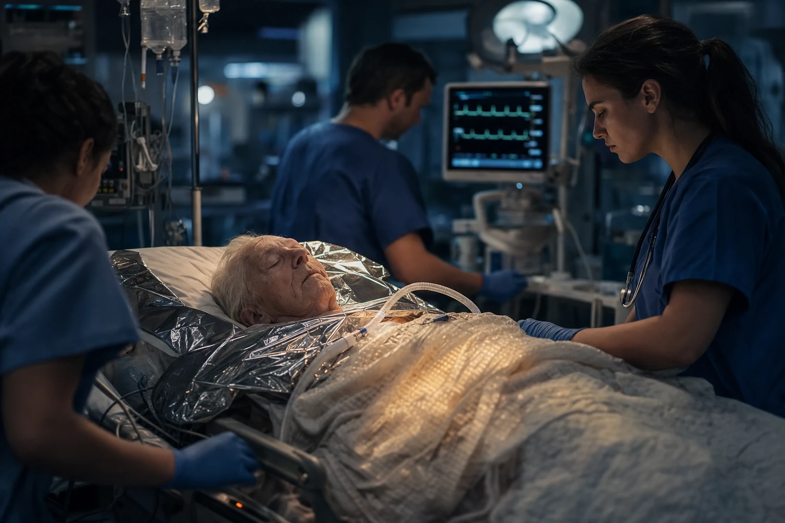
Hypothermia is defined as a core body temperature below 35 degrees Celsius, measured at a central site with a low-reading thermometer. Two staging systems coexist and the candidate must know both, because the examiner may quote either. The standard medical classification divides hypothermia by core temperature into mild, moderate, severe, and profound, and each stage carries a characteristic clinical picture.[1]
The standard medical staging of hypothermia
The Swiss staging system (the HT or Durrer classification) is the field scale used by prehospital services, avalanche rescue, and the Wilderness Medical Society, and it grades the patient by the level of consciousness and the vital signs rather than by a thermometer that may be unavailable on a mountain. It maps directly onto the rewarming decision and is the staging the ERC and WMS guidelines cite.[1][2]
The Swiss (Durrer) field staging — HT I to HT V
Hypothermia is further divided into primary (environmental exposure in an otherwise healthy person — the stranded walker, the immersion victim) and secondary (a patient whose thermoregulation has failed because of illness, drugs, or trauma — the septic, the myxoedematous, the intoxicated, the injured). The distinction matters because secondary hypothermia is a marker of the underlying disease, and the resuscitation must treat the cause in parallel with the temperature. [1]
Epidemiology and risk
Hypothermia is a disease of the vulnerable and the exposed. The classic ANZ and UK case is the elderly person found indoors on a cold floor, dehydrated, on sedatives, with a urinary tract infection or a fractured neck of femur — the hypothermia is secondary and the mortality is high. The outdoor cases are the stranded bushwalker, the immersion victim, the avalanche burial, and the alcohol-intoxicated young man asleep in the open. The risk factors cluster into three groups: increased heat loss (immersion, wet clothing, cold environment, burns, the large surface-area-to-mass ratio of the infant and the elderly), impaired thermogenesis (exhaustion, malnutrition, hypothyroidism, hypoadrenalism, sepsis), and impaired behaviour or awareness (alcohol, opioids, sedatives, dementia, head injury).[1]
[1]Pathophysiology — the mechanism
Heat is lost by radiation (about 60 per cent in the resting adult), convection, conduction, evaporation from the skin, and the warming and humidification of inspired air. The healthy response to cold is peripheral vasoconstriction and shivering. Shivering increases metabolic heat production two- to five-fold but is energetically expensive and fails as the core falls, ceasing at about 30 to 32 degrees — a patient who has stopped shivering is by definition at least moderately hypothermic. As the core falls further the cardiovascular, neurological, respiratory, and metabolic systems fail in a predictable sequence.[1]
The myocardium becomes cold and irritable. The sinus node slows, the conduction system delays, and atrial fibrillation appears in the moderate range; below 28 degrees the risk of ventricular fibrillation rises sharply, and the cold ventricle will fibrillate in response to a seemingly trivial stimulus — rough movement, a central line, an endotracheal tube, a premature defibrillation. Once fibrillating, the cold heart is refractory to defibrillation and to antiarrhythmic drugs, which is the physiological basis of the modified ALS algorithm.[3] The central nervous system is progressively depressed — ataxia and confusion in the mild range, coma in the moderate, and areflexia and fixed dilated pupils in the severe. The candidate must remember that fixed dilated pupils are not a sign of brain death in hypothermia; the pupils can dilate at low temperature and still recover with rewarming.
Two biochemical consequences are examined. The coagulation cascade is temperature-dependent, so the hypothermic patient is functionally coagulopathic — but the laboratory measures the clotting at 37 degrees, warming the sample, and so the prothrombin time and the activated partial thromboplastin time read falsely normal. The Fellowship candidate who trusts a normal coagulation profile in a cold, bleeding trauma patient has fallen for the trap. The serum potassium rises as cells lyse in profound hypothermia, and a potassium at or above 10 mmol per litre reflects irreversible cell death and is incompatible with survival — it is a recognised criterion to cease resuscitation.[2][4]
Clinical presentation
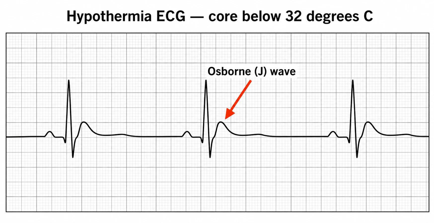
The presentation tracks the core temperature. The mild case (32 to 35 degrees) is awake, cold, pale, and shivering vigorously, with tachycardia, tachypnoea, fine motor clumsiness, and apathy or poor judgement — the "umbles": stumbles (ataxia), mumbles (slurred speech), fumbles (clumsy hands), and grumbles (irritability or confusion). The moderate case (28 to 32 degrees) has stopped shivering, is drowsy or confused, bradycardic with a slow respiratory rate, hyporeflexic, and shows the Osborne J wave on the ECG. The severe case (below 28 degrees) is unconscious, profoundly bradycardic, hypotensive or in shock, may have fixed and dilated pupils and absent reflexes, and is at imminent risk of ventricular fibrillation or asystole.[1]
[1]The history must establish the likely core temperature (the environment, the duration, the wet clothing), the mechanism (an immersion, an avalanche, a fall, a collapse indoors), and the potential precipitants and mimics — alcohol and drugs, a thyroid or adrenal failure, sepsis, hypoglycaemia, and occult trauma. A buried avalanche victim raises the separate question of asphyxia against hypothermia: an airway packed with snow and a burial under 35 minutes points to asphyxia, while a free airway and a long burial point to hypothermia.[2]
Differential diagnosis
The differential of hypothermia is the differential of the cold, collapsed patient, and the candidate must distinguish the primary environmental case from the secondary causes that both mimic and coexist with it. Every hypothermic patient is searched for a precipitant, because the temperature is often the consequence, not the cause. [1]
Primary environmental hypothermia
- Exposure to cold: immersion, avalanche, the stranded walker, the elderly on a cold floor
- A core below 35 degrees with a compatible history and no other obvious cause
- Remove from the cold, dry and insulate, and rewarm on the staged ladder; search for an underlying precipitant
Sepsis
- A systemic infection with vasodilation, septic encephalopathy, and impaired thermogenesis
- Hypothermia in sepsis is itself a poor-prognostic sign; the lactate is high and the white count deranged
- Treat the infection and the sepsis in parallel with rewarming; blood cultures and early antibiotics
Myxoedema coma
- Long-standing hypothyroidism, a trigger (infection, cold, opioid), and a hypothermic, bradycardic, hyponatraemic patient
- Slow-relaxing reflexes, a thyroidectomy scar, an elevated thyroid-stimulating hormone
- Intravenous levothyroxine and hydrocortisone (coexisting adrenal insufficiency) alongside rewarming; passive rewarming only — active rewarming can precipitate cardiovascular collapse
Adrenal crisis
- A primary or secondary adrenal failure, the steroid-dependent patient who has stopped the tablets, abdominal pain and hypotension
- Hyponatraemia, hyperkalaemia, hypoglycaemia, and a low cortisol
- Hydrocortisone 100 mg intravenously and fluid resuscitation in parallel with rewarming
Hypoglycaemia
- A low blood glucose producing confusion, coma, and hypothermia; the diabetic on insulin or a sulphonylurea, the alcoholic with depleted glycogen
- A capillary glucose below 3 mmol per litre (check at the bedside on arrival)
- Glucose 25 g intravenously (50 mL of 50 per cent) and recheck; thiamine in the malnourished or alcoholic
Opioid and sedative overdose
- Alcohol, opioids, benzodiazepines: the patient found cold, comatose, and bradypnoeic
- Miosis and a reduced respiratory rate in the opioid case; the history of intake
- Naloxone titrated for the opioid, supportive care and rewarming; the overdose is both the precipitant and a coexistent diagnosis
Trauma and TBI
- A head injury, a spinal cord injury, an exposure with immobilisation; "the patient is not hypothermic because of the cold room but because of the injury"
- External signs of injury, a focused neurological examination, the secondary survey
- Manage the trauma in parallel; warm fluids, warm blood, and a warmed bay; hypothermia worsens bleeding
Bedside assessment
The assessment runs in parallel with the resuscitation, and it begins with the low-reading thermometer — the single most important and most often forgotten piece of equipment. A standard oral or tympanic device floors at about 34 degrees and will report a profoundly hypothermic patient as 34; the candidate who records "34.0" in a cold patient and is satisfied has missed the diagnosis. A core temperature is measured at the rectum, the bladder, or the oesophagus with a probe that reads to at least 25 degrees (and ideally lower), and it is the number that drives every subsequent decision.[1][2]
TheABCDE is applied with the constant proviso of gentle handling. Airway and breathing — assess the airway and the respiratory rate (a cold patient breathes slowly and may apnoeic); preoxygenate with warmed humidified oxygen, and intubate gently only if the airway is unprotected or the breathing is failing, accepting that intubation itself can provoke fibrillation. Circulation — apply the cardiac monitor (the J wave, the bradycardia, the atrial fibrillation), establish access, and check the pulse for a full minute because the cold bradycardia is slow and easily missed; in the severe case, feel for a pulse and confirm with bedside ultrasound before declaring an arrest, because a cold heart may have a faint output invisible to a hurried finger. Disability — document the Glasgow Coma Scale, reproduced because it is examined and because it is the bedside prognostic anchor: eye opening (4 spontaneous, 3 to speech, 2 to pain, 1 none), verbal response (5 oriented, 4 confused, 3 inappropriate words, 2 incomprehensible sounds, 1 none), and motor response (6 obeys commands, 5 localises pain, 4 withdraws, 3 abnormal flexion, 2 abnormal extension, 1 none), for a maximum of 15; check the pupils and the glucose, and remember that a fixed dilated pupil is not brain death in hypothermia. Exposure — remove the wet clothing, dry and insulate the patient, and complete the secondary survey for the precipitant and the trauma. [1]
[1]Investigations
The investigations follow the resuscitation and never delay oxygen and rewarming. The electrocardiogram is the highest-yield single test and carries the eponymous sign of hypothermia — the Osborne wave, or J wave, a positive deflection at the junction of the QRS complex and the ST segment, seen below about 32 degrees and growing with the depth of hypothermia. The ECG also shows a sinus bradycardia, a prolonged PR and QT interval, atrial fibrillation in the moderate range, and, in the severe case, ventricular fibrillation or a slow, broad complex that can mimic a Brugada pattern. The J wave is the sign the examiner shows on a data-viva strip.[1]
The key investigations and their traps
The blood gas shows a metabolic and respiratory acidosis and is interpreted uncorrected — the "corrected for temperature" value is a mathematical fiction, and the candidate acts on the uncorrected value that reflects what the patient's tissues actually see. The electrolytes and glucose are essential: hypoglycaemia is common and correctable, and a rising potassium is the central prognostic marker. The coagulation profile, the full blood count, the troponin, and the creatine kinase are sent in the moderate and severe case — the troponin for a myocardial injury that may have caused the collapse, the creatine kinase for a rhabdomyolysis, and the coagulation profile interpreted with the caveat above. A chest radiograph looks for the aspiration and the precipitant pneumonia; a computed tomography of the head is reserved for the patient who fails to recover consciousness after rewarming or who has a head-injury mechanism. An alcohol and drug screen identifies the coexistent overdose. In the cardiac arrest, the HOPE score (Hypothermia Outcome Prediction after Extracorporeal life support) integrates the age, the sex, the asystole versus VF, the serum potassium, the core temperature, and the CPR duration to estimate the probability of survival after extracorporeal rewarming, and supports the decision to commence or to cease ECMO.[5]
Resuscitation — the first ten minutes
The resuscitation rests on three principles that the candidate must state aloud in any hypothermia station: handle the patient gently, remove the ongoing heat loss, and rewarm in a staged fashion matched to the severity. Rough handling — dragging a patient, a vigorous roll onto the spinal board, a forceful intubation — can precipitate ventricular fibrillation in a cold, irritable myocardium, and the patient is moved smoothly and with the minimum of disturbance. The wet clothing is removed (cut off, to avoid movement), the patient is dried and insulated in warm blankets, and the resuscitation bay is warmed.[1][2]
Oxygen is given warmed and humidified, the cardiac monitor is attached from the outset, and intravenous or intraosseous access is established. A capillary glucose is checked immediately and hypoglycaemia corrected with glucose 25 g intravenously (50 mL of 50 per cent). In the malnourished or the alcoholic patient a thiamine 100 to 300 mg intravenously is given before the glucose, and a naloxone trial is considered in the comatose patient with a respiratory depression. The patient who is shivering and cardiovascularly stable (mild hypothermia) needs no more than passive rewarming; the patient who has stopped shivering, who is bradycardic, or who is unconscious is escalated to active rewarming and continuous monitoring. The precipitant is sought and treated in parallel — the sepsis with antibiotics, the myxoedema with levothyroxine and hydrocortisone, the overdose with the antidote. [1]
Definitive management — the rewarming ladder
The rewarming method is chosen by the severity and is built on a four-rung ladder, from the least to the most invasive. The rate of rewarming is matched to the patient: too fast in the severe case can precipitate rewarming shock and arrhythmia, and too slow in the arrest is futile.[1][2]
The four rungs of the rewarming ladder
P-A-I-E
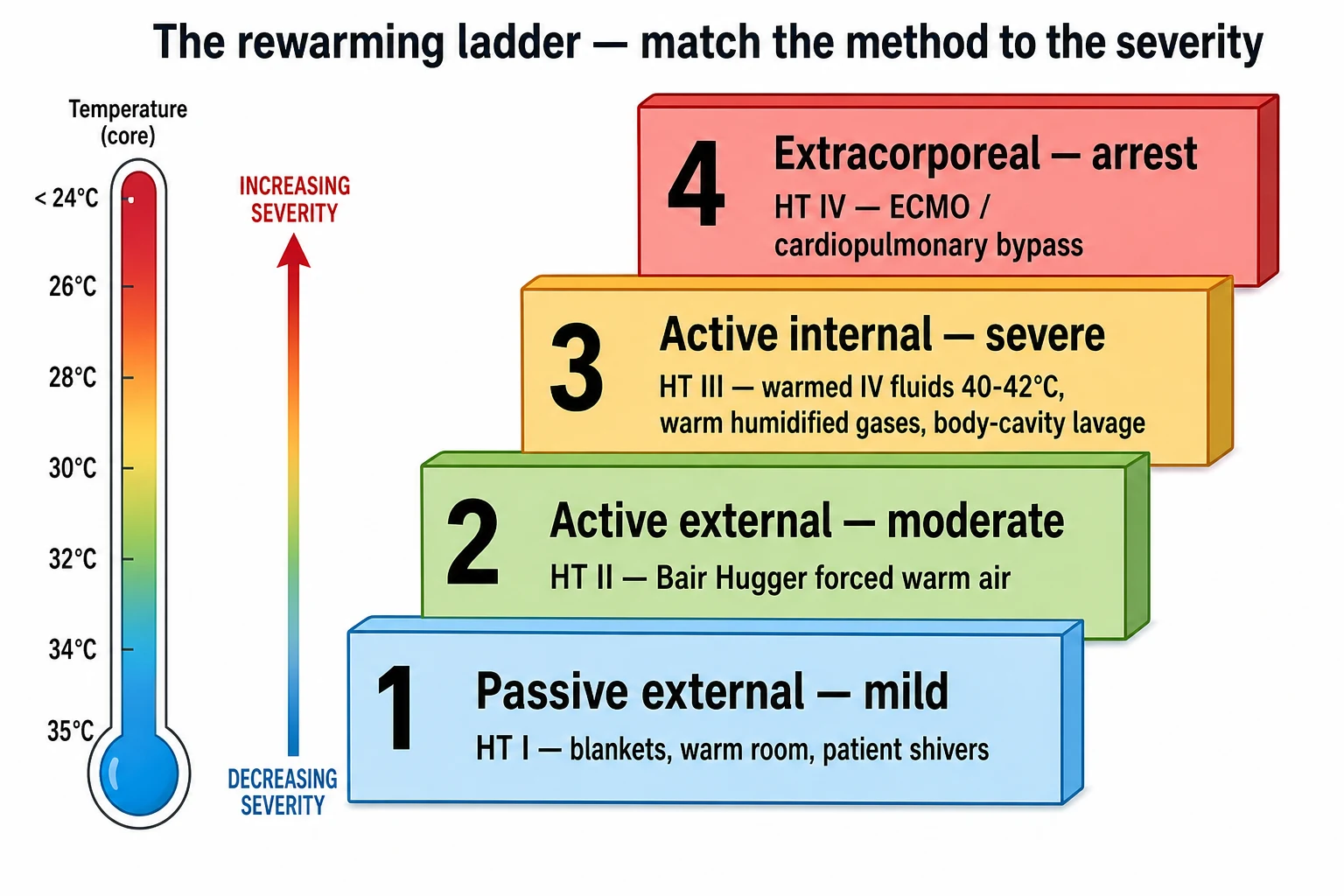
Passive external rewarming is the foundation for the mild case: remove the wet clothing, dry and insulate the patient in warm blankets in a warm room, and let the shivering generate the heat. It is effective only in the patient who can shiver and who is cardiovascularly stable, and it achieves a rise of 0.5 to 2 degrees per hour. Active external rewarming adds the Bair Hugger (forced warm air at 43 degrees), warmed blankets, and a radiant heater, and is the standard for the moderate case; the trunk is warmed before the limbs to limit the afterdrop — the continued fall in core temperature as cold, acidotic peripheral blood returns to the circulation when the limbs are rewarmed first. Active internal rewarming delivers heat directly: warmed intravenous crystalloid at 40 to 42 degrees (each litre of room-temperature fluid lowers the core by about 0.25 degrees, so all fluids are warmed), warm humidified inspired gases, and body-cavity lavage through bladder, gastric, pleural, or peritoneal catheters; it is indicated in the severe, unconscious patient and in any patient in or near arrest. Extracorporeal rewarming with veno-arterial ECMO or cardiopulmonary bypass is the gold standard for the hypothermic cardiac arrest: it rewarms the blood directly to 37 to 38 degrees, it provides circulatory support, and it is the method with the best survival in arrest, validated in the Danish and the subsequent European series.[4]
[1]Adjunctive drug doses follow the clinical picture. Morphine 2.5 to 5 mg intravenously is titrated for the pain and the distress of vigorous shivering (shivering is exhausting and painful); ondansetron 4 mg intravenously for the nausea of rewarming; antibiotics for a confirmed aspiration or pneumonia — but not prophylactically; levothyroxine and hydrocortisone 100 mg intravenously for the suspected myxoedema coma with coexistent adrenal insufficiency; and glucose for the hypoglycaemia. In the trauma patient every unit of blood and every fluid is warmed, and the coagulopathy is anticipated and treated with warm blood products, not with a normal-looking coagulation profile.[1]
Modified advanced life support
The hypothermic cardiac arrest is the single most modified algorithm in emergency medicine, and the candidate who runs a standard ALS loop on a core below 30 degrees fails the station. The principles, codified in the ERC 2021 and the WMS guidelines, rest on the physiology of the cold heart: it is refractory to defibrillation, it is unresponsive to adrenaline and amiodarone, and it tolerates ischaemia far longer than the warm heart — which is why prolonged cardiopulmonary resuscitation is appropriate and why the patient is not declared dead until rewarmed.[2][3]
[1]The sequence is as follows. Confirm the arrest carefully (a full minute for the pulse, and bedside ultrasound), because chest compressions on a cold but perfusing heart can precipitate fibrillation. Begin high-quality chest compressions and ventilate with warmed humidified oxygen. If the rhythm is ventricular fibrillation, deliver a single biphasic shock at 150 to 200 joules; if fibrillation persists, defer further shocks and withhold adrenaline and amiodarone until the core temperature is above 30 degrees, because below that the drugs are ineffective and accumulate. Once the core is above 30 degrees, resume standard ALS with adrenaline 1 mg every 3 to 5 minutes (some guidelines extend the interval to 6 to 10 minutes to limit accumulation) and amiodarone 300 mg after the third shock. Continue prolonged cardiopulmonary resuscitation — the hypothermic myocardium tolerates ischaemia for hours, and CPR for several hours is appropriate where rewarming is under way or in transit to ECMO. Activate extracorporeal rewarming early — the patient in arrest is transferred to an ECMO-capable centre, with CPR maintained in transit, because ECMO rewarming is the intervention that changes survival. Do not pronounce death until the core is at least 32 degrees and the patient is unresponsive to the full algorithm — the rule of "no one is dead until warm and dead".[3][4]
Subtypes and scenarios
The Fellowship case is often a scenario. The elderly person found indoors — secondary hypothermia on a background of sepsis, a fracture, or a stroke, with comorbidity and polypharmacy; rewarm passively or with active external, treat the precipitant, and prognosticate cautiously because the mortality is high. The immersion victim — a drowning overlay, with the primary respiratory failure of aspiration and the hypothermia of cold water; resuscitate toward oxygenation first and rewarm in parallel, and apply the modified ALS. The avalanche burial — distinguish asphyxial death (a packed airway, a short burial under 35 minutes) from hypothermic arrest (a free airway, a long burial, a cold core), because the asphyxial case is not salvageable and the hypothermic case may be. The trauma patient — hypothermia as the first leg of the trauma triad of death; warm the bay, warm the fluids and blood, and treat the temperature as an injury. The intoxicated young person — alcohol and opioids cause both the exposure and the hypothermia, and the overdose is treated alongside the rewarming.[1][2]
Complications and pitfalls
The complications of hypothermia and its rewarming are predictable and examined. The afterdrop is the continued fall in core temperature after removal from the cold, as cold peripheral blood returns to the core; it is limited by warming the trunk first and by gentle handling. Rewarming shock is the vasodilation and the relative hypovolaemia of rewarming, requiring fluid support and vasopressors in the severe case. Ventricular fibrillation precipitated by movement or by premature defibrillation is the feared event, and it is the reason for gentle handling and the single-shock rule. Rhabdomyolysis with an acute kidney injury, and a disseminated intravascular coagulation, are recognised late complications. Aspiration pneumonia complicates the comatose patient.[1]
The pitfalls are the inverse of the management. Using a standard thermometer and missing the severe case is the commonest error. Rough handling that precipitates fibrillation is the second. Running a standard ALS algorithm — multiple shocks, early adrenaline, early cessation of CPR — on a core below 30 degrees is the third and the one the examiner most often tests. Trusting the normal coagulation profile in a bleeding trauma patient is the fourth. Treating the corrected blood gas rather than the patient is the fifth. Calling death too early, before rewarming to at least 32 degrees, is the sixth — the failure that the "warm and dead" rule exists to prevent. Forgetting the precipitant — the sepsis, the myxoedema, the adrenal crisis, the overdose — is the discharge pitfall. [1]
Prognosis and disposition
The prognosis of hypothermia spans the spectrum. The mild case recovers fully with passive rewarming and is discharged once normothermic and the precipitant is addressed. The moderate case is admitted to a monitored bed for active rewarming and observation. The severe case and any arrest are admitted to the intensive care unit, with extracorporeal rewarming for the arrest in an ECMO-capable centre. The two prognostic tools the examiner expects are the HOPE score, which estimates the survival probability after extracorporeal rewarming from the age, the sex, the rhythm, the potassium, the core temperature, and the CPR duration, and the serum potassium — a level at or above 10 mmol per litre in accidental hypothermia reflects massive cell lysis and is incompatible with survival, a recognised criterion to cease resuscitation.[4][5]
The prognostic anchors in the hypothermic arrest
The rule that overrides every other is that no one is dead until warm and dead. A patient in asystole at a core of 24 degrees may recover neurologically intact after prolonged CPR and extracorporeal rewarming, and the candidate resuscitates fully, uses the cluster of prognostic factors (the HOPE score, the potassium, the mechanism, the downtime), and does not cease until the core is at least 32 degrees and the algorithm has failed.[4]
Special populations
The elderly patient is the classic ANZ case — secondary hypothermia, comorbidity, polypharmacy, and a high mortality; the precipitant is sought and treated, and the rewarming is gentle. The child has a large surface-area-to-mass ratio (losing heat fast) but also a robust diving reflex and a tolerance of hypothermia that makes the paediatric submersion the archetype of "dead until warm and dead"; resuscitate fully and prognosticate late. The avalanche victim is triaged by the asphyxia-versus-hypothermia distinction. The pregnant patient is resuscitated as the non-pregnant, with manual left uterine displacement and a perimortem caesarean at four minutes of arrest beyond 20 weeks; the fetal survival tracks the maternal rewarming. The trauma patient is warmed aggressively as part of damage control, with warmed fluids and blood and a warmed bay, because hypothermia worsens the coagulopathy and the bleeding. [1]
Evidence and regional guidelines
The evidence base for hypothermia is built on the Brown review (NEJM 2012) — the definitive contemporary reference, codifying the definition, the staging, the rewarming ladder, and the modified ALS; the Wilderness Medical Society 2019 guidelines (Dow et al), which set the out-of-hospital evaluation and treatment and the staging-to-rewarming mapping; the European Resuscitation Council 2021 guidelines (Lott et al), which set the modified ALS for the hypothermic arrest; the Danish Praesto Fjord series (Wanscher et al, 2012), which demonstrated that circulatory arrest from hypothermia is survivable with extracorporeal rewarming; and the HOPE score (Pasquier et al, 2019), which gave a validated prognostic tool for the hypothermic arrest after ECMO.[1][2][3][4][5]
ANZ practice note. The Australian Resuscitation Council and the New Zealand Resuscitation Council (ANZCOR) govern the Australasian practice, and their guidance aligns with the ERC and the ILCOR consensus. The classic ANZ case is the elderly person found indoors, but the immersion and the alpine case (the New Zealand skier, the Tasmanian bushwalker) are common. ANZ practice follows the staged rewarming ladder, the modified ALS (single shock, drugs withheld below 30 degrees, prolonged CPR, rewarm to at least 32 degrees before calling death), and an early transfer to an ECMO-capable centre for the hypothermic arrest — the retrieval services (RFDS, air ambulance) are experienced in the prolonged transport with CPR in transit. The "no one is dead until warm and dead" rule is applied strictly, and the public-health discharge (warm clothing, heating, the home-safety review for the elderly, the surf-safety for the immersion case) is part of the survivor's care. [1]
Exam-exhaustive expansion — staging, the ECG, arrhythmia drugs, and the rewarming ladder
The Fellowship station turns on a small number of discriminating facts that the examiner probes by name: the Swiss HT staging and its rewarming mapping, the temperature-mapped ECG, the antiarrhythmic drugs that work and the ones that do not, and the ED approach as a protocol. This section assembles them for the viva.[1][2][3]
The Swiss staging in operational detail (HT I to HT V)
The Swiss (Durrer) scale is the field staging the candidate must recite cold — it maps the level of consciousness and the vital signs to a rewarming strategy without a thermometer, and it is the staging the ERC and the WMS guidelines cite.[2]
HT I — 35 to 32 degrees
clear consciousness, shivering
- Conscious and shivering, tachycardia, ataxia, the "umbles"; a coordinated, ambulant patient
- Passive external rewarming — dry, insulate, warm room, warm sweet drinks; the patient generates the heat by shivering
- Rate of rise 0.5 to 2 degrees per hour; observe and treat the precipitant
- Disposition: discharge once normothermic and the cause addressed
HT II — 32 to 28 degrees
altered consciousness, no shivering
- Impaired consciousness, shivering has ceased, bradycardia, slowed respirations, the Osborne J wave appears
- Active external rewarming — the forced warm air blanket (Bair Hugger) at 43 degrees over the trunk, warmed blankets
- Warm the trunk BEFORE the limbs to limit the afterdrop; rate 1 to 2 degrees per hour
- Disposition: admit to a monitored bed with continuous cardiac monitoring
HT III — 28 to 24 degrees
unconscious, vital signs present
- Unconscious, profound bradycardia, hypotension, areflexia, the risk of ventricular fibrillation with any stimulus
- Active INTERNAL rewarming — warmed IV fluids at 40 to 42 degrees, warmed humidified oxygen, body-cavity lavage
- Prepare the transfer to an ECMO-capable centre; intubate gently if the airway is unprotected (GCS below 8)
- Disposition: ICU; have ECMO on the horizon if the core keeps falling or arrest occurs
HT IV — 24 to 13 degrees
no vital signs, apparent death
- Apparent death — no palpable pulse, may be in ventricular fibrillation or asystole; the patient who looks dead but is not
- Cardiopulmonary resuscitation plus extracorporeal rewarming (ECMO or cardiopulmonary bypass) is the standard
- Handle with extreme gentleness; confirm the arrest with a 60-second pulse check and ultrasound before compressions
- Disposition: ECMO centre with CPR maintained in transit — no one is dead until warm and dead
HT V — below 13 degrees
irreversible, fatal
- Irreversible hypothermia — a core below about 13 degrees with a fatal mechanism (a fatal avalanche, a prolonged ice-water submersion)
- Not salvageable; the cooling has passed the threshold of cellular recovery
- Distinguished from HT IV by the mechanism and the duration — a witnessed short down-time with a cold core is still HT IV
- The staging the candidate must not confuse: HT IV is salvageable, HT V is not
The rewarming decision by Swiss stage — a one-glance protocol
1
2
3
4
5
The ECG of hypothermia — temperature-mapped
The ECG tracks the falling core in a predictable sequence, and the candidate must be able to map each change to a temperature band.[1]
ECG changes mapped to the core temperature
Arrhythmia management in the hypothermic heart
The cold myocardium is electrically irritable but pharmacologically refractory. The standard antiarrhythmic algorithm fails, and the candidate must know which drugs work, which do not, and which have been retired.[2][3]
Amiodarone
first-line for VF above 30 degrees
- The preferred antiarrhythmic for ventricular fibrillation in hypothermia once the core is above 30 degrees — 300 mg IV after the third shock
- Below 30 degrees it is withheld because the cold myocardium is refractory and the drug accumulates in the slow peripheral circulation
- Resume at standard or extended intervals (every 6 to 10 minutes) once the core is above 30 degrees
- Supported by the ERC 2021 and the WMS 2019 guidelines
Lidocaine
ineffective in hypothermia
- Largely INEFFECTIVE in the hypothermic myocardium — animal and clinical data show a poor conversion of the cold VF
- Not recommended as the first-line agent in the hypothermic ventricular fibrillation
- If it is used, the doses are spaced widely and the effect is unreliable below 30 degrees
- Avoid in favour of amiodarone
Bretylium
historical, no longer available
- Once the agent of choice for the hypothermic VF on the strength of the early animal work, it is no longer manufactured or available in most jurisdictions
- Mentioned in the older texts and the exam questions — the candidate must know it has been retired
- No role in the current practice
- A trap answer the examiner may offer
Magnesium
may help the refractory VF
- Case reports and series suggest the magnesium sulphate may terminate the ventricular fibrillation refractory to defibrillation in hypothermia
- A reasonable adjunct (1 to 2 g IV) for the refractory VF that persists despite rewarming and a single shock
- Not a substitute for the rewarming and the extracorporeal support
- The evidence is limited but the toxicity is low — a recognised rescue option
Procainamide and class Ia
avoid
- The class Ia antiarrhythmics are avoided — they worsen the conduction delay and the QT prolongation and may precipitate asystole in the cold heart
- The hypothermic QT is already prolonged; further prolongation is dangerous
- No role in the management of the hypothermic arrhythmia
- Avoid alongside the other QT-prolonging agents
The ED approach — the first ten minutes as a protocol
The ED approach to the hypothermic patient is the ABCDE with three modifications: a gentle handling to avoid VF, a warmed everything, and a low-reading core temperature from the first minute.[1][2]
The ED approach to the hypothermic patient — the first ten minutes
1
2
3
4
5
6
7
8
Rewarming strategies — the methods compared
The four rungs of the ladder differ in the rate, the indication, the equipment, and the principal pitfall.[1][2]
Passive external
HT I, 0.5 to 2 degrees/h
- Method: dry, insulate, warm room, warm oral fluids; relies on the endogenous shivering
- For the conscious shivering patient (HT I, mild 35 to 32)
- No special equipment; the foundation for every case
- Pitfall: ineffective once the shivering ceases; the patient who stops shivering needs the escalation
Active external
HT II, 1 to 2 degrees/h
- Method: forced warm air blanket (the Bair Hugger) at 43 degrees over the trunk, warmed blankets, a radiant heater
- For the altered-consciousness, non-shivering patient (HT II, moderate 32 to 28)
- Widely available; the workhorse of the moderate case
- Pitfall: the afterdrop — warm the TRUNK before the limbs to limit the cold peripheral blood returning to the core
Active internal
HT III, 2 to 4 degrees/h
- Method: warmed IV fluids at 40 to 42 degrees, warmed humidified oxygen (42 to 46 degrees), gastric and bladder lavage, pleural and peritoneal lavage
- For the unconscious patient with the vital signs (HT III, severe 28 to 24) and any near-arrest
- Delivers the heat directly to the core; the combination therapy is the norm
- Pitfall: the under-dosing — a single-modality internal rewarming is slow; combine the fluids, the gases, and the lavage
Extracorporeal (ECMO and CPB)
HT IV, 1 to 2 degrees/h to 37
- Method: veno-arterial ECMO or cardiopulmonary bypass, rewarming the blood to 37 to 38 degrees while providing the circulatory support
- For the hypothermic cardiac arrest (HT IV) and the refractory severe case
- The gold standard for the arrest; the best survival, validated in the European series
- Pitfall: a late activation — call the ECMO centre EARLY and maintain the CPR in transit; the transfer, not the cannulation, is usually the rate-limiting step
The modified ALS — step by step
The hypothermic arrest is the most modified algorithm in the resuscitation syllabus, and the candidate must be able to walk the examiner through it.[3][6]
The modified ALS for the hypothermic cardiac arrest
1
2
3
4
5
6
7
Prognostication — the HOPE score and the potassium
[1] [1]Landmark evidence
Brown, Brugger, Boyd, Paal — Accidental Hypothermia (NEJM 2012)
New England Journal of Medicine
PMID 23150960
Key finding
The definitive contemporary review, codifying the definition (a core below 35 degrees), the dual staging (the standard medical mild/moderate/severe/profound and the Swiss HT I to V), the four-rung rewarming ladder, and the modified ALS (a single shock, drugs withheld below 30 degrees, a prolonged CPR, rewarm to at least 32 degrees before pronouncing the death).
Practice change
The reference the examiner expects by name — the staging, the ladder, and the modified algorithm are all in this paper. Know it cold.
Wanscher et al — Danish Praesto Fjord boating accident (Resuscitation 2012)
Resuscitation
PMID 22634431
Key finding
The case series of the circulatory arrest survivors from a single boating accident, rewarmed on the cardiopulmonary bypass, demonstrating that a profound hypothermic circulatory arrest is survivable with an intact neurology after the extracorporeal rewarming — the empirical foundation of the no-one-is-dead-until-warm-and-dead rule.
Practice change
The paper that proved the rule. The candidate who can quote the Praesto Fjord series answers the why-do-you-keep-going question with the evidence.
Pasquier et al — The HOPE score (Resuscitation 2019)
Resuscitation
PMID 30940473
Key finding
A validated prognostic score (the Hypothermia Outcome Prediction after Extracorporeal life support) estimating the survival with a good neurological outcome after the extracorporeal rewarming for the hypothermic cardiac arrest, from the age, the sex, the mechanism, the serum potassium, the core temperature, and the CPR duration.
Practice change
The decision tool for the ECMO cannulation — a high HOPE score supports the full resuscitation, a negligible score with a potassium at or above 10 supports ceasing.
More clinical pearls — the high-yield cluster
[1] [1] [1] [1] [1] [1] [1] [1]SAQ — Severe accidental hypothermia (HT III) in a stranded bushwalker: active internal rewarming and ECMO transfer
10 minutes · 10 marks
A 67-year-old man is brought to a regional emergency department by ambulance after being found collapsed in a Tasmanian national park in winter, having been missing for approximately 18 hours in wet clothing at an ambient temperature of 2 degrees Celsius. On arrival he is unconscious (GCS 8, E2V2M4), pale and cold. Heart rate 42 in sinus bradycardia, BP 78/44, respiratory rate 8 and shallow, SpO2 90 per cent on 15 L oxygen via a non-rebreather mask. A low-reading bladder probe records a core temperature of 28.2 degrees Celsius. The ECG shows sinus bradycardia with prominent Osborne J waves and a prolonged QT interval. Capillary glucose is 3.1 mmol/L. The nearest ECMO-capable centre is a 90-minute aeromedical retrieval.
SAQ — Hypothermic cardiac arrest (HT IV) in a paediatric ice-water submersion: modified ALS and the warm-and-dead rule
10 minutes · 10 marks
A 14-year-old boy is pulled from an icy lake after a submersion of an estimated 25 minutes; bystander CPR is in progress. On arrival he is in cardiac arrest with asystole on the monitor and no palpable pulse, ventilated with a bag-valve-mask. A low-reading oesophageal probe records a core temperature of 25.5 degrees Celsius. Capillary glucose is 5.8 mmol/L. The venous gas (uncorrected) shows pH 7.08, PaCO2 8.2 kPa, lactate 8.4 mmol/L, potassium 6.2 mmol/L. The retrieval service advises that the nearest ECMO-capable centre is 70 minutes away by rotary wing.
Exam pearls
- Definition: hypothermia is a core temperature below 35 degrees Celsius; measure it with a low-reading thermometer.
- Staging: know both the standard medical (mild 32 to 35, moderate 28 to 32, severe 24 to 28, profound below 24) and the Swiss HT I to V; the cessation of shivering marks the transition to moderate.
- The J wave (Osborne wave) is the ECG hallmark, a positive deflection at the J point below about 32 degrees.
- Handle gently — the cold, irritable myocardium fibrillates with rough movement, intubation, and central lines.
- Modified ALS: one shock, then defer shocks and drugs until the core is above 30 degrees; prolonged CPR; rewarm to at least 32 degrees before calling death; ECMO for the arrest.
- The coagulation profile is falsely normal (the lab warms the sample); the patient is coagulopathic in vivo.
- A potassium at or above 10 mmol per litre is incompatible with survival — a criterion to cease resuscitation.
- Interpret the blood gas uncorrected; treat the patient, not the corrected number.
- No one is dead until warm and dead — resuscitate fully and prognosticate from the cluster of factors, not a single time cutoff. [1]
Red flags
[1]References
- [1]Brown DJA, Brugger H, Boyd J, Paal P. Accidental hypothermia N Engl J Med, 2012.PMID 23150960
- [2]Dow J, Giesbrecht GG, Danzl DF, et al. Wilderness Medical Society Clinical Practice Guidelines for the Out-of-Hospital Evaluation and Treatment of Accidental Hypothermia: 2019 Update Wilderness Environ Med, 2019.PMID 31740369
- [3]Lott C, Truhlář A, Alfonzo A, et al. European Resuscitation Council Guidelines 2021: Cardiac arrest in special circumstances Resuscitation, 2021.PMID 33773826
- [4]Wanscher M, Agersnap L, Ravn J, et al. Outcome of accidental hypothermia with or without circulatory arrest: experience from the Danish Præstø Fjord boating accident Resuscitation, 2012.PMID 22634431
- [5]Pasquier M, Hugli O, Paal P, et al. Hypothermia outcome prediction after extracorporeal life support for hypothermic cardiac arrest patients: An external validation of the HOPE score Resuscitation, 2019.PMID 30940473
- [6]Nolan JP, Soar J, Wenzel V, Paal P. Cardiopulmonary resuscitation and management of cardiac arrest Nat Rev Cardiol, 2012.PMID 22665327