EM · Paediatric rashes and febrile illness
Paediatric rashes and febrile illness
Also known as Febrile rash · Paediatric exanthem · Meningococcal rash · Kawasaki disease · Henoch-Schonlein purpura · IgA vasculitis
The febrile child with a rash is a high-stakes presentation because a small number of rashes signal time-critical disease while the majority are benign viral exanthems. The Fellowship candidate must read rash morphology first: a non-blanching petechial or purpuric rash with fever is meningococcal septicaemia until proven otherwise and earns ceftriaxone 50 mg per kilogram intravenously immediately, while a fever of five days or more with bilateral non-purulent conjunctivitis, a polymorphous rash, a strawberry tongue and swollen hands is Kawasaki disease, treated with intravenous immunoglobulin 2 g per kilogram and aspirin within ten days of fever onset to prevent coronary artery aneurysm. Measles descends from the hairline after a prodrome of fever, coryza, cough and conjunctivitis with Koplik spots; chickenpox is a vesicular rash in crops at different stages; erythema multiforme is a target rash with at most one mucosal site; and Henoch-Schonlein purpura is a palpable purpura on the lower limbs with abdominal pain, arthritis and renal involvement. The differential is the viral exanthem, the drug reaction and the autoimmune vasculitis, distinguished by morphology, tempo, distribution and toxicity. ACEM-primary, globally tagged.
On this page & tools
Your progress
Saved locally on this device.
5 MCQs with explanations
Target exams
Red flags
Related topics
- Paediatric fever and serious bacterial illness (the febrile child in the emergency department)
- Paediatric sepsis and septic shock (the septic child in the emergency department)
- The sick child and paediatric resuscitation
- Paediatric respiratory distress — croup, bronchiolitis and acute asthma
- Sepsis and septic shock — the emergency department approach
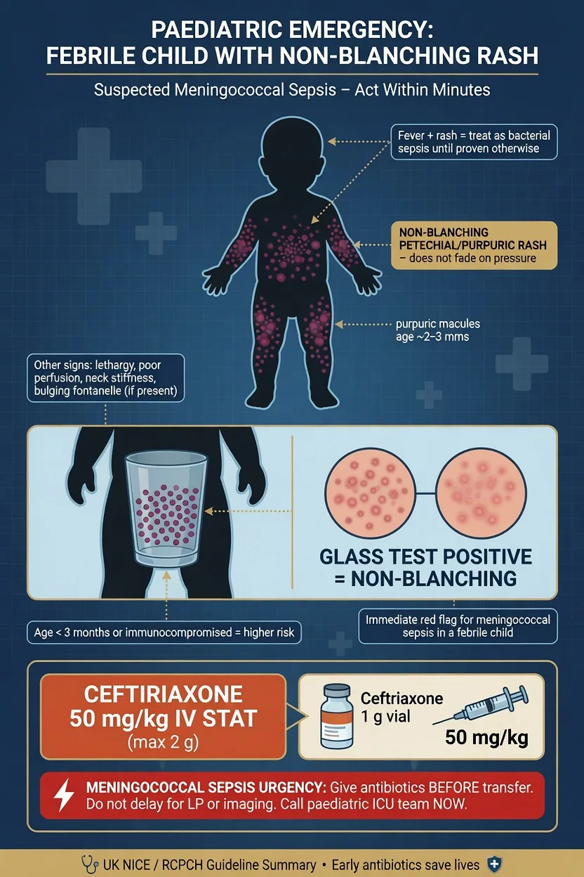
The febrile child with a rash is one of the most discriminating presentations in paediatric emergency medicine because the same symptom cluster spans trivial viral exanthems and three time-critical diagnoses — meningococcal septicaemia, Kawasaki disease and herpes simplex infection of the neonate. The Fellowship candidate's task is not to memorise every rash but to apply a morphology-led framework that separates the dangerous from the benign at the bedside, reproduce the named diagnostic criteria for Kawasaki and Henoch-Schonlein purpura, and give the right drug at the right dose in the right window. The single decisive act is the blanching test: a non-blanching petechial or purpuric rash in a febrile child is meningococcal disease until proven otherwise.[1]
Definition and classification
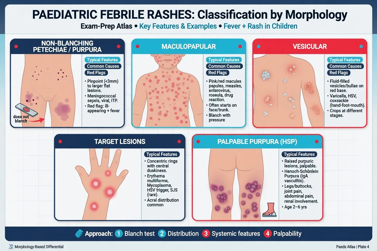
A febrile rash syndrome is any acute febrile illness in which a cutaneous eruption is part of the disease. Classification is by rash morphology, because morphology predicts mechanism and danger far better than fever height or duration. Five morphology groups carry the examinable core. Petechial and purpuric rashes (non-blanching) indicate extravasation of red cells through a damaged vessel wall or thrombocytopenia — meningococcal septicaemia, Henoch-Schonlein purpura, idiopathic thrombocytopenic purpura and leukaemia. Vesicular rashes indicate epidermal cytopathology — varicella, herpes simplex, hand-foot-mouth disease. Maculopapular and erythematous rashes (blanching) are the commonest group and span the viral exanthems, measles, rubella, roseola, fifth disease, scarlet fever, Kawasaki and drug eruptions. Target lesions indicate the erythema multiforme spectrum. Palpable purpura indicates leucocytoclastic vasculitis, of which Henoch-Schonlein purpura is the archetype. The morphology group, combined with the fever pattern and the associated features, generates the diagnosis.[1][4]
Erythematous (blanching)
- Diffuse confluent erythema that blanches under pressure; "sunburn-like" quality
- Causes: viral exanthems (roseola prodrome, fifth disease, measles prodrome), scarlet fever, toxic shock syndrome, Kawasaki disease
- Danger signal: confluent "sunburn" rash with mucosal erythema and hypotension = toxic shock
- Management: identify trigger; TSS demands immediate fluid resuscitation and source control
Maculopapular (blanching)
- Discrete macules and papules that may coalesce; the commonest morphology in paediatrics
- Causes: measles (descending from hairline), rubella (fine pink, Forchheimer spots), roseola (HHV-6, rash as fever breaks), EBV (especially after ampicillin), drug eruptions, parvovirus B19
- Danger signal: Koplik spots + descending progression + prodrome of 3 Cs = measles (notifiable, airborne isolation)
- Management: supportive for viral; stop drug for drug eruption; isolate suspected measles immediately
Vesicular / bullous
- Fluid-filled lesions on an erythematous base; may evolve through papule-vesicle-pustule-crust
- Causes: varicella (crops at different stages), herpes simplex (grouped vesicles, neonatal disseminated), hand-foot-mouth disease (coxsackie A16 — palms, soles, oral mucosa), bullous impetigo, SSSS (diffuse superficial peeling)
- Danger signal: neonate under 28 days with vesicles = HSV; give aciclovir 20 mg/kg every 8 hours and full septic workup
- Management: aciclovir for HSV and high-risk varicella; supportive for uncomplicated HFM; flucloxacillin for impetigo
Petechial / purpuric (non-blanching)
- Pinpoint (petechiae) or larger (purpura) lesions that do not blanch — extravasated red cells
- Causes: meningococcal septicaemia (rapidly progressive), Henoch-Schonlein purpura (palpable, lower limbs), idiopathic thrombocytopenic purpura (isolated thrombocytopenia, otherwise well), leukaemia (pallor, bruising, hepatosplenomegaly), traumatic, viral (mild)
- Danger signal: fever + rapidly evolving petechiae/purpura + toxicity = meningococcal — give ceftriaxone 50 mg/kg immediately
- Management: treat meningococcal empirically; FBC and coagulation for all non-blanching rashes; bone marrow for suspected leukaemia
Urticarial (wheals)
- Raised erythematous wheals with central pallor that migrate and individual lesions resolve within 24 hours
- Causes: IgE-mediated allergy (food, drug, sting), viral infection (serum-sickness-like), serum sickness, drug reaction, autoimmune
- Danger signal: urticaria PLUS stridor, wheeze, hypotension or abdominal pain = anaphylaxis — IM adrenaline 0.01 mg/kg
- Management: antihistamine; stop culprit drug; adrenaline for anaphylaxis; exclude underlying infection in serum-sickness-like reactions
Desquamating
- Peeling or scaling of the epidermis, often beginning at flexures, peri-orally, or periungually
- Causes: SSSS (perioral/flexural superficial peeling), Kawasaki (periungual desquamation in week 2-3), scarlet fever (fine desquamation after rash fades), toxic shock syndrome, toxic epidermal necrolysis (full-thickness skin detachment)
- Danger signal: widespread epidermolysis with skin tenderness and mucosal involvement = SJS/TEN — burn-unit care
- Management: treat the underlying disease — IVIG and aspirin for Kawasaki, anti-staphylococcal antibiotic for SSSS, penicillin for scarlet fever
Pathophysiology
Four mechanisms generate fever and rash, and the candidate who knows the mechanism can predict the morphology and the danger. Vasculitis of small vessels from immune-complex deposition produces palpable purpura — immunoglobulin-A complexes in Henoch-Schonlein purpura deposit in dermal capillaries and the renal mesangium, causing the palpable lower-limb purpura, the abdominal pain and the glomerulonephritis. Endothelial injury with microvascular thrombosis and disseminated intravascular coagulation produces the rapidly evolving petechiae and purpura of meningococcal septicaemia, where endotoxin damages the endothelium and the consumptive coagulopathy drives purpura fulminans with skin necrosis. Viral cytopathic and cell-mediated mechanisms generate the typical exanthems: measles virus replicates in endothelial cells and the rash is a T-cell-mediated reaction; varicella-zoster virus produces intra-epidermal vesicles that evolve in crops because viraemia is intermittent. Drug and infection-triggered hypersensitivity generates erythema multiforme, where cytotoxic T-cells attack keratinocytes to produce the target lesion, and its severe mimics Stevens-Johnson syndrome and toxic epidermal necrolysis. Kawasaki disease is a medium-vessel vasculitis driven by marked immune activation that preferentially targets the coronary arteries.[1][2]
The morphology-led approach — the framework
The approach to the febrile rash runs morphology first, danger assessment second, diagnosis third. Press a clear glass or a finger against the rash: blanching rashes are overwhelmingly benign; non-blanching rashes are dangerous until proven otherwise. In parallel, assess toxicity through the paediatric assessment triangle — appearance, work of breathing and circulation to the skin — because a toxic-appearing child with any rash earns the septic bundle immediately. Record the fever duration, because five days is the Kawasaki threshold, and examine the distribution, the staging and the mucous membranes, which distinguish Kawasaki, erythema multiforme and measles. Estimate the weight before any drug is drawn up, using the Broselow tape or the formula weight in kilograms equals age in years plus four, times two.[1]
[1] [1]Meningococcal disease — the archetype petechial emergency
Meningococcal disease is the febrile rash the candidate cannot miss. Neisseria meningitidis colonises the nasopharynx and, in the small fraction who develop invasive disease, seeds the bloodstream and the meninges. The classic presentation is a febrile child with a rapidly evolving petechial or purpuric rash that progresses to large purpuric patches and, in the worst case, to purpura fulminans with skin necrosis, disseminated intravascular coagulation and shock. Early meningococcal rash can be maculopapular and blanching, which is the trap — a febrile child with a viral-looking rash and any perfusion abnormality is still managed as meningococcal until proven otherwise. The signs of decompensated sepsis are cool mottled peripheries, a capillary refill over three seconds, tachycardia, altered mental state and, as a late and pre-terminal sign, hypotension. Management is the paediatric sepsis bundle applied without delay: airway, breathing and high-flow oxygen, intravenous or intra-osseous access after two failed attempts or 90 seconds, blood cultures if they do not delay the antibiotic, and ceftriaxone 50 mg per kilogram intravenously as a single stat dose (maximum 2 g) given within the first hour because time-to-antibiotic tracks mortality.[1]
Treat shock with a 10 mL per kilogram bolus of balanced crystalloid reassessed after each aliquot, escalating to vasopressors and paediatric intensive care for refractory shock; give fresh frozen plasma for disseminated intravascular coagulation. Add dexamethasone 0.15 mg per kilogram every six hours before or with the antibiotic when meningitis is the suspected focus. Notify public health from the emergency department and arrange chemoprophylaxis of household and kissing contacts — rifampicin orally for two days, or a single dose of ciprofloxacin, or ceftriaxone intramuscularly — because the secondary attack rate in close household contacts is up to a thousand times that of the general population.[1]
Kawasaki disease — the five-day fever
Kawasaki disease is a medium-vessel vasculitis of childhood whose danger lies not in the acute illness but in the coronary artery aneurysms it produces when treatment is delayed. The diagnosis is clinical, made from the American Heart Association criteria: fever for five days or more plus at least four of the five principal features — bilateral non-purulent conjunctivitis, polymorphous rash, oral changes (strawberry tongue, cracked and fissured lips, oropharyngeal erythema), peripheral extremity changes (erythema and oedema of the palms and soles in the acute phase, periungual desquamation in the second and third weeks) and cervical lymphadenopathy of at least 1.5 centimetres. The diagnosis can also be made with fewer features if echocardiography shows coronary artery disease, and incomplete Kawasaki is the particular trap in infants under six months, who present with unexplained fever for seven days or more and fewer criteria but the same coronary risk.[2]
Kawasaki diagnostic criteria — the five principal features (fever 5 days or more plus 4 of 5)
The treatment is time-critical. Intravenous immunoglobulin 2 g per kilogram as a single infusion over 10 to 12 hours plus aspirin — high-dose 30 to 50 mg per kilogram per day in the acute febrile phase, stepping down to low-dose 3 to 5 mg per kilogram per day as the antiplatelet agent once the fever resolves — given within ten days of fever onset reduces the coronary artery aneurysm rate from approximately 25 per cent untreated to 3 to 5 per cent. An echocardiogram is obtained at diagnosis and repeated at six to eight weeks. Refractory Kawasaki, defined as persistent or recrudescent fever 36 hours or more after the first immunoglobulin infusion, is treated with a repeat infusion, then infliximab, systemic corticosteroids or anakinra under specialist guidance.[2]
[1]Measles — the descending maculopapular rash
Measles is a highly contagious viral illness that has re-emerged in under-vaccinated populations and is notifiable worldwide. After an incubation of 10 to 14 days the prodrome is fever with the three Cs — coryza, cough and conjunctivitis — and the pathognomonic Koplik spots, grey-white grains on an erythematous buccal mucosa opposite the lower molars, which appear a day or two before the rash. The maculopapular rash then descends from the hairline behind the ears, spreading to the face, trunk and limbs, and may coalesce. The child is unwell and contagious from the prodrome until four days after the rash appears. Management is supportive, with vitamin A 200,000 international units as a single dose for the malnourished or severe case, strict airborne isolation, and public-health notification with contact tracing. The differential from a drug rash and from other viral exanthems rests on the prodrome, the Koplik spots and the descending progression.[3][4]
Chickenpox — the vesicular rash in crops
Varicella is a universal childhood illness characterised by a pruritic vesicular rash that appears in crops at different stages of evolution simultaneously — papules, vesicles, pustules and crusts all present at once — distributed centripetally on the trunk, face and scalp, with mucosal involvement. Fever is mild and the child is usually not toxic. The diagnosis is clinical and management is supportive: hydration, antipyretics and oral antihistamine for the itch, with attention to secondary bacterial skin infection (group A streptococcus and staphylococcus), the commonest complication. Aciclovir is reserved for the high-risk groups — immunocompromised, neonates, adolescents, pregnancy and chronic skin or lung disease — at 10 mg per kilogram intravenously every eight hours, or oral 20 mg per kilogram four times daily for the uncomplicated at-risk case. Aspirin is avoided in varicella and influenza because of the association with Reye syndrome; paracetamol is the antipyretic of choice.[4]
[1]Erythema multiforme — the target rash
Erythema multiforme is an acute, self-limited immune-mediated reaction defined by typical target lesions — at least three concentric zones of colour change, with a dark central zone, a pale oedematous ring and an outer erythematous ring — distributed symmetrically on the acral sites (palms, soles, extensor surfaces), with at most a single mucosal site involved. The commonest triggers are herpes simplex virus infection, recurrent in the classic minor form, and Mycoplasma pneumoniae. Management is supportive, with treatment of the identified trigger — suppressive aciclovir for the recurrent herpes-associated form, antibiotics for proven Mycoplasma — and topical steroids or antihistamines for symptomatic relief. The critical distinction is from Stevens-Johnson syndrome and toxic epidermal necrolysis, the severe drug-induced mimics in which atypical targets, detachment of skin, involvement of two or more mucosal sites and systemic toxicity are present; management of those is withdrawal of the culprit drug, burn-unit wound care, fluid and nutritional support and urgent ophthalmology.[5]
Henoch-Schonlein purpura — the palpable purpura
Henoch-Schonlein purpura, now termed immunoglobulin-A vasculitis, is the commonest vasculitis of childhood, peaking between three and fifteen years and often following an upper respiratory infection. The EULAR/PRINTO/PRES criteria require the mandatory finding of palpable purpura or petechiae predominantly on the lower limbs and buttocks, plus at least one of diffuse abdominal pain, arthritis or arthralgia, renal involvement (haematuria or proteinuria), or histological evidence of immunoglobulin-A deposition on biopsy. The tetrad is therefore palpable lower-limb purpura, colicky abdominal pain, arthritis and renal involvement, but the four do not need to coincide.[6]
EULAR/PRINTO/PRES criteria for Henoch-Schonlein purpura (IgA vasculitis)
Management is supportive: hydration, simple analgesia and non-steroidal anti-inflammatory drugs for joint pain and abdominal pain, with systemic corticosteroids reserved for severe gastrointestinal or renal disease. The two complications the candidate must hold are intussusception — which in Henoch-Schonlein purpura is atypically ileo-ileo, caused by a bowel-wall haematoma acting as a lead point, so the classic ultrasound target sign may be absent and the child presents with severe colicky abdominal pain, vomiting and a palpable mass — and renal involvement, which dictates blood-pressure and urinalysis monitoring for six to twelve months because end-stage renal failure develops in a small minority years later.[6]
Staphylococcal scalded skin syndrome — the desquamating rash
Staphylococcal scalded skin syndrome (SSSS) is a toxin-mediated exfoliative dermatosis of young children, typically under five years, caused by exfoliative toxin-producing strains of Staphylococcus aureus (phage group II, types 71 and 55). The toxin cleaves desmoglein 1 in the stratum granulosum, producing superficial intra-epidermal splitting at the level of the granular layer — a split so superficial that the skin appears scalded. The focus is usually a localised staphylococcal infection — conjunctivitis, otitis media, umbilical or nasopharyngeal infection, or an impetiginous skin lesion — and the child presents with fever, irritability and diffuse skin tenderness. The rash evolves through three stages: first, a scarlatiniform erythema that is tender to touch and accentuated in the flexures and peri-orally; then, within 24 to 48 hours, large flaccid bullae that rupture easily leaving a moist, glistening, scalded-appearing surface; and finally, desquamation in large sheets beginning around the mouth, eyes, neck, axillae and groins. The Nikolsky sign is positive — gentle lateral pressure on apparently normal skin induces shearing and peeling. Crucially, the mucous membranes are spared because desmoglein 3 compensates in mucosa — this is the key distinction from Stevens-Johnson syndrome and toxic epidermal necrolysis, which involve the mucosa.[7]
[1]Management is anti-staphylococcal antibiotic — flucloxacillin 50 mg per kilogram intravenously (or clindamycin 10 mg per kilogram every eight hours if staphylococcal resistance or toxin suppression is desired), analgesia, careful fluid and electrolyte management because the extensive skin loss mimics a partial-thickness burn, and emollient wound care. Corticosteroids are avoided. Most children recover within 7 to 14 days with complete re-epithelialisation because the split is superficial and the basal layer is preserved. Mortality is under 5 per cent in children (compared with over 50 per cent in adults with comorbidity). Isolation is required because the exfoliative toxin is contagious.[7]
Toxic shock syndrome — the toxin-mediated emergency
Toxic shock syndrome (TSS) is an acute, toxin-mediated, multi-system illness caused by the superantigenic exotoxins of Staphylococcus aureus (toxic shock syndrome toxin-1, TSST-1) and, in streptococcal TSS, the pyrogenic exotoxins of group A streptococcus. Superantigens bypass conventional antigen presentation and directly bridge the T-cell receptor V-beta region to MHC class II, activating 5 to 30 per cent of all T-cells and generating a catastrophic cytokine cascade — interleukin-1, interleukin-2, tumour necrosis factor alpha and gamma-interferon — that produces capillary leak, hypotension and multi-organ failure. Staphylococcal TSS is associated with retained tampons, nasal packing after surgery, and focal staphylococcal infections; streptococcal TSS is associated with invasive group A streptococcal infection, particularly cellulitis, necrotising fasciitis and empyema.[8]
CDC case definition of toxic shock syndrome (all six criteria)
Management is aggressive: remove the source (remove tampon or nasal pack, debride necrotic tissue), aggressive fluid resuscitation because capillary leak produces massive fluid shifts — often 100 to 200 mL per kilogram in the first 24 hours — guided by central venous pressure monitoring, empiric antibiotics covering both staphylococcal and streptococcal causes (clindamycin 10 mg per kilogram every eight hours to suppress toxin synthesis plus flucloxacillin 50 mg per kilogram plus vancomycin 15 mg per kilogram for MRSA), intravenous immunoglobulin 1 to 2 g per kilogram to neutralise circulating superantigen in severe disease, and vasopressors (noradrenaline) for refractory shock. Streptococcal TSS has a significantly higher mortality (30 to 70 per cent) than staphylococcal TSS (under 5 per cent). All suspected cases are admitted to paediatric intensive care.[8]
[1]Scarlet fever — the sandpaper streptococcal rash
Scarlet fever is a toxin-mediated exanthem caused by group A beta-haemolytic streptococcus (Streptococcus pyogenes) producing erythrogenic exotoxin (usually type A, B or C). It follows streptococcal pharyngitis or, less commonly, a wound or skin infection. After a 1- to 4-day incubation the child presents with fever, sore throat, headache and abdominal pain. The rash appears 12 to 48 hours after the fever, beginning on the neck, chest and axillae and spreading to the trunk and limbs, sparing the face (which may show circumoral pallor). The rash is fine, punctate, sandpaper-like and palpable — the texture is the key. Pastia lines — linear petechial streaks in the skin folds of the axillae, groin and antecubital fossae — are highly characteristic. The tongue evolves from a white furry coating through to a strawberry tongue (red, swollen papillae projecting through a white coat) and then raspberry tongue (bright red, denuded). The rash fades over 4 to 5 days and is followed by fine desquamation of the trunk, and sheet-like peeling of the hands and feet in the second and third weeks — the desquamination that confuses it with Kawasaki.[9]
Management is phenoxymethylpenicillin (oral penicillin V) for 10 days, or a single dose of benzathine penicillin G intramuscularly, or amoxicillin for 10 days; clarithromycin or clindamycin for the penicillin-allergic child. The child can return to school 24 hours after starting antibiotics. Complications — acute rheumatic fever and post-streptococcal glomerulonephritis — are prevented by completing the antibiotic course. The IDSA 2012 guideline for group A streptococcal pharyngitis reinforces a clinical score (Centor or McIsaac) supplemented by a rapid antigen test or throat swab before antibiotics.[9]
[1]Roseola infantum — the rash when the fever breaks
Roseola infantum (exanthem subitum, sixth disease) is caused by human herpesvirus 6 (HHV-6, and less commonly HHV-7) and is the classic cause of febrile convulsions in infants aged six months to three years. The clinical pattern is unmistakable: three to five days of high fever — often 39 to 40 degrees — in a remarkably well-appearing child, followed by the abrupt defervescence and the simultaneous appearance of a fine pink maculopapular rash that begins on the trunk and spreads to the face and limbs. The rash appears when the fever has resolved — the temporal relationship is the diagnostic clue. Management is supportive: antipyretics, hydration and parental reassurance. Febrile convulsions occur in 10 to 15 per cent. The rare complication is HHV-6 encephalitis in the immunocompromised child. No isolation is required. The clinical diagnosis is made by the fever pattern — the rash appearing as the fever breaks — and the well-looking child. [1]
Fifth disease — the slapped cheek
Erythema infectiosum (fifth disease) is caused by parvovirus B19, a single-stranded DNA virus that targets erythroid precursor cells via the P-antigen (globoside) receptor. The classic presentation in a child is bright red confluent erythema of both cheeks — the slapped-cheek appearance — sparing the perioral area and the nasal bridge, followed 1 to 4 days later by a lacy, reticular, pink maculopapular rash on the trunk and extremities that may fluctuate with environmental temperature (recrudescing after a warm bath or sunlight exposure). Fever is mild or absent. Arthralgia and arthritis of the small joints are common in adults but uncommon in children. The clinical importance of parvovirus B19 lies in three settings: pregnancy — transplacental infection causes fetal hydrops and intrauterine death in up to 10 per cent of infections in the first 20 weeks, so any pregnant contact requires serology and obstetric referral; chronic haemolytic anaemia (sickle cell disease, hereditary spherocytosis) — the virus suppresss erythropoiesis producing a life-threatening transient aplastic crisis; and immunocompromise — persistent infection producing chronic pure red cell aplasia. Management of uncomplicated fifth disease is supportive; the child is no longer infectious once the rash appears.[11]
[1]Rubella — the Forchheimer rash
Rubella (German measles, third disease) is a togavirus infection rendered rare by universal vaccination but still relevant in the under-vaccinated and in the returning traveller. After a 14- to 21-day incubation, the prodrome is mild — low-grade fever, malaise, lymphadenopathy — and the rash appears as fine pink maculopapules beginning on the face and spreading caudally to the trunk and limbs within 24 hours, fading by the third day in the order it appeared. Tender postauricular, suboccipital and cervical lymphadenopathy is the hallmark. Forchheimer spots — small rose-coloured petechiae on the soft palate — are a helpful but non-specific sign. The disease is mild in children, but congenital rubella syndrome — cataracts, cardiac defects (patent ductus arteriosus, pulmonary artery stenosis), sensorineural deafness, growth retardation and purpura — is devastating in the first trimester. Management is supportive; the diagnosis is confirmed by IgM serology or rising IgG titres, the case is notifiable, and pregnant contacts require urgent serology. All suspected cases are isolated with droplet precautions.[10]
Hand-foot-mouth disease — the coxsackie vesicle
Hand-foot-mouth disease (HFMD) is caused by coxsackievirus A16 and enterovirus 71, transmitted by the faeco-oral and respiratory routes, peaking in summer and autumn in children under 10. The child presents with mild fever, sore mouth and refusal to eat. The rash is vesicular or vesiculo-pustular affecting the palms, soles, buttocks and perioral area, with painful oral ulcers on the tongue, buccal mucosa and palate that may cause significant dehydration due to poor oral intake in young children. The lesions are oval or elliptical, parallel to the skin lines, and are typically smaller and more peripheral than varicella. Enterovirus 71 is associated with more severe disease — meningoencephalitis, acute flaccid paralysis, brainstem encephalitis and pulmonary oedema — and has caused large outbreaks with fatalities in the Asia-Pacific region. Management is supportive: hydration (the principal concern), oral analgesia with paracetamol and topical anaesthetic mouthwash, and isolation until the lesions dry. The child is infectious from the prodrome until all lesions have crusted. No antibiotic is indicated. The main pitfall is confusing HFMD with foot-and-mouth disease of cattle — it is unrelated. [1]
Infectious mononucleosis and the ampicillin rash
Epstein-Barr virus infection in the child or adolescent presents with fever, tonsillar pharyngitis with exudate, generalised lymphadenopathy and fatigue. The rash itself may be maculopapular and blanching in 5 to 15 per cent of untreated cases, but the classic exam point is the ampicillin rash — a diffuse maculopapular eruption occurring in 80 to 100 per cent of patients with EBV who receive ampicillin or amoxicillin. The rash is not a true IgE-mediated allergy to penicillin — it is a virus–drug interaction that does not preclude future penicillin use in most cases. The patient is managed supportively with rest, hydration and paracetamol; avoid ampicillin and amoxicillin when infectious mononucleosis is suspected. Splenomegaly is present in over 50 per cent and mandates avoidance of contact sport for at least four weeks to prevent splenic rupture. The differential includes group A streptococcal pharyngitis, which coexists in up to 30 per cent — a throat swab is warranted. [1]
Idiopathic thrombocytopenic purpura — the well child with petechiae
Immune thrombocytopenia (ITP) of childhood, formerly idiopathic thrombocytopenic purpura, is an autoimmune destruction of platelets by IgG antibodies, typically following a viral infection 1 to 4 weeks earlier. The classic presentation is a well-appearing child with sudden onset of petechiae and bruising — the child looks entirely well, which is the key distinction from meningococcal disease and leukaemia. The platelet count is typically below 20 times 10 to the 9 per litre; the haemoglobin, white cell count and film are otherwise normal. Wet purpura — bullous haemorrhage on the buccal mucosa — indicates higher bleeding risk. The principal danger is intracranial haemorrhage, which occurs in under 0.5 per cent. The ASH 2019 guideline recommends: for the child with no or mild bleeding (skin only), observation alone regardless of platelet count; for the child with significant bleeding, a single dose of IVIG 0.8 to 1 g per kilogram or a short course of corticosteroid — either is first-line. Anti-D immunoglobulin is an alternative in Rh-positive non-splenectomised children. Splenectomy is reserved for chronic, refractory ITP. A bone marrow aspirate is NOT required before treatment in typical cases but is performed if atypical features are present — anaemia, neutropenia, or a smear with blasts.[12]
[1]Leukaemia — the hidden dangerous petechiae
Acute lymphoblastic leukaemia, the commonest childhood malignancy, may present with petechiae, purpura and bruising indistinguishable morphologically from ITP at first glance. The discriminating features are the other cytopenias — anaemia with pallor and lethargy, neutropenia with recurrent or severe infection, and the systemic features of fever, bone pain, hepatosplenomegaly and lymphadenopathy. A full blood count showing pancytopenia or a blood film with circulating blasts mandates urgent paediatric haematology referral and a bone marrow aspirate. The emergency physician's role is to recognise the abnormal count, avoid steroids (which can mask the diagnosis and alter the marrow), treat any infection or coagulopathy, and transfer urgently. Never give corticosteroids to a child with suspected leukaemia before the bone marrow is sampled — this is a cardinal rule because steroids lyse lymphoblasts and render the diagnosis impossible. [1]
Urticarial rashes — the wheals that move
Urticaria (hives) is characterised by raised erythematous wheals with central pallor that are intensely pruritic and individual lesions resolve within 24 hours — the last point is critical: a rash that looks urticarial but stays in the same place for more than a day is not simple urticaria. The mechanism is mast-cell degranulation with histamine release producing dermal oedema. Three aetiologies dominate paediatric practice. IgE-mediated allergy to food (peanut, egg, milk), drug (penicillin, non-steroidal anti-inflammatory drugs), latex or insect sting presents with acute urticaria, often with angio-oedema and anaphylaxis — the child with urticaria PLUS stridor, wheeze, hypotension or abdominal pain needs intramuscular adrenaline 0.01 mg per kilogram (0.01 mL per kilogram of 1 in 1000) immediately. Viral infection is the commonest cause of acute urticaria in children under five — the rash resolves with the virus. Serum-sickness-like reaction presents with urticaria or_target-like lesions plus fever, arthralgia and lymphadenopathy, classically triggered by cefaclor but also by other antibiotics — it resolves on stopping the drug. Management of uncomplicated urticaria is a non-sedating antihistamine (cetirizine) and stopping the culprit agent. Chronic urticaria — daily wheals for more than six weeks — warrants referral and an autoimmune workup.[5]
[1]Differential diagnosis
The differential of the febrile rash splits into the self-limiting mimics, the dangerous mimics and the immune-mediated mimics, distinguished by morphology, tempo, distribution, mucosal involvement and toxicity. The Fellowship candidate must hold the benign list and the dangerous list at once — to recognise the trivial and not to dismiss the dangerous behind it. [1]
Viral exanthem
- Blanching, maculopapular, diffuse; the child looks well between fever spikes
- Roseola (HHV-6) — high fever in a well infant then rash as fever breaks; fifth disease — slapped-cheek then lacy reticular rash; hand-foot-mouth — vesicles on palms, soles and oral mucosa
- Coryza, cough, normal hydration and activity
- Supportive; antipyretics; safety-net; the diagnosis of exclusion once meningococcal and Kawasaki are ruled out
Drug reaction
- Blanching maculopapular or urticarial rash days to weeks after a new drug — antibiotics (penicillins, sulfonamides), anticonvulsants, non-steroidal anti-inflammatory drugs
- May be simple morbilliform, or fixed drug eruption, or the severe Stevens-Johnson syndrome
- Itch is common; mucosal involvement and skin detachment signal the severe end
- Stop the culprit drug; supportive; treat Stevens-Johnson as a burn
Meningococcal septicaemia
- Non-blanching petechial or purpuric rash evolving rapidly, with purpura fulminans and disseminated intravascular coagulation in the worst case
- Toxic child, cold mottled peripheries, prolonged capillary refill, altered mental state, hypotension late
- Ceftriaxone 50 mg per kilogram intravenously immediately, fluid resuscitation, paediatric intensive care
- The single febrile rash the candidate cannot miss
Kawasaki disease
- Fever five days or more plus four of five principal features — conjunctivitis, rash, strawberry tongue, extremity change, lymphadenopathy
- Not an infection — a medium-vessel vasculitis; the danger is coronary artery aneurysm
- Intravenous immunoglobulin 2 g per kilogram and aspirin within ten days
- Refer urgently to paediatrics; do not wait for confirmation
Autoimmune / vasculitic
- Henoch-Schonlein purpura — palpable purpura on the lower limbs plus abdominal pain, arthritis, renal involvement
- Systemic lupus erythematosus and juvenile idiopathic arthritis may present with fever and rash in the older child
- Immunoglobulin-A vasculitis is the commonest paediatric vasculitis; biopsy shows IgA deposition
- Supportive; corticosteroids for severe gut or renal disease; long-term urinalysis and blood-pressure follow-up
Other infectious exanthems
- Scarlet fever — sandpaper rash, Pastia lines, streptococcal pharyngitis; infectious mononucleosis with ampicillin rash; dengue and rickettsial disease in the returning traveller
- Distinguished by the exposure history, the throat, the travel and the specific morphology
- Targeted antibiotic or supportive care as indicated
- A travel and contact history is part of every febrile-rash assessment
Bedside assessment
The bedside assessment runs morphology, toxicity and focus in parallel. Begin with the paediatric assessment triangle across the room — appearance, work of breathing, circulation to the skin — because a toxic child earns the septic bundle before the rash is examined. Perform the glass test on any rash to determine blanching. Examine the distribution (lower limbs and buttocks for Henoch-Schonlein purpura, acral for erythema multiforme, centripetal for varicella, descending from the hairline for measles), the staging (varicella crops at different stages simultaneously), the desquamation (Kawasaki and scarlet fever), and the mucous membranes (strawberry tongue of Kawasaki, Koplik spots of measles, the single mucosal site of erythema multiforme versus the multiple sites of Stevens-Johnson). Record the fever duration against the five-day Kawasaki threshold. Palpate the abdomen for the Henoch-Schonlein mass and examine every joint. Estimate the weight before any drug, using the Broselow tape or the age-formula.[1][2]
Investigations
Investigation is driven by morphology and toxicity, not applied blanket. For the toxic child or any non-blanching rash, send blood cultures, a full blood count and coagulation screen, a venous gas with the lactate, C-reactive protein and a meningococcal blood polymerase-chain-reaction test; the lumbar puncture is deferred in shock but never the antibiotic. For suspected Kawasaki, send a full blood count, C-reactive protein and erythrocyte sedimentation rate, albumin, liver enzymes and a urinalysis — the sterile pyuria and the raised inflammatory markers support the diagnosis — and obtain an echocardiogram to detect coronary artery aneurysm. For Henoch-Schonlein purpura, send a urinalysis for haematuria and proteinuria, a blood pressure, renal function and coagulation to exclude thrombocytopenia; biopsy is reserved for atypical cases. Measles and varicella are clinical diagnoses confirmed by polymerase-chain-reaction or serology when public-health notification demands it. Erythema multiforme is clinical; biopsy is reserved when Stevens-Johnson is in question.[2][6]
Immediate management and resuscitation
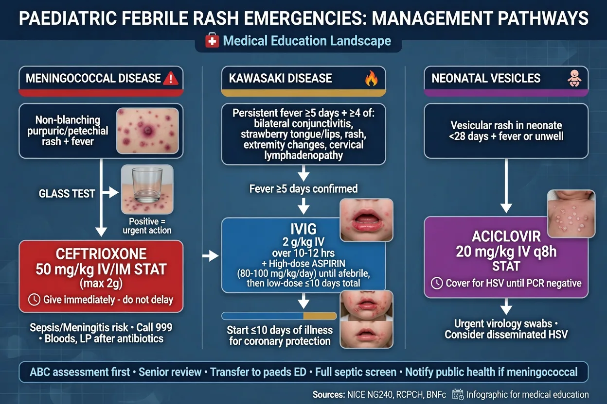
The immediate management is determined by toxicity and morphology. For the toxic or shocked child, apply the paediatric sepsis bundle — airway, high-flow oxygen, intra-osseous access after two failed intravenous attempts or 90 seconds, a 10 mL per kilogram balanced-crystalloid bolus reassessed after each aliquot, and the empiric antibiotic within the first hour. The empiric antibiotic for suspected meningococcal disease is ceftriaxone 50 mg per kilogram intravenously or intramuscularly as a single stat dose (maximum 2 g); add vancomycin 15 mg per kilogram if pneumococcal meningitis with penicillin resistance is plausible, and aciclovir 20 mg per kilogram every eight hours if herpes simplex encephalitis is in the differential. Give dexamethasone 0.15 mg per kilogram with the antibiotic when meningitis is the focus. For Kawasaki, the time-critical bundle is intravenous immunoglobulin 2 g per kilogram plus aspirin, not a resuscitation bundle, and the child is admitted to paediatrics rather than intensive care unless shocked. Antipyresis uses paracetamol 15 mg per kilogram every four to six hours for distress, never aspirin in the child with a possible viral illness.[1][2]
[1]The structured ABCDE approach to the febrile rash
The candidate who applies a structured approach to every febrile rash will not miss the dangerous one behind the trivial one. The method is resuscitation first (ABCDE), then morphology identification, then targeted treatment of life-threatening causes empirically, then isolation and disposition. [1]
ABCDE approach to the febrile child with a rash
A — Airway
Assess patency. Rarely compromised unless anaphylaxis with upper airway angio-oedema (prepare for IM adrenaline and intubation) or SJS/TEN with airway mucosal sloughing.
B — Breathing
High-flow oxygen via mask if SpO2 under 94% or any respiratory distress. Assess work of breathing, rate, recession. Severe varicella pneumonia or measles pneumonia may require ventilatory support.
C — Circulation
The critical step. Look for shock: capillary refill over 3 seconds, mottled peripheries, cool extremities, tachycardia, weak pulse. Hypotension is late and pre-terminal in children. Establish IV/IO access after two failed attempts or 90 seconds. Give balanced crystalloid 10 mL per kilogram boluses, reassessed after each. Escalate to noradrenaline for refractory shock. Check capillary glucose.
D — Disability
AVPU/GCS, pupils, capillary glucose (hypoglycaemia is common in the septic child and worsens outcome). Meningococcal meningitis may present with altered mental state, photophobia, neck stiffness, bulging fontanelle.
E — Exposure and rash assessment
Fully undress the child. Examine the entire skin surface, scalp, ears, behind the knees, buttocks, palms, soles, and all mucous membranes. Perform the glass test for blanching. Assess Nikolsky sign if desquamating. Record distribution, morphology, staging, desquamation, mucosal involvement.
Identify rash type
Classify into one of six morphologies: erythematous, maculopapular, vesicular/bullous, petechial/purpuric, urticarial, desquamating. The morphology group generates the differential.
Treat life-threatening causes empirically
Do not wait for confirmation. Non-blanching petechiae with fever: ceftriaxone 50 mg/kg. Kawasaki features: IVIG 2 g/kg and aspirin. Desquamating with mucosal sparing: flucloxacillin for SSSS. Desquamating with mucosal involvement: drug withdrawal and burn-unit for SJS/TEN. Toxic shock: clindamycin, flucloxacillin, IVIG, aggressive fluids. Neonate with vesicles: aciclovir.
Isolate and dispose
Airborne isolation for suspected measles; droplet for meningococcal and rubella until 24 hours of antibiotics; contact for SSSS and varicella. Admit the toxic, the meningococcal, the Kawasaki, the SSSS, the TSS and the neonate. Safety-net the discharged viral exanthem.
Trial evidence — the randomised data that changed practice
IVIG in Kawasaki disease — the Newburger 1986 and 1991 trials
New England Journal of Medicine
PMID 2426590 and 1709446
Population: Children with acute Kawasaki disease meeting diagnostic criteria
Key finding
The 1986 trial established that a single course of intravenous immunoglobulin (400 mg/kg/day for 4 days) plus aspirin reduced the coronary artery aneurysm rate from 20% in the aspirin-alone group to 4% when echocardiography was assessed at 7 weeks. The 1991 trial compared a single 2 g/kg infusion over 10 hours with four daily 400 mg/kg infusions and found the single high-dose infusion was more effective (fewer aneurysms, shorter fever), establishing the 2 g/kg single infusion as the standard of care.
ASH 2019 guideline — ITP in children: observation over treatment
Blood Advances
PMID 31794604
Population: Children with newly diagnosed immune thrombocytopenia
Key finding
For children with newly diagnosed ITP and no bleeding or mild bleeding (skin only), the guideline strongly recommends observation regardless of platelet count, because the risk of intracranial haemorrhage is under 0.5% and treatment with IVIG or corticosteroid carries morbidity. For significant bleeding, a single dose of IVIG 0.8 to 1 g/kg is first-line because it raises the platelet count more rapidly than corticosteroid.
Dangerous rashes — the five you cannot miss
The Fellowship candidate must hold five dangerous rashes at immediate recall. Each has a pathognomonic morphology or criterion and a time-critical treatment. Missing any one is an exam failure and a patient harm event. [1]
Meningococcal
- Non-blanching petechial or purpuric rash, rapidly progressive; child toxic or in shock
- Ceftriaxone 50 mg/kg IV/IM immediately (max 2 g); fluid resuscitation 10 mL/kg boluses; do NOT delay for LP or imaging
- Notify public health; chemoprophylaxis of household contacts (rifampicin, ciprofloxacin or ceftriaxone)
- Case fatality 5 to 10 per cent even with optimal care
Kawasaki
- Fever 5+ days plus 4 of 5: bilateral non-purulent conjunctivitis, polymorphous rash, oral changes (strawberry tongue, cracked lips), extremity changes, cervical lymphadenopathy
- IVIG 2 g/kg over 10-12 hours plus aspirin 30-50 mg/kg/day then 3-5 mg/kg/day, within 10 days
- Echocardiogram at diagnosis and 6-8 weeks; lifelong cardiology follow-up if aneurysms present
- Reduces coronary aneurysm from 25% to 3-5%; leading cause of acquired heart disease in children if missed
SSSS
- Young child (under 5), diffuse erythema, skin tenderness, flaccid bullae, perioral and flexural desquamation; Nikolsky positive; mucosa SPARED
- Flucloxacillin 50 mg/kg IV (or clindamycin for toxin suppression); analgesia; fluid and electrolyte management; emollient wound care
- NO corticosteroids; isolate (contagious exfoliative toxin)
- Split is superficial (granular layer); re-epithelialises in 7-14 days; mortality under 5% in children
Toxic shock
- Fever, diffuse sunburn-like macular erythroderma, hypotension, multi-organ involvement (GI, muscular, mucosal, renal, hepatic, haematological, CNS); desquamation of palms and soles at 1-2 weeks
- Remove source (tampon, nasal packing); aggressive fluids 100-200 mL/kg in first 24 hours; clindamycin + flucloxacillin + vancomycin; IVIG 1-2 g/kg; noradrenaline for refractory shock
- Staphylococcal TSS mortality under 5%; streptococcal TSS mortality 30-70%
- Superantigen bypasses antigen presentation; activates 5-30% of all T-cells
SJS/TEN
- Atypical target lesions, skin detachment (SJS under 10% body surface area; TEN over 30%), two or more mucosal sites, skin tenderness; usually drug-triggered (sulfonamides, anticonvulsants, NSAIDs)
- Withdraw culprit drug immediately; burn-unit wound care; fluid and nutritional support; urgent ophthalmology (corneal scarring); do NOT routinely use corticosteroids or IVIG (evidence conflicting)
- Mortality 10% for SJS, 30% for TEN; Stevens-Johnson is the severe mimic of erythema multiforme
- Distinct from erythema multiforme: EM has typical targets and one mucosal site at most
Complications and pitfalls
The complications of a missed febrile rash are the failure mode of the presentation. Untreated meningococcal disease progresses to septic shock, purpura fulminans, disseminated intravascular coagulation, limb amputation and death, with a case-fatality of 5 to 10 per cent even with optimal care. Untreated Kawasaki grows coronary artery aneurysms that thrombose and infarct the child years later, the leading cause of acquired heart disease in children in the developed world. Missed measles causes pneumonia, encephalitis and the delayed subacute sclerosing panencephalitis. The pitfalls cluster on five errors. First, attributing every febrile rash to a viral exanthem before excluding meningococcal and Kawasaki — every febrile child gets the glass test and the fever-duration check. Second, giving aspirin for a viral illness — Reye syndrome. Third, delaying the antibiotic for the lumbar puncture or the scan in suspected meningococcal disease. Fourth, missing incomplete Kawasaki in the febrile infant under six months with fewer than four criteria. Fifth, labelling Stevens-Johnson syndrome as erythema multiforme because of a target rash, missing the multiple mucosal sites and skin detachment that demand burn-unit care.[1][2][5]
Prognosis and disposition
The prognosis of the febrile rash follows the diagnosis. Viral exanthems recover within days and the child is discharged with a safety-net. Meningococcal disease is admitted to paediatric intensive care if shocked, with a 5 to 10 per cent mortality and a survivor cohort carrying skin, limb and neurologic sequelae; chemoprophylaxis of contacts and public-health notification are completed from the emergency department. Kawasaki is admitted for intravenous immunoglobulin, aspirin and an echocardiogram, with paediatric cardiology follow-up for at least six to eight weeks and lifelong cardiology surveillance if aneurysms are present. Measles and varicella are managed in isolation at home unless complicated. Henoch-Schonlein purpura is admitted for severe abdominal or renal disease and discharged with a structured plan for urinalysis and blood-pressure monitoring for six to twelve months. Erythema multiforme is managed outpatient unless the trigger is severe or Stevens-Johnson is suspected.[2][6]
Special populations
The neonate under 28 days with any vesicular rash has herpes simplex until proven otherwise — aciclovir 20 mg per kilogram every eight hours, a full septic workup and admission. The immunocompromised or neutropenic child runs severe varicella and atypical rashes and is managed with broad empiric cover and early aciclovir. The asplenic or complement-deficient child is at catastrophic risk of meningococcal disease and receives ceftriaxone on arrival at any febrile presentation. The pregnant adolescent with varicella risks maternal pneumonia and congenital varicella syndrome and receives intravenous aciclovir. The under-vaccinated child brings measles and Haemophilus influenzae type b back into the differential, and both are notifiable on suspicion. The returning traveller expands the differential to dengue, rickettsial disease and viral haemorrhagic fevers, where the non-blanching rash with thrombocytopenia and fever is managed with isolation and infectious-disease input.[1][3]
Evidence and regional guidelines
The contemporary frameworks are the American Heart Association Kawasaki guideline (McCrindle and colleagues, 2017), which sets the diagnostic criteria, the intravenous immunoglobulin and aspirin regimen and the echocardiographic follow-up used throughout this topic,[2] and the EULAR/PRINTO/PRES criteria for Henoch-Schonlein purpura (Ozen and colleagues, 2010), which define the immunoglobulin-A vasculitis classification.[6] The meningococcal reference (Rosenstein and colleagues, New England Journal of Medicine, 2001) underpins the ceftriaxone dose, the shock management and the chemoprophylaxis of contacts.[1] Measles is covered by the Moss and Griffin Lancet review (2012) and the emergency-department management of measles and varicella (Cavallaro and colleagues, 2025).[3][4] Erythema multiforme and its distinction from Stevens-Johnson syndrome rest on the Sokumbi and Wetter review (2012).[5]
The staphylococcal scalded skin syndrome and the toxin-mediated exfoliative dermatoses are covered by the Stanley and Amagai review in the New England Journal of Medicine (2006), which explains the desmoglein 1 cleavage and the mucosal-sparing mechanism.[7] Toxic shock syndrome — both staphylococcal and streptococcal — is reviewed by Lappin and Ferguson in the Lancet Infectious Diseases (2009), which sets out the superantigen mechanism and the management framework.[8] The IDSA guideline for group A streptococcal pharyngitis (Shulman and colleagues, 2012) governs the diagnosis and antibiotic management of scarlet fever.[9] Rubella is covered by the Banatvala and Brown Lancet review (2004),[10] and human parvoviruses — including parvovirus B19 and its complications in pregnancy, haemolytic anaemia and immunocompromise — by the Qiu, Soderlund-Venermo and Young review in Clinical Microbiology Reviews (2017).[11] Immune thrombocytopenia management follows the American Society of Hematology 2019 guideline (Neunert and colleagues), which established observation over treatment for the well child with skin-only bleeding.[12] The landmark randomised trials of intravenous immunoglobulin in Kawasaki disease — Newburger and colleagues in the New England Journal of Medicine (1986 and 1991) — established the single 2 g per kilogram infusion that reduced coronary artery aneurysms from 25 per cent to 3 to 5 per cent.[13][14]
ANZ practice note. The Royal Children's Hospital Melbourne clinical practice guidelines govern Australasian management of the febrile child, Kawasaki disease, meningococcal infection and Henoch-Schonlein purpura, and align with the NICE feverish-child framework. Meningococcal disease and measles are notifiable in all Australian states and New Zealand; chemoprophylaxis of meningococcal contacts uses rifampicin or single-dose ciprofloxacin, and the public-health unit is engaged from the emergency department. Weight-based dosing uses the Broselow tape or the formula weight equals age in years plus four, times two. [1]
Clinical pearls — the exam-ready detail
[1] [1] [1] [1] [1] [1] [1] [1] [1] [1] [1] [1] [1] [1] [1] [1] [1] [1]Exam pearls
- A non-blanching petechial or purpuric rash with fever is meningococcal until proven otherwise — ceftriaxone 50 mg per kilogram immediately.
- Fever of five days or more plus four of five principal features is Kawasaki — intravenous immunoglobulin 2 g per kilogram and aspirin within ten days to prevent coronary artery aneurysm.
- Never give aspirin to a child with varicella or influenza — Reye syndrome.
- Varicella rash is vesicular in crops at different stages; measles descends from the hairline after Koplik spots.
- Palpable purpura on the lower limbs plus abdominal pain, arthritis and renal involvement is Henoch-Schonlein purpura; its intussusception is atypically ileo-ileo.
- Erythema multiforme has typical targets and at most one mucosal site; Stevens-Johnson has atypical targets, two or more mucosal sites and skin detachment.
- Incomplete Kawasaki in the infant under six months with seven days of fever is the classic miss.
- Weight before drug — Broselow tape, or weight in kilograms equals age in years plus four, times two.
- SSSS spares the mucosa; Stevens-Johnson involves it — the Nikolsky sign is positive in both, so the mucosa is the discriminator.
- Scarlet fever: sandpaper rash, Pastia lines, strawberry tongue — 10 days of penicillin V or amoxicillin to prevent rheumatic fever.
- Roseola (HHV-6): three to five days of high fever in a well infant, then rash as the fever breaks — the commonest cause of febrile convulsions in infants.
- Strawberry tongue appears in three diseases: Kawasaki, scarlet fever and toxic shock — the tongue looks the same; the rest of the picture discriminates.
- Toxic shock: fever, sunburn rash, hypotension, multi-organ — remove the source, aggressive fluids, clindamycin and IVIG.
- Ampicillin rash in EBV is not true penicillin allergy — but never give amoxicillin for a sore throat that might be mono.
- Parvovirus B19 in pregnancy: up to 10 per cent fetal loss — check IgG and IgM in any pregnant contact of a slapped-cheek child.
- ITP: well child, isolated thrombocytopenia, no blasts on film — observe if skin-only bleeding; IVIG 0.8 to 1 g per kilogram only for significant bleeding.
- Enterovirus 71 HFMD causes brainstem encephalitis and pulmonary oedema — any neurological sign with HFMD needs urgent escalation.
- Urticarial wheals that persist in the same place for over 24 hours are not simple allergy — think erythema multiforme, serum sickness, urticarial vasculitis or Kawasaki.
- Measles needs airborne isolation (R-nought 12 to 18); meningococcal and rubella need droplet; varicella and SSSS need contact precautions.
- Never give steroids before a bone marrow in suspected ITP — if the child has leukaemia, steroids lyse lymphoblasts and obscure the diagnosis. [1]
Short answer questions
SAQ — Febrile child with a rapidly evolving purpuric rash
10 minutes · 10 marks
A 3-year-old boy presents to the emergency department with a six-hour history of fever and irritability, and a rapidly spreading purpuric rash over the trunk and limbs. He is lethargic, with cool mottled peripheries, capillary refill of five seconds, heart rate 160 and blood pressure 70 over 40. The rash is non-blanching on the glass test.
SAQ — Fever for six days with conjunctivitis and swollen hands
10 minutes · 10 marks
A 4-year-old girl is brought to the emergency department with fever for six days that has not responded to antibiotics or antipyretics. Her mother reports red eyes, a sore mouth and swollen painful hands. On examination she is irritable, with bilateral non-purulent conjunctivitis, cracked red fissured lips, a strawberry tongue, an erythematous maculopapular trunk rash, and erythema and oedema of the palms and soles.
Red flags
[1]References
- [1]Rosenstein NE, Perkins BA, Stephens DS, Popovic T, Hughes JM. Meningococcal disease N Engl J Med, 2001.PMID 11333996
- [2]McCrindle BW, Rowley AH, Newburger JW, et al. Diagnosis, Treatment, and Long-Term Management of Kawasaki Disease: A Scientific Statement for Health Professionals From the American Heart Association Circulation, 2017.PMID 28356445
- [3]Moss WJ, Griffin DE. Measles Lancet, 2012.PMID 21855993
- [4]Cavallaro SC, Shihabuddin BS, Brown AM. Emergency department management of measles, mumps, rubella, and varicella in pediatric patients Pediatr Emerg Med Pract, 2025.PMID 41092047
- [5]Sokumbi O, Wetter DA. Clinical features, diagnosis, and treatment of erythema multiforme: a review for the practicing dermatologist Int J Dermatol, 2012.PMID 22788803
- [6]Ozen S, Pistorio A, Iusan SM, et al. EULAR/PRINTO/PRES criteria for Henoch-Schönlein purpura, childhood polyarteritis nodosa, childhood Wegener granulomatosis and childhood Takayasu arteritis: Ankara 2008. Part II: Final classification criteria Ann Rheum Dis, 2010.PMID 20413568
- [7]Stanley JR, Amagai M. Pemphigus, bullous impetigo, and the staphylococcal scalded-skin syndrome N Engl J Med, 2006.PMID 17065642
- [8]Lappin E, Ferguson AJ. Gram-positive toxic shock syndromes Lancet Infect Dis, 2009.PMID 19393958
- [9]Shulman ST, Bisno AL, Clegg HW, et al. Clinical practice guideline for the diagnosis and management of group A streptococcal pharyngitis: 2012 update by the Infectious Diseases Society of America Clin Infect Dis, 2012.PMID 22965026
- [10]Banatvala JE, Brown DW. Rubella Lancet, 2004.PMID 15064032
- [11]Qiu J, Soderlund-Venermo M, Young NS. Human Parvoviruses Clin Microbiol Rev, 2017.PMID 27806994
- [12]Neunert C, Terrell DR, Arnold DM, et al. American Society of Hematology 2019 guidelines for immune thrombocytopenia Blood Adv, 2019.PMID 31794604
- [13]Newburger JW, Takahashi M, Beiser AS, et al. The treatment of Kawasaki syndrome with intravenous gamma globulin N Engl J Med, 1986.PMID 2426590
- [14]Newburger JW, Takahashi M, Burns JC, et al. A single intravenous infusion of gamma globulin as compared with four infusions in the treatment of acute Kawasaki syndrome N Engl J Med, 1991.PMID 1709446