EM · Paediatric respiratory distress
Paediatric respiratory distress — croup, bronchiolitis and acute asthma
Also known as Croup · Laryngotracheobronchitis · Bronchiolitis · Paediatric asthma · Stridor in children · Wheeze in children
Paediatric respiratory distress — the three exam pillars of croup (subglottic viral oedema, inspiratory stridor, barking cough, dexamethasone 0.15 mg/kg orally with nebulised adrenaline 1:1000 at 0.5 mL/kg max 5 mL for moderate to severe), bronchiolitis (RSV bronchiolar obstruction, wheeze, hypoxia, supportive care with no routine bronchodilator or steroid), and acute asthma (salbutamol 2.5 to 5 mg nebulised, ipratropium, early steroid, magnesium sulfate 40 mg/kg), distinguished from the can't-miss mimics of foreign body aspiration, epiglottitis and bacterial tracheitis. The Poiseuille r-to-the-fourth rationale, the Westley croup score reproduced, weight-based dosing before any drug, and the escalation ladder from humidified oxygen through high-flow and non-invasive ventilation to intubation. ACEM-primary, globally tagged.
On this page & tools
Your progress
Saved locally on this device.
Target exams
Red flags
Related topics
- The sick child and paediatric resuscitation
- Upper airway obstruction in the emergency department
- Acute severe asthma
- Respiratory failure (type 1 and type 2)
- Paediatric sepsis and septic shock (the septic child in the emergency department)
- Non-invasive ventilation in the emergency department (CPAP and BiPAP)
Paediatric respiratory distress is one of the commonest and highest-stakes presentations an emergency physician manages, and it tests three skills at once: the structured visual assessment across the room, the weight-based drug dosing, and the single most important discriminator in paediatric airway medicine — stridor versus wheeze. The three exam pillars are croup (viral subglottic oedema, inspiratory stridor, barking cough), bronchiolitis (viral bronchiolar obstruction, wheeze, first episode under two years) and acute asthma (reversible lower-airway obstruction, recurrent wheeze, one year and over). The framework — recognise the level and the severity, keep the child calm, treat the cause, escalate before exhaustion — applies to all three.[1][5][7]
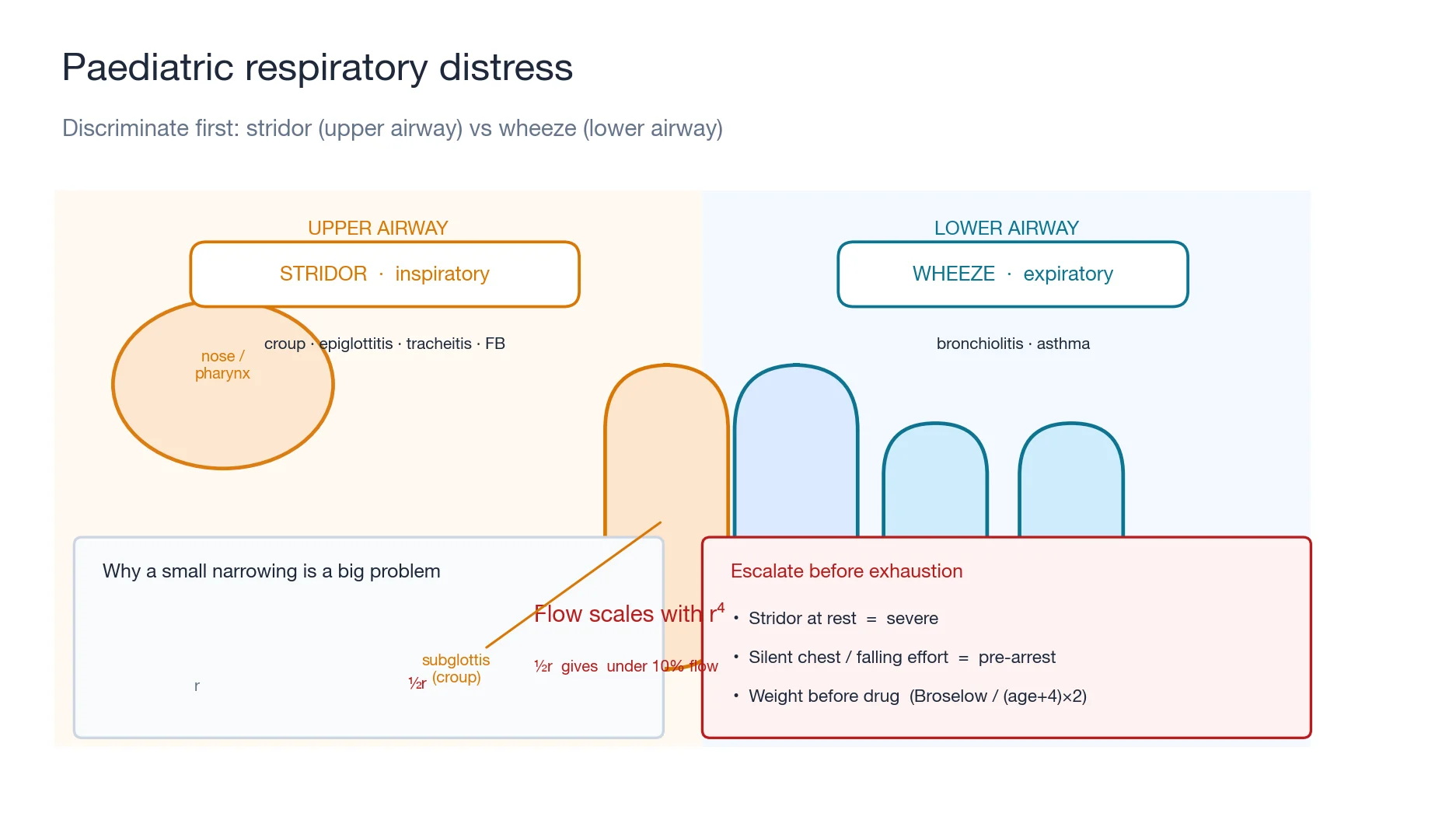
The framework — stridor or wheeze, then severity
The first decision is anatomical. Stridor localises the obstruction to the upper airway (the nose to the subglottis): an inspiratory stridor means an extrathoracic lesion (croup, epiglottitis), a biphasic stridor means a fixed glottic or subglottic lesion, and a stridor at rest means a severe obstruction with critically narrowed flow. Wheeze localises the obstruction to the lower airway (the bronchi and bronchioles): bronchiolitis in the first-time wheezer under two, asthma in the recurrent wheezer over one. The second decision is severity, judged visually — respiratory rate, recession, accessory-muscle use, colour, and conscious level — and never by a single number. The trajectory matters most: the improving croup, the tiring bronchiolitic, and the silent chest of severe asthma all look different over ten minutes than over one. [1]
[1]Why a small narrowing is a big problem in the child — Poiseuille's law
Airflow through a tube is proportional to the fourth power of the radius (Poiseuille's law), so a fifty per cent reduction in airway radius reduces flow by over ninety per cent. The paediatric airway is already narrow — the infant cricoid is around four millimetres in diameter — so the same millimetre of mucosal oedema that barely troubles an adult can halve the airway radius of a toddler. This is why croup, epiglottitis and bacterial tracheitis are so much more dangerous in children than in adults, and why a small amount of subglottic oedema produces a disproportionate and rapidly escalating work of breathing.[1][9]
Differential diagnosis — the causes, distinguished
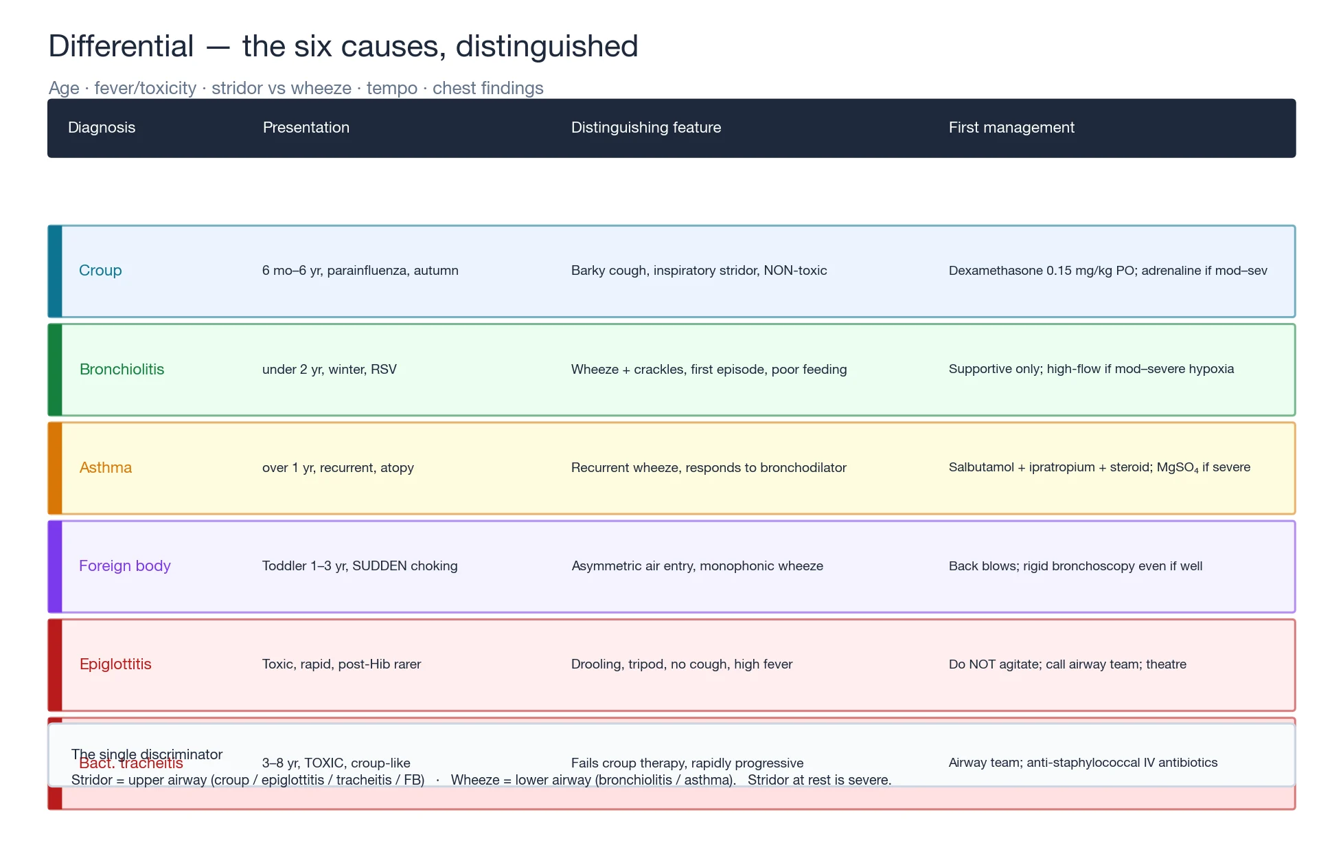
The causes of paediatric respiratory distress are grouped by the level of obstruction and the tempo, and the identification of the cause directs the specific therapy. [1]
Croup
- 6 mo to 6 yr, peak 1 to 2 yr, autumn parainfluenza
- Barking cough, inspiratory stridor, low fever, NON-toxic
- Dexamethasone 0.15 mg/kg PO; nebulised adrenaline if moderate to severe
- Most discharge within hours; benign course
Bronchiolitis
- Under 2 yr, peak 2 to 6 mo, winter RSV
- Coryza, wheeze, crackles, poor feeding, apnoea in small infants
- SUPPORTIVE only — oxygen, feeds, suction; no routine bronchodilator or steroid
- Admit if hypoxia, apnoea, poor feeding
Acute asthma
- Recurrent wheeze, age 1 yr and over, atopy, viral trigger
- Wheeze, prolonged expiration, recession, speech limited
- Salbutamol 2.5 to 5 mg neb + ipratropium + steroid; MgSO4 40 mg/kg if severe
- Escalate to PICU if silent chest, exhaustion, rising CO2
Foreign body
- Toddler 1 to 3 yr, SUDDEN choking, eating/playing
- Asymmetric air entry, monophonic wheeze, or asymptomatic
- Back blows / chest thrusts if choking; rigid bronchoscopy definitive
- Refer for bronchoscopy even if well — delayed = lobar collapse
Epiglottitis
- Toxic, rapid, drooling, tripod posture, no cough
- High fever, distressed, soft stridor — post-Hib vaccine, rarer
- DO NOT agitate — call anaesthetist + ENT; theatre airway; ceftriaxone
- Airway secured in theatre, then PICU
Bacterial tracheitis
- 3 to 8 yr, TOXIC with a croup-like cough, rapidly progressive
- High fever, looks septic, soft stridor, does NOT respond to croup therapy
- Call airway team early; controlled airway; anti-staphylococcal IV antibiotics
- Masks as croup — the trap of the toxic stridulous child
Common causes by age — the second discriminator after stridor or wheeze
Age is the second discriminator after the level of obstruction, because the causative organism and the airway anatomy change across the paediatric age bands. A confident age-stratified differential narrows the working diagnosis within seconds of meeting the child and guards against the dangerous habit of treating every wheeze as asthma or every cough as croup.[1][5][12]
Infant (0 to 12 mo)
- Bronchiolitis is the number-one cause — RSV, winter, first wheeze, crackles, apnoea in the ex-preterm
- Pertussis — paroxysmal cough, whoop, post-tussive vomiting, apnoea in the unimmunised infant
- Congenital airway — laryngomalacia (inspiratory stridor from birth, worse with feeding and crying, settles at rest)
- Vascular ring, tracheo-oesophageal fistula, congenital heart disease — biphasic stridor, feeding difficulty
- Pneumonia — viral (RSV) most common; bacterial rare under one month (group B strep)
Toddler (1 to 3 yr)
- Croup — parainfluenza, barking cough, inspiratory stridor; the classic toddler cause
- Foreign body aspiration — sudden choking, asymmetric air entry, monophonic wheeze
- Asthma emerging — recurrent viral wheeze in the atopic child (first episodes overlap bronchiolitis)
- Pertussis still possible in the under-immunised toddler
- Bacterial tracheitis and epiglottitis peak here into the early school years
Child (4 to 12 yr)
- Asthma — the dominant cause of wheeze; atopy, exertional and viral triggers
- Community-acquired pneumonia — Streptococcus pneumoniae, and atypical Mycoplasma in the school-age child
- Bacterial tracheitis — 3 to 8 yr, the toxic mimic of croup
- Epiglottitis — now rare post-Hib vaccine but lethal when missed
- Functional or large-airway causes (vocal-cord dysfunction) in the older anxious child
The age-stratified logic is anatomical and immunological. The infant airway is small and floppy (laryngomalacia is the commonest congenital stridor), the toddler explores the world by mouth (foreign body) and the subglottis is at its narrowest (croup), and the school-age child has outgrown the structural susceptibility but accumulates an atopic and exposure burden (asthma, Mycoplasma pneumonia).[12][13]
AGE-IC — the age-stratified causes of paediatric respiratory distress
AGE-IC
The recurrent wheezer over one year — the dominant cause in the school-age child; reversible, atopic, viral- or exertion-triggered
The toddler 6 mo to 6 yr — barking cough, parainfluenza, subglottic oedema; the classic upper-airway cause
The toxic stridulous child — high fever, septic, rapidly progressive; call the airway team and do not agitate
Bronchiolitis (RSV, number one), pertussis (whoop, apnoea), congenital airway (laryngomalacia, vascular ring) — the under-12-month band
The toddler with sudden choking and asymmetric air entry — rigid bronchoscopy even if the child looks well

Croup — diagnosis and management
Croup (acute laryngotracheobronchitis) is the commonest cause of stridor in children, peaking between one and two years and driven by parainfluenza virus in autumn. Viral inflammation of the subglottic mucosa produces oedema in the narrowest part of the paediatric airway, and the Poiseuille effect converts a small swelling into inspiratory stridor, a barking cough and a hoarse voice. The child is afebrile or low-grade fever and non-toxic, the distinction from bacterial tracheitis and epiglottitis. Symptoms characteristically worsen at night and improve with cool air.[1][3]
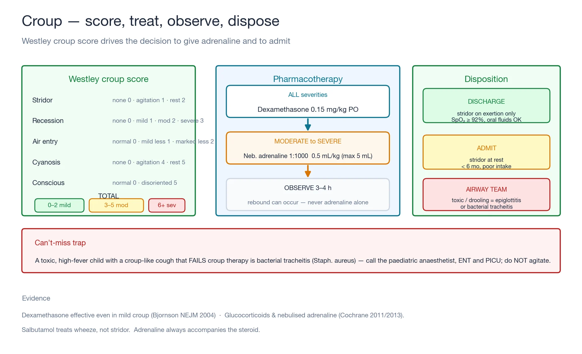
Severity is scored with the Westley croup score, which drives the decision to give adrenaline and to admit: [1]
| Westley component | 0 | 1 | 2 | 3 | 4 | 5 |
|---|---|---|---|---|---|---|
| Level of consciousness | Normal | — | — | — | — | Disoriented |
| Cyanosis | None | — | — | — | With agitation | At rest |
| Stridor | None | With agitation | At rest | — | — | — |
| Air entry | Normal | Mild decrease | Marked decrease | — | — | — |
| Recession | None | Mild | Moderate | Severe | — | — |
Management. Every child with croup receives dexamethasone 0.15 mg/kg orally as a single dose — the 2004 Bjornson trial proved efficacy even in mild disease, and corticosteroids reduce croup scores within six hours, shorten stay and cut return visits.[2][3] For moderate to severe croup (stridor at rest, marked recession, or any cyanosis) add nebulised adrenaline 1:1000 at 0.5 mL/kg to a maximum of 5 mL; onset is within minutes, the effect lasts one to two hours, and the child must be observed for at least three to four hours because rebound can occur.[4] Humidified oxygen is for the hypoxic or distressed child and is a comfort measure, not a therapy for obstruction. Always co-give the steroid with the adrenaline — adrenaline alone wears off and the obstruction recurs.
[1] [1]Discharge criteria after croup treatment
Bronchiolitis — diagnosis and management
Bronchiolitis is a viral lower-airway illness of infants under two years (peak two to six months), most often respiratory syncytial virus, presenting with coryza, wheeze, crackles, poor feeding and, in small or ex-preterm infants, apnoea. The bronchioles are inflamed, oedematous and plugged with mucus and necrotic debris, producing air-trapping, ventilation-perfusion mismatch and hypoxia. The diagnosis is clinical; the role of the emergency physician is to support the child through a self-limiting illness and to identify the minority who escalate.[5]
Management is supportive, and the evidence is clear that bronchodilators, corticosteroids, antibiotics and chest physiotherapy do not change the course. The 2014 American Academy of Pediatrics guideline recommends against their routine use: a bronchodilator may be trialled in a severely distressed child and stopped if there is no objective response, but it should not be continued by default.[5] Oxygen is given to maintain SpO₂ at 90 to 92 per cent or above (threshold varies by unit); nasogastric feeds support the infant who is too breathless to feed; gentle nasal suction clears secretions. Hydration, monitoring and patience are the therapy.
[1]Escalation to PICU is for recurrent or significant apnoea, persistent hypoxaemia below 90 per cent despite oxygen, a rising carbon dioxide on gas, or severe distress with exhaustion. The ex-preterm infant under six months corrected age and the child with congenital heart disease or chronic lung disease are higher risk and warrant a lower threshold to admit and to escalate.[5][6]
Acute asthma — diagnosis and management
Acute asthma is reversible lower-airway obstruction in the child over one year with a history of recurrent wheeze or atopy, triggered most often by a viral upper-respiratory infection. Bronchospasm, mucosal oedema and mucus produce expiratory wheeze, prolonged expiration, hyperinflation and recession. Severity is graded by the response to treatment and the markers of fatigue, and the dangerous sign is the silent chest with poor air entry — the obstruction is so complete that no sound is made, and the child is tiring. [1]
| Feature | Moderate | Severe | Life-threatening |
|---|---|---|---|
| SpO₂ (air) | at or above 92 per cent | below 92 per cent | below 92 per cent plus a danger sign |
| Speech / feeding | sentences | words or unable | exhausted, altered |
| Work of breathing | mild recession | marked recession, accessory muscle | silent chest, poor air entry |
| RR / HR | raised | above age threshold | bradycardia or irregular |
| Conscious level | normal | agitated | drowsy, confused |
Management. Give oxygen to keep SpO₂ at 94 per cent or above. The cornerstone is nebulised salbutamol 2.5 mg for a child under five years and 5 mg for five years and over, driven by oxygen and repeated every twenty minutes for three doses (an equivalent regimen via a spacer — four to ten puffs — suits the milder child who can cooperate). Add ipratropium bromide 250 micrograms nebulised every twenty minutes for three doses in the severe attack; the Cochrane evidence supports the combination of anticholinergic and beta-agonist for the initial treatment of acute asthma in children.[7] Give a systemic corticosteroid early — prednisolone 1 to 2 mg/kg orally to a maximum of 40 to 60 mg, or hydrocortisone 4 mg/kg intravenously (or methylprednisolone 1 mg/kg) if severe or unable to tolerate oral. For the severe attack unresponsive to the first hour of therapy, add magnesium sulfate 40 mg/kg intravenously over twenty minutes; the MAGICIAN trial informs current paediatric practice on intravenous magnesium in acute severe asthma.[10] Intravenous salbutamol is the next tier and is a PICU decision.
[1]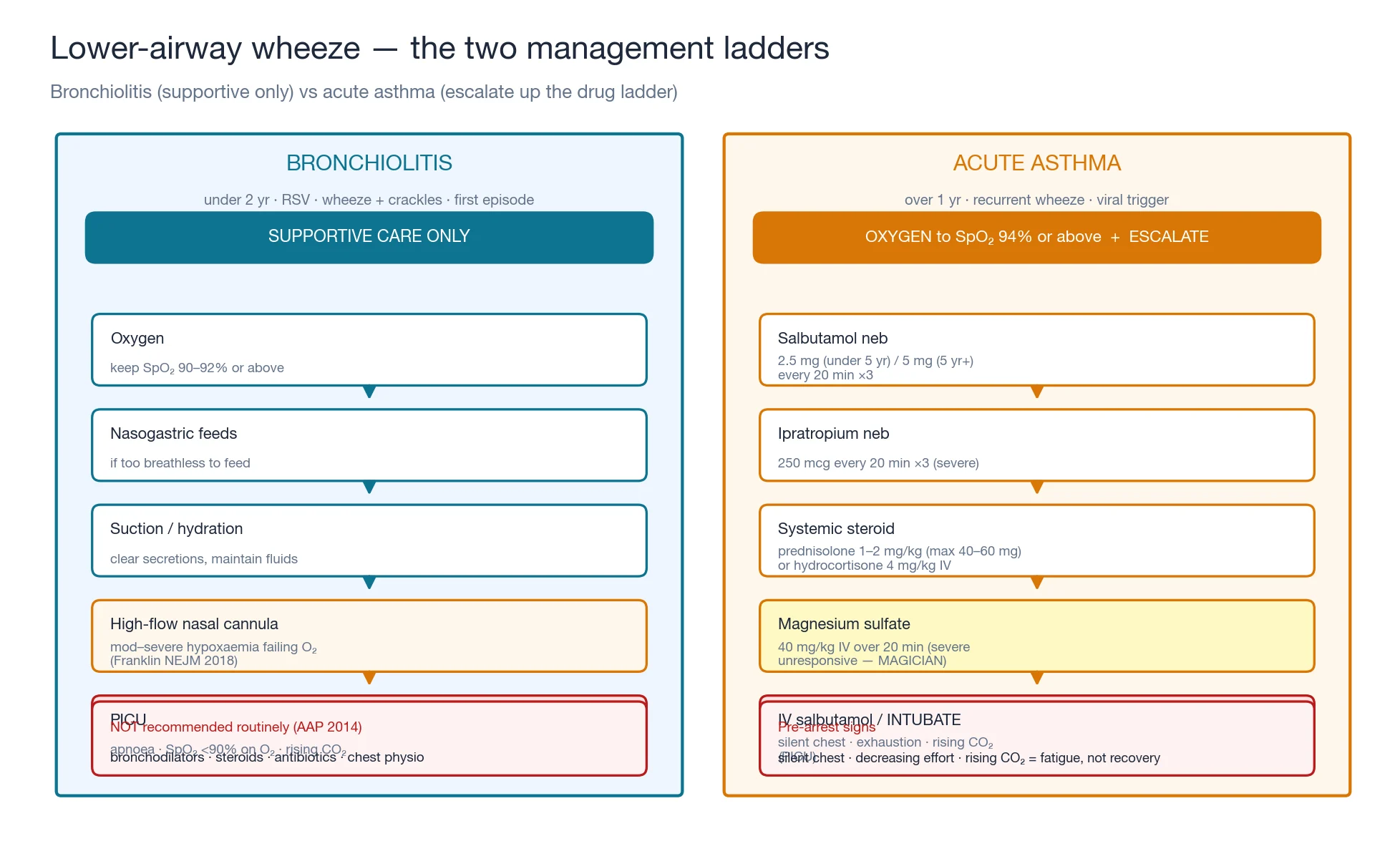
Foreign body aspiration — sudden choking
An inhaled foreign body classically presents in a toddler one to three years with a sudden choking episode while eating or playing, followed by cough, asymmetric air entry, or a monophonic wheeze. The trap is the delayed presentation: the child who choked days ago and now has lobar collapse or recurrent pneumonia on one side. A high index of suspicion is the single most important diagnostic tool. Inspiratory and expiratory chest radiographs (or a lateral neck film) may show hyperinflation of the obstructed lobe (air-trapping behind a ball-valve obstruction) but are often normal. Rigid bronchoscopy under general anaesthetic is the gold standard for both confirmation and removal, and a child with a convincing history should be referred even if well and even if the radiograph is normal.[8]
If the child is choking and conscious, give back blows and chest thrusts for an infant under one year, or abdominal thrusts (the Heimlich manoeuvre) for a child over one year. If the obstruction is complete and the child becomes unconscious, begin cardiopulmonary resuscitation and check the airway before each breath. Do not perform a blind finger sweep. [1]
Pertussis — the paroxysmal cough and the apnoeic infant
Pertussis (whooping cough) is a Bordetella pertussis toxin-mediated illness whose hallmark is the paroxysmal cough — runs of five to ten rapid coughs ending in an inspiratory whoop in the older child, or in apnoea, cyanosis and post-tussive vomiting in the young infant who cannot generate the whoop. It targets the under-immunised (the infant too young to have completed the primary course), the waning-immunity adolescent, and the adult whose acellular-vaccine immunity has faded. The diagnosis is clinical: a catarrhal prodrome of one to two weeks, then a paroxysmal phase of two to six weeks, then a convalescent phase — confirmed by nasopharyngeal PCR.[11]
Management. Give a macrolide — azithromycin 10 mg/kg orally on day one then 5 mg/kg daily for four more days (or clarithromycin 7.5 mg/kg twice daily for seven days) — which shortens infectivity and improves symptoms only if given within the first three weeks; later, it is for infection control, not symptom relief. Admit the infant under six months for apnoea monitoring — pertussis is a leading infectious cause of infant death, and the apnoea can be life-threatening and unpredictable. Prophylax the household and close contacts. The Fellowship trap is the afebrile, well-looking infant with paroxysmal cough and intermittent apnoea — admit and monitor.[11]
[1]Pneumonia in children — bacterial, viral and atypical
Childhood pneumonia presents with fever, tachypnoea and respiratory distress, often with crackles, bronchial breathing or recession; the chest radiograph shows consolidation. The organism is age-stratified: viral (RSV, influenza, parainfluenza) dominates at every age and is especially common under five years; bacterial — Streptococcus pneumoniae (the commonest bacterial cause at every age), nontypeable Haemophilus influenzae, and in the neonate group B streptococcus and Gram-negative bacilli; atypical — Mycoplasma pneumoniae in the school-age child with a prominent dry cough and a radiograph that looks worse than the chest.[12]
Severity and the threshold to treat. The BTS 2011 paediatric guideline uses the WHO tachypnoea threshold (a respiratory rate at or above 50 breaths per minute under one year, or at or above 40 over one year with chest indrawing) as the bedside marker of pneumonia, and stratifies management by severity: oral amoxicillin for mild community-acquired pneumonia in the child over six months, intravenous therapy (benzylpenicillin or amoxicillin, plus a macrolide if atypical infection is suspected) for the severe or the failing-to-respond case, and oxygen for SpO₂ below 92 per cent.[12]
Viral pneumonia
- RSV, influenza, parainfluenza; often coexists with bronchiolitis
- Diffuse crackles, wheeze, low-grade fever, a prodromal coryza
- Radiograph: bilateral interstitial infiltrates and hyperinflation
- Supportive — oxygen, hydration; antibiotics only if bacterial superinfection
Bacterial pneumonia
- Streptococcus pneumoniae is number one; Haemophilus, and staphylococcal post-influenza
- High fever, focal signs, unwell, sometimes pleuritic
- Radiograph: lobar consolidation with or without an effusion
- Amoxicillin orally (benzylpenicillin IV if severe); add a macrolide if atypical overlap
Atypical (Mycoplasma)
- School-age child (5 to 15 yr), prominent dry cough, low fever, protracted
- Extrapulmonary: rash (erythema multiforme), myalgia, gastrointestinal symptoms
- Radiograph: patchy and multifocal, worse than the clinical picture
- Macrolide (azithromycin, clarithromycin) — no cell wall, so a beta-lactam fails
Congenital and structural causes — the stridor from birth
Not all stridor is croup. Laryngomalacia is the commonest congenital cause of stridor — a flaccid supraglottis that prolapses inward on inspiration, producing an inspiratory stridor from the first weeks of life that worsens with feeding, crying and upper-respiratory infections and settles when the child is calm or asleep. It is benign in over 90 per cent and resolves by 18 to 24 months, but the severe case causes feeding difficulty, failure to thrive and obstructive apnoea and needs supraglottoplasty.[13] The discriminating feature from croup is the chronicity (present from birth, not an acute illness) and the absence of fever or toxicity.
The other structural causes — vocal-cord paralysis (often after cardiac surgery; bilateral causes stridor and aphonia), subglottic stenosis (often post-intubation, a fixed biphasic stridor), vascular ring (a double aortic arch compressing the trachea, causing biphasic stridor and feeding difficulty, with the radiograph showing a right-sided aortic arch), laryngeal webs and cysts, and tracheo-oesophageal fistula — all need flexible laryngoscopy or bronchoscopy and a paediatric ENT and cardiology referral. The role of the emergency physician is to recognise the chronic, afebrile, non-toxic stridor as structural and to refer, rather than to treat it as croup.[13]
[1]Epiglottitis and bacterial tracheitis — the toxic stridulous child
These two conditions are the can't-miss mimics of croup, and both demand the senior airway team before any intervention. [1]
Acute epiglottitis (supraglottitis) is a rapidly progressive, septic illness with high fever, drooling, an inability to swallow secretions, a muffled voice, a tripod posture and a soft or absent stridor. It has become rare in children since the Haemophilus influenzae type b vaccine but remains lethal when missed. The cardinal rule is do not agitate: no tongue depressor, no lying flat, no cannulation that upsets the child. Call the paediatric anaesthetist, the ENT surgeon and PICU immediately, and the airway is secured by an inhalational induction in theatre with the surgeon scrubbed for a front-of-neck access; ceftriaxone 50 to 75 mg/kg intravenously is given after the airway is secured.[9]
Bacterial tracheitis (bacterial laryngotracheobronchitis) is the trap of the topic: a child of three to eight years with a croup-like cough and stridor who is toxic, high-fever and rapidly progressive, and does not respond to croup therapy. The organism is usually Staphylococcus aureus (also streptococci, Haemophilus). The mucosal membranes of the trachea slough and can obstruct the airway with a cast. Management is the same disciplined approach: do not agitate, call the airway team, secure the airway by a controlled induction with a surgical airway prepared, give anti-staphylococcal intravenous antibiotics (vancomycin or clindamycin with a third-generation cephalosporin), and admit to PICU. The child who looks toxic with a croup score that does not fit, or who fails standard croup treatment, is bacterial tracheitis until proven otherwise.[9]
[1]Bedside assessment — across the room, then close
Begin with visual assessment across the room: the work of breathing (recession, accessory-muscle use, head bobbing in the infant, nasal flare), the colour (pallor, mottling, cyanosis), and the conscious level (alert, agitated suggesting hypoxia, or drowsy suggesting hypercapnia and exhaustion). Count the respiratory rate over a full minute against the age-specific threshold (a rough guide: a rate above 60 in a neonate, above 50 in an infant, above 40 in a one- to five-year-old, and above 30 over five years is abnormal). Measure SpO₂. Then approach and auscultate: confirm an upper-airway stridor by listening over the mouth with the stethoscope, listen for wheeze and crackles, and check for asymmetry of air entry (foreign body). Estimate the weight before giving any drug — with a Broselow tape, or the formula weight in kilograms equals age in years plus four, times two. Watch the trend, because the single most useful observation is whether the child is improving or tiring over ten minutes.[1][5]
The components of increased work of breathing — read them individually
The work of breathing is a composite of mechanical signs, each of which localises and grades the effort the child is making to move air. Read each component individually, because the pattern tells you where the obstruction is and how hard the child is working: [1]
Sign
- Retractions (recession)
- Nasal flaring
- Grunting
- Accessory-muscle use
- Head bobbing
- Seesaw (paradoxical) breathing
- Tracheal tug
What it means
- Subcostal, intercostal and suprasternal recession — the chest wall is drawn in; worse in the infant with compliant ribs
- The infant tries to lower nasal resistance — a sign of moderate to severe distress
- Forced expiration against a partly closed glottis (auto-PEEP) — the body making its own CPAP; pneumonia, atelectasis, pulmonary oedema
- Sternocleidomastoid and scalene recruitment — severe lower-airway obstruction (asthma, bronchiolitis)
- The head falls forward with each expiration, the neck muscles acting as an accessory pump — severe infant distress, pre-exhaustion
- The abdomen moves out as the chest moves in — fatigue, impending respiratory failure, neuromuscular weakness
- Inspiration draws the trachea downward — upper-airway obstruction, severe distress
Normal respiratory rate by age — the threshold you must know
Count the respiratory rate over a full minute in a settled child (it rises with crying and fever). The thresholds below are a pragmatic guide; the trend matters more than any single value, and a falling rate in a tiring child is deterioration, not improvement. [1]
The first 15 minutes of the paediatric respiratory distress
0 min — observe across the room
Stay at the doorway. Read the work of breathing (recession, nasal flare, accessory-muscle use, head bobbing, grunting), the colour (pallor, mottling, cyanosis), and the conscious level (alert, agitated means hypoxia, drowsy means hypercapnia). Decide stridor or wheeze, and toxic or not. Keep the child on the lap of the carer — do not separate.
0 to 5 min — oxygen and the weight
Give humidified oxygen to target (92 per cent for croup and bronchiolitis, 94 per cent for asthma) by the least upsetting method. Estimate the weight NOW — a Broselow tape, or (age in years plus four) times two — before any drug. Count the respiratory rate over a full minute; measure SpO₂. Call senior paediatric and anaesthetic help for any stridor at rest, a toxic appearance, or an exhaustion sign.
5 to 10 min — focused examination and the score
Auscultate: confirm the stridor over the mouth, listen for wheeze and crackles, and check the symmetry of air entry (foreign body). Look in the mouth only if epiglottitis and tracheitis are NOT suspected — never agitate the toxic stridulous child. Apply the Westley croup score or the asthma severity grade. Take a capillary glucose.
10 to 15 min — targeted therapy
Croup: dexamethasone 0.15 mg/kg orally, add nebulised adrenaline 1:1000 at 0.5 mL/kg to a maximum of 5 mL if moderate to severe. Asthma: salbutamol 2.5 mg (under 5 yr) or 5 mg nebulised, ipratropium 250 mcg for three doses if severe, prednisolone 1 to 2 mg/kg orally or hydrocortisone 4 mg/kg IV. Bronchiolitis: oxygen, nasal suction, feed support — no routine drugs. Reassess every 10 to 15 minutes; the trend decides the disposition.
15 min onward — escalate or disposition
Escalate to high-flow nasal cannula, then NIV, then intubation for exhaustion, a silent chest, a rising CO₂, refractory hypoxaemia, or a falling conscious level. The paediatric arrest is asphyxial — secure the airway before the arrest, not after. Discharge only the child whose trajectory is improving and who meets the safety-net criteria.
Investigations — mostly clinical
Paediatric respiratory distress is diagnosed and managed clinically, and over-investigation agitates the child and adds little. A chest radiograph is reserved for an uncertain diagnosis, a suspected foreign body (inspiratory and expiratory films, or a lateral neck), suspected pneumothorax or consolidation, or failure to respond as expected — and shows the steeple sign of croup on the anteroposterior neck, the thumb sign of epiglottitis on the lateral neck, the hyperinflation and peribronchial cuffing of bronchiolitis and asthma, or the asymmetric hyperinflation of a foreign body. A viral panel or RSV test informs cohorting, infection control and palivizumab decisions, not acute management. A venous or capillary blood gas is reserved for the severe or deteriorating child, where a rising carbon dioxide signals fatigue. Check a capillary glucose. The Westley score and the asthma severity grade remain the functional bedside scores; both are reproduced above. [1]
Resuscitation and the escalation ladder
Airway and breathing come first, with the child kept calm on the carer's lap and in the position they choose. Give humidified oxygen to the target saturation (92 per cent or above for croup and bronchiolitis, 94 per cent or above for asthma). Call for senior paediatric and anaesthetic help early for any stridor at rest, any toxic appearance, or any sign of exhaustion. Place intravenous or intraosseous access once the child is stabilised, not as a first manoeuvre in the distressed toddler, because cannulation upsets the child and worsens the work of breathing — use intraosseous access after two failed attempts if drugs are urgent. Escalate the respiratory support in a ladder: low-flow oxygen, then high-flow nasal cannula, then non-invasive ventilation (continuous positive airway pressure or bi-level), then intubation. The trigger to intubate is exhaustion, a silent chest, a rising carbon dioxide, refractory hypoxaemia, or a decreasing conscious level — paediatric arrest is asphyxial, and the intubation should happen before, not after, the arrest. Prepare the difficult paediatric airway equipment (a cuffed tube sized by age, a videolaryngoscope, a supraglottic airway, and a surgical-airway kit) whenever escalation is anticipated. [1]
Complications and pitfalls
The untreated or mismanaged child progresses to respiratory failure and an asphyxial cardiac arrest — the commonest pattern of paediatric arrest. Severe asthma may complicate with pneumothorax or pneumomediastinum. The complications of intubation and the difficult airway are oesophageal intubation, main-stem intubation, barotrauma and the failed airway. The pitfalls are the inverse of the framework: failing to score the croup and so under-treating it; giving nebulised adrenaline without dexamethasone (the adrenaline wears off and the obstruction recurs); under-treating mild croup when dexamethasone has proven efficacy even in mild disease; over-using bronchodilators, steroids and antibiotics in bronchiolitis against the evidence; not recognising the toxic tracheitis masquerading as croup; missing the foreign body with its sudden onset and asymmetric signs; applying adult drug doses instead of weight-based paediatric doses; chasing collapsing peripheral veins instead of placing an intraosseous needle; and not escalating to high-flow, non-invasive ventilation or PICU in time.[2][5][9]
Prognosis and disposition
The prognosis is excellent for all three pillars when the framework is applied. Croup resolves within days, and most children are discharged within hours of dexamethasone and a period of observation; admit if stridor persists at rest, if under six months, if poor oral intake, or if there are social or transport concerns. Bronchiolitis is self-limiting over one to two weeks; admit for hypoxia, apnoea, poor feeding or dehydration, and discharge the rest with a clear safety-net. Acute asthma usually responds within one to two hours; admit if symptoms persist, if the response is poor, or if the child has recurrent returns, and reserve PICU for the severe or life-threatening attack. The disposition is decided by the trajectory, not the snapshot — a child who is improving can go home, and a child who is tiring must not. [1]
Special populations
The ex-preterm infant under six months corrected age with bronchiolitis carries a high risk of apnoea and warrants a low threshold to admit and to escalate. The child with congenital heart disease, bronchopulmonary dysplasia or immunocompromise is at higher severity, may be eligible for palivizumab prophylaxis, and needs infectious-disease input. The first-time wheezer in a young child must be distinguished — bronchiolitis (viral, crackles, poor feeding) versus asthma (response to a bronchodilator, atopy, recurrent). The non-verbal or developmentally delayed child is assessed by observation alone. In the rural or remote setting, give dexamethasone before retrieval and apply explicit retrieval criteria, because the time to definitive care is long. The family is part of the assessment and the plan — keep the carer present, explain the trajectory, and give a written safety-net on discharge. [1]
The landmark trials and guidelines — what the evidence says
Bjornson 2004 — oral dexamethasone for mild croup (NEJM)
N Engl J Med
PMID 15385657
Key finding
A randomised, placebo-controlled trial of 720 children with mild croup (Westley score <3). A single oral dose of dexamethasone 0.6 mg/kg reduced the return to medical care within seven days from 17 per cent (placebo) to 7 per cent, shortened sleep disturbance, and reduced parental anxiety.
Practice change
The trial that established corticosteroids for ALL severities of croup, including mild — the foundation of the universal-dexamethasone standard.
Russell 2011 — glucocorticoids for croup (Cochrane)
Cochrane Database Syst Rev
PMID 21249651
Key finding
A meta-analysis of 38 trials in over 4300 children. Corticosteroids (dexamethasone or budesonide) reduced the Westley croup score at six hours, halved the rate of return visits and re-presentation, and cut the need for adrenaline. Benefit was seen across mild, moderate and severe disease.
Practice change
The definitive synthesis — corticosteroids are the standard of care for croup at every severity, with no meaningful safety signal.
Bjornson 2013 — nebulised epinephrine for croup (Cochrane)
Cochrane Database Syst Rev
PMID 24114291
Key finding
A meta-analysis of eight trials. Nebulised adrenaline (epinephrine) gave a clinically and statistically significant improvement in the Westley score at 30 minutes compared with placebo, with an onset within minutes and a duration of one to two hours.
Practice change
The evidence for adrenaline as the rescue agent in moderate to severe croup — fast, effective, and the reason to observe for three to four hours for rebound.
Ralston 2014 — AAP bronchiolitis guideline (Pediatrics)
Pediatrics
PMID 25349312
Key finding
The American Academy of Pediatrics clinical practice guideline. It recommends AGAINST the routine use of bronchodilators, corticosteroids, antibiotics and chest physiotherapy in bronchiolitis; a trial of a bronchodilator may be stopped if there is no objective clinical response.
Practice change
The definitive statement that bronchiolitis is supportive care only — the single most testable fact in paediatric respiratory medicine.
Franklin 2018 — early high-flow in bronchiolitis (NEJM)
N Engl J Med
PMID 29562151
Key finding
A multicentre randomised trial of 202 infants with bronchiolitis and hypoxaemia failing standard oxygen. Early high-flow nasal cannula reduced the rate of escalation to intubation and NIV (12 per cent versus 31 per cent with standard oxygen plus NIV rescue), with no difference in adverse events.
Practice change
High-flow nasal cannula is now the standard step between low-flow oxygen and NIV or intubation in moderate to severe bronchiolitis.
MAGICIAN 2026 — IV magnesium in paediatric asthma (PLOS ONE)
PLOS ONE
PMID 42234723
Key finding
A multicentre randomised controlled trial of intravenous magnesium sulfate in children with severe acute asthma unresponsive to standard therapy. It informs the contemporary role and safety of intravenous magnesium as the second-line agent before IV salbutamol and intubation.
Practice change
Intravenous magnesium sulfate 40 mg/kg over 20 minutes is the evidence-based second-line agent in the severe paediatric asthma attack unresponsive to bronchodilators and steroids.
Harris 2011 — BTS paediatric pneumonia guideline (Thorax)
Thorax
PMID 21903691
Key finding
The British Thoracic Society guideline for community-acquired pneumonia in children. It endorses the WHO tachypnoea threshold for diagnosis, oral amoxicillin as the first-line in the ambulant child over six months, and severity-stratified IV therapy and oxygen targets.
Practice change
The UK and ANZ standard for paediatric pneumonia — the bedside tachypnoea threshold and the amoxicillin-first empiric choice.
Evidence and regional guidelines
The contemporary evidence base is strong and convergent. The American Academy of Pediatrics bronchiolitis guideline (Ralston 2014) established supportive care as the only proven therapy.[5] The Bjornson trial (NEJM 2004) proved a single dose of oral dexamethasone effective even in mild croup, and the Russell and Bjornson Cochrane reviews consolidated corticosteroids and nebulised adrenaline as the croup pharmacotherapy.[2][3][4] The Franklin trial (NEJM 2018) defined the role of early high-flow in bronchiolitis.[6] The Griffiths Cochrane review supports combined ipratropium and salbutamol for severe paediatric asthma, and the MAGICIAN trial informs intravenous magnesium.[7][10] Regional practice follows SIGN/BTS and GINA for asthma, the AAP and NICE for bronchiolitis, and the ANZ high-flow and retrieval consensus.
ANZ practice note. Australasian emergency practice follows the Paediatric Research in Emergency Departments International Collaborative (PREDICT) guidance for croup and bronchiolitis, with humidified high-flow nasal cannula widely adopted for moderate to severe bronchiolitis and the early involvement of paediatric retrieval for rural and remote presentations. Dexamethasone 0.15 mg/kg orally is standard for croup, and the dose of nebulised adrenaline is 1:1000 at 0.5 mL/kg to a maximum of 5 mL. Weight-based dosing with a Broselow tape or the age formula is the defence against the dosing error. [1]
Exam pearls
- Dexamethasone 0.15 mg/kg orally — works even in mild croup (Bjornson NEJM 2004); always give it with adrenaline.
- Nebulised adrenaline 1:1000 at 0.5 mL/kg (max 5 mL) for moderate to severe croup; observe at least three to four hours for rebound.
- Bronchiolitis is supportive care only — no routine bronchodilator, steroid, antibiotic or chest physio (AAP 2014).
- Salbutamol 2.5 mg (under 5 yr) or 5 mg (5 yr and over) nebulised; add ipratropium 250 mcg ×3 for severe; MgSO₄ 40 mg/kg IV next.
- The toxic child with a croup-like cough is bacterial tracheitis — call the airway team.
- Stridor at rest is severe; a silent chest or exhaustion is pre-arrest.
- Weight before drug — Broselow tape or weight = (age + 4) × 2.
- Westley croup score: 0 to 2 mild, 3 to 5 moderate, 6 and above severe.
- A sudden choking story with asymmetric signs is an inhaled foreign body — rigid bronchoscopy, even if the child looks well.
- Pertussis: paroxysmal cough plus whoop plus post-tussive vomiting; the infant under 6 mo presents with apnoea and cyanosis — admit for monitoring, give azithromycin 10 mg/kg then 5 mg/kg.
- The pertussis macrolide only changes the course if given in the first 3 weeks (catarrhal or early paroxysmal); after that it is for infection control, not symptom relief.
- Paediatric pneumonia: the WHO tachypnoea threshold — RR at or above 50 under 1 yr, at or above 40 over 1 yr with chest indrawing; the first-line is oral amoxicillin (BTS 2011).
- Mycoplasma pneumonia: a school-age child, dry cough, multifocal radiograph worse than the chest — treat with a macrolide (no cell wall, a beta-lactam fails).
- Laryngomalacia: inspiratory stridor from birth, worse with feeding and crying, settling at rest and asleep — benign in 90 per cent, refer for laryngoscopy.
- Dexamethasone for croup: 0.15 to 0.6 mg/kg, a single dose, by mouth, IM or IV — preferred over prednisolone for its half-life and single-dose evidence.
- Grunting is auto-PEEP — the child generating its own CPAP; head bobbing and seesaw breathing are exhaustion signs that call for escalation.
- Count the RR over a full minute in a settled child — at or above 60 in a neonate, 50 in an infant, 40 in a 1 to 5 yr old, and 30 over 5 yr is abnormal; a falling rate in a tiring child is failure, not recovery.
- The SpO₂ target: 92 per cent or above for croup and bronchiolitis, 94 per cent or above for asthma; a fall below 92 per cent in asthma is a life-threatening feature.
- Biphasic stridor means a fixed glottic or subglottic lesion (severe); stridor at rest means severe obstruction — senior airway help now.
- The paediatric arrest is asphyxial — intubate before, not after, the arrest; the trigger is exhaustion, a silent chest, a rising CO₂, or refractory hypoxaemia. [1]
Exam practice
SAQ — Bronchiolitis with apnoea in the ex-preterm infant
10 minutes · 10 marks
A 4-month-old male infant (born at 29 weeks, corrected age 3 months, weight 5.2 kg) is brought to the emergency department in mid-winter with three days of coryza and an increasing work of breathing. Today he has taken only half his usual bottle feeds, and the mother reports two brief episodes where he stopped breathing and went blue around the lips before self-resolving. On arrival he is alert but tired-looking: temperature 37.2, respiratory rate 68, SpO2 87 per cent in room air, heart rate 170, with moderate subcostal and intercostal recession, nasal flaring, and a diffuse expiratory wheeze with coarse crackles throughout both lung fields.
SAQ — Acute severe asthma with a quiet chest in a six-year-old
10 minutes · 10 marks
A 6-year-old girl (weight 22 kg) with known asthma on salbutamol as needed and fluticasone 100 micrograms twice daily is brought to the emergency department by ambulance with worsening wheeze and breathlessness over eight hours despite ten puffs of salbutamol via a spacer at home. She has a viral upper respiratory tract infection. On arrival she is unable to complete sentences, sitting upright and using her accessory muscles; respiratory rate 42, heart rate 142, SpO2 89 per cent in room air, blood pressure 98/64 with a palpable pulsus paradoxus. The chest is quiet with reduced air entry and a soft expiratory wheeze bilaterally. She is agitated.
Red flags
[1]References
- [1]Smith DK, McDermott AJ, Sullivan JF. Croup: Diagnosis and Management Am Fam Physician, 2018.PMID 29763253
- [2]Bjornson CL, Klassen TP, Williamson J, et al. A randomized trial of a single dose of oral dexamethasone for mild croup N Engl J Med, 2004.PMID 15385657
- [3]Russell KF, Liang Y, O'Gorman K, et al. Glucocorticoids for croup Cochrane Database Syst Rev, 2011.PMID 21249651
- [4]Bjornson C, Russell K, Vandermeer B, et al. Nebulized epinephrine for croup in children Cochrane Database Syst Rev, 2013.PMID 24114291
- [5]Ralston SL, Lieberthal AS, Meissner HC, et al. Clinical practice guideline: the diagnosis, management, and prevention of bronchiolitis Pediatrics, 2014.PMID 25349312
- [6]Franklin D, Babl FE, Schibler AS, et al. A Randomized Trial of High-Flow Oxygen Therapy in Infants with Bronchiolitis N Engl J Med, 2018.PMID 29562151
- [7]Griffiths B, Ducharme FM. Combined inhaled anticholinergics and short-acting beta2-agonists for initial treatment of acute asthma in children Cochrane Database Syst Rev, 2013.PMID 23966133
- [8]Cole S, Prowse S, Speggiorin S, et al. Rigid bronchoscopy is the gold standard management of inhaled foreign bodies in children: myth or maxim? Breathe (Sheff), 2026.PMID 42344147
- [9]Akhavan M, Fleyshman N, Kaji AH, et al. Ear, Nose, Throat: Beyond Pharyngitis: Retropharyngeal Abscess, Peritonsillar Abscess, Epiglottitis, Bacterial Tracheitis, and Postoperative Tonsillectomy Emerg Med Clin North Am, 2021.PMID 34215408
- [10]Schuh S, Zemek R, Gouin S, et al. Magnesium in acute pediatric asthma in the emergency department (MAGICIAN)-A multicentre randomized controlled clinical trial protocol PLoS One, 2026.PMID 42234723
- [11]Cherry JD. Adult pertussis in the pre- and post-vaccine eras: lifelong vaccine-induced immunity? Expert Rev Vaccines, 2014.PMID 24985069
- [12]Harris M, et al. British Thoracic Society guidelines for the management of community acquired pneumonia in children: update 2011 Thorax, 2011.PMID 21903691
- [13]Hazkani I, et al. Congenital Laryngomalacia: Pathophysiology, Clinical Spectrum, and Holistic Management Otolaryngol Clin North Am, 2026.PMID 41963137