EM · Pelvic inflammatory disease
Pelvic inflammatory disease
Also known as PID · Acute salpingitis · Upper genital tract infection
Pelvic inflammatory disease is the ascending infection of the upper female genital tract — endometritis progressing to salpingitis, oophoritis, parametritis and sometimes a tubo-ovarian abscess. It is a clinical syndrome with polymicrobial aetiology (Neisseria gonorrhoeae, Chlamydia trachomatis, Mycoplasma genitalium, anaerobes and bacterial-vaginosis-associated organisms). The Fellowship candidate must hold the clinical diagnosis (the minimum CDC criteria of cervical motion, uterine or adnexal tenderness, sharpened by fever, discharge and raised inflammatory markers), the mandatory beta-hCG to exclude ectopic pregnancy, the outpatient and inpatient antibiotic regimens, the admission criteria, the tubo-ovarian abscess pathway, the partner treatment, and the 72-hour review — and must never forget the long-term sequelae of tubal infertility, ectopic pregnancy and chronic pelvic pain. ACEM-primary, globally tagged.
On this page & tools
Your progress
Saved locally on this device.
Target exams
Red flags
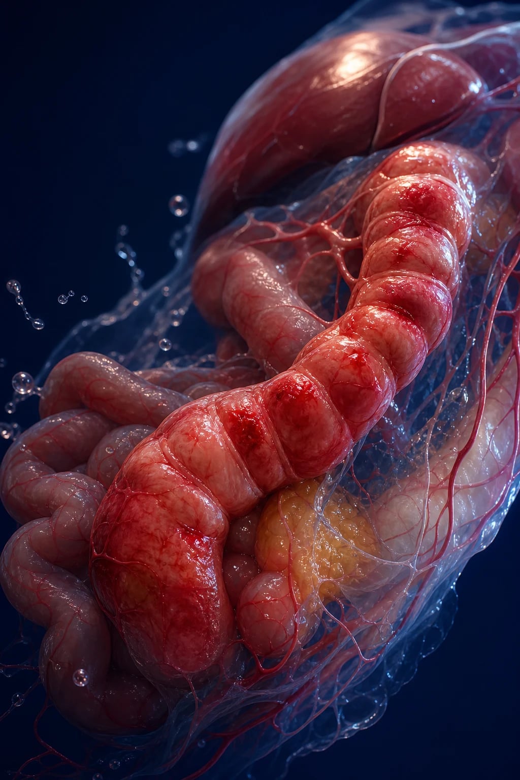
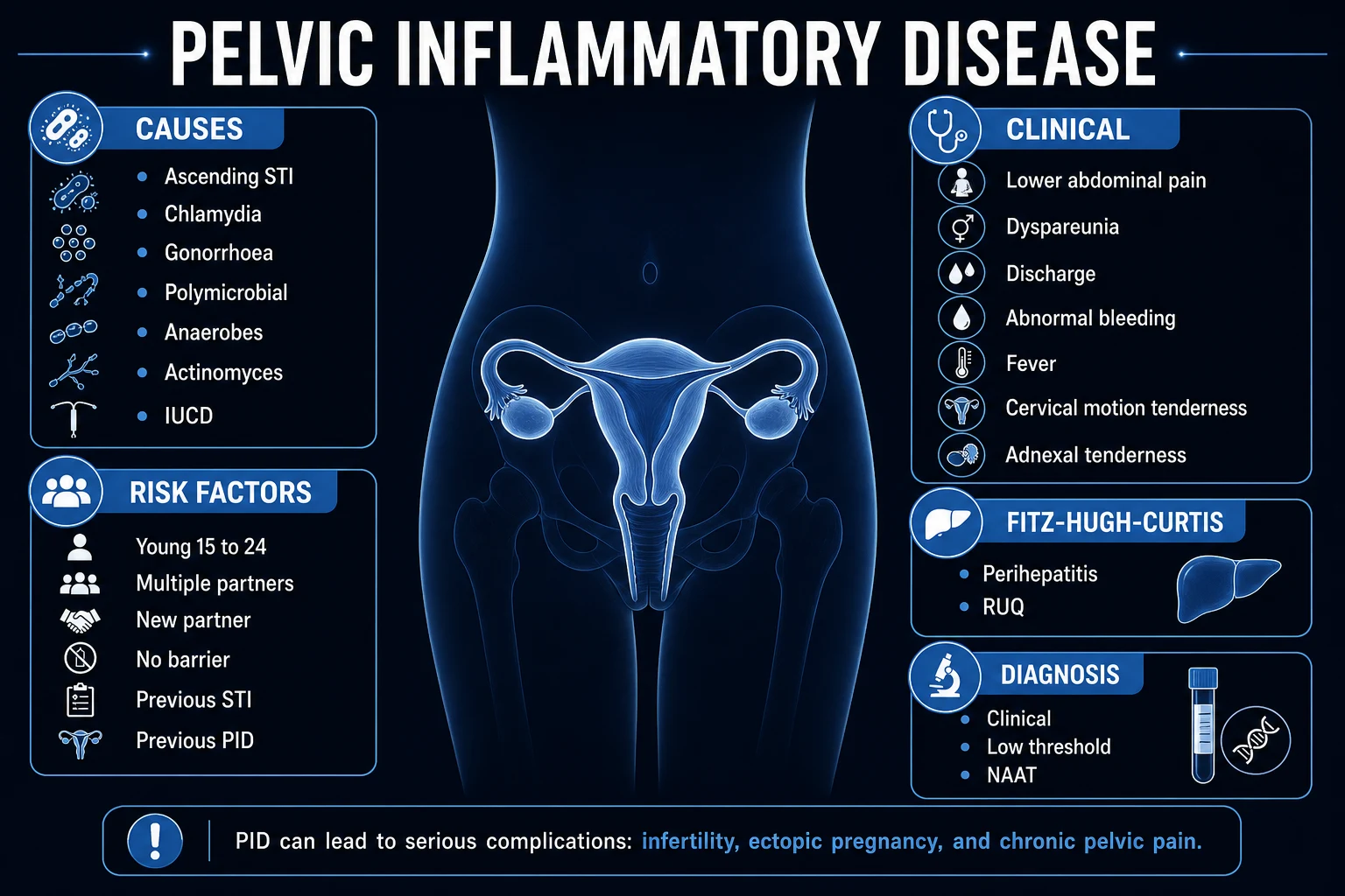
Pelvic inflammatory disease (PID) is the ascending infection of the upper female genital tract — from the vagina and cervix through the endometrium (endometritis) into the fallopian tubes (salpingitis), the ovaries (oophoritis), the parametrium (parametritis), and in some women the peritoneum and a tubo-ovarian abscess. It is a clinical syndrome, not a single microbiological diagnosis, and its aetiology is polymicrobial: Neisseria gonorrhoeae, Chlamydia trachomatis and Mycoplasma genitalium alongside the anaerobes and bacterial-vaginosis-associated organisms that flourish once the cervical barrier is breached. The Fellowship candidate must make the diagnosis on clinical grounds, must exclude ectopic pregnancy before anything else, must choose the outpatient or inpatient antibiotic regimen correctly, must recognise a tubo-ovarian abscess, and must arrange partner treatment and the 72-hour review that prevent the reinfection driving the long-term sequelae.[1][2]
Definition and epidemiology
Pelvic inflammatory disease is the spectrum of upper-genital-tract inflammation produced by ascending infection from the lower tract. It spans endometritis, salpingitis, oophoritis and parametritis, and it complicates into a tubo-ovarian abscess or diffuse peritonitis in the severe case. The syndrome is most common in sexually active women aged 15 to 25 years. The recognised risk factors are multiple or new sexual partners, a partner with a sexually transmitted infection, a prior episode of PID, age under 25, the absence of barrier contraception, bacterial vaginosis, and recent uterine instrumentation including intrauterine device insertion — the device elevates PID risk only in the first three weeks after placement, after which the risk returns to baseline. PID is the commonest preventable cause of tubal factor infertility, which is why the ED management is not merely an antibiotic course but the start of a fertility-preserving pathway.[1]
Pathophysiology
The cervical mucus normally bars vaginal organisms from the upper tract. This barrier breaks at menstruation, after instrumentation, after pregnancy loss, or in the dysbiosis of bacterial vaginosis, allowing organisms to ascend through the endometrium into the tubes. The tubal mucosa becomes inflamed and oedematous, the ciliated epithelium fails, and a purulent exudate (pyosalpinx) distends the tube. If the fimbriated end seals, the distended tube can rupture into the ovary or the peritoneal cavity, seeding a tubo-ovarian abscess. The infection is polymicrobial: the classic sexually transmitted pathogens (N. gonorrhoeae, C. trachomatis, M. genitalium) are joined by anaerobes (Prevotella, Peptostreptococcus), bacterial-vaginosis-associated organisms (Gardnerella vaginalis), genital mycoplasmas and enteric Gram-negatives. It is the tubal scarring from this inflammation — not the acute episode itself — that drives the long-term sequelae of infertility, ectopic pregnancy and chronic pelvic pain.[1][2]
Clinical presentation
The classic presentation is bilateral lower abdominal pain of gradual onset over days, with dyspareunia, an abnormal vaginal discharge, postcoital or intermenstrual bleeding, a low-grade fever and dysuria. Bimanual examination demonstrates the cardinal findings: cervical motion tenderness (the reproducible pain as the cervix is moved — the so-called chandelier sign), uterine tenderness and bilateral adnexal tenderness, with a mucopurulent cervical discharge and, in some, a palpable adnexal fullness or mass suggesting a tubo-ovarian abscess. Atypical and subtle presentations are common and dangerous: mild or even absent pain, the "silent" PID that inflames and scars the tubes without symptoms, right upper quadrant pleuritic pain (Fitz-Hugh–Curtis perihepatitis), or a tubo-ovarian abscess presenting with sepsis and peritonism. A high index of suspicion is therefore maintained for any sexually active woman with lower abdominal pain, abnormal bleeding, or unexplained vaginal discharge.[1]
Differential diagnosis
Ectopic pregnancy
- Unilateral pain with vaginal bleeding; the beta-hCG positive; can be haemodynamically unstable
- Adnexal mass or free fluid on ultrasound; absent bilateral adnexal pattern
- Pelvic ultrasound localises the pregnancy; laparoscopy if ruptured
- Surgical or methotrexate management; the beta-hCG is the decisive test
Acute appendicitis
- Periumbilical pain migrating to the right iliac fossa; anorexia, nausea, low-grade fever
- Migratory pain, RIF peritonism, McBurney tenderness; a normal pelvic examination
- Raised inflammatory markers; CT or ultrasound confirms the inflamed appendix
- Surgical appendicectomy; the right-sided focal peritonism and the migration distinguish it
Ovarian torsion
- Sudden severe unilateral pain with nausea and vomiting; often a prior similar self-resolving episode
- A tender adnexal mass; the pain disproportionate to the examination
- Pelvic ultrasound with Doppler shows reduced or absent ovarian venous and arterial flow
- Urgent laparoscopy for detorsion and oophoropexy; the surgical clock governs salvage
Ovarian cyst rupture or haemorrhage
- Sudden unilateral pain, often at ovulation or after intercourse; usually self-limiting
- Normal cervical motion tenderness; no fever, no discharge
- Ultrasound shows free fluid in the pouch of Douglas, sometimes a collapsing cyst
- Analgesia and observation; surgery only if haemodynamically unstable
Urinary tract infection or cystitis
- Dysuria, frequency, urgency; suprapubic rather than adnexal pain
- No cervical motion tenderness, no adnexal tenderness, no discharge
- Urinalysis shows nitrites and leucocytes; culture grows the organism
- Oral antibiotics (trimethoprim or nitrofurantoin) per the local guideline
The remaining differentials to hold are endometriosis (cyclical, chronic, deep dyspareunia), a degenerating or septic pelvic mass, functional chronic pelvic pain, and the rare but lethal causes such as a septic abortion or postpartum endometritis. The unifying rule: in any woman of reproductive age with lower abdominal pain, ectopic pregnancy is excluded first by a beta-hCG, and the surgical mimics (appendicitis, torsion) are separated from PID by the history, the migration and lateralisation of the pain, the pelvic examination, and the imaging. [1]
Bedside assessment
The assessment runs the ABCDE first, because most women with PID are haemodynamically stable but the ruptured tubo-ovarian abscess or the late presentation presents as sepsis with peritonism. The history captures the sexual risk (number of partners, a new partner, a partner with symptoms, prior sexually transmitted infection or PID, contraception), the last menstrual period, the character and lateralisation of the pain, and any recent instrumentation or pregnancy. The abdominal examination seeks peritonism and the migrating right iliac fossa pain of appendicitis. The bimanual and speculum examination reproduces the cervical motion, uterine and adnexal tenderness, samples the discharge, and palpates for an adnexal mass; the right upper quadrant is examined for the pleuritic tenderness of Fitz-Hugh–Curtis perihepatitis. Vitals are charted for the temperature threshold of 38.3°C.[1]
Investigations
Investigations confirm the syndrome, exclude the mimics, and sample the causative organisms. The urine or serum beta-hCG is mandatory in any woman of reproductive potential with lower abdominal pain — it excludes ectopic pregnancy and, if positive, redirects management. Nucleic-acid amplification testing (NAAT) for N. gonorrhoeae and C. trachomatis is taken from the endocervix, a self-taken vaginal swab, or a first-void urine; testing for M. genitalium is added where available, given its emerging role and macrolide-resistance problem. A full sexually-transmitted-infection screen adds HIV and syphilis serology, and considers hepatitis B and C and herpes simplex where the picture warrants. The urinalysis excludes a urinary tract infection; the full blood count, CRP and ESR are supportive (a raised ESR or CRP and the fever sharpen the specificity of the CDC criteria); and the saline wet-mount microscopy of vaginal fluid demonstrates the abundant white cells that are a sensitive bedside marker of upper-tract inflammation. The pelvic ultrasound identifies a tubo-ovarian abscess (a complex adnexal mass with internal debris or a dilated pyosalpinx) and excludes the surgical mimics; transvaginal ultrasound is more sensitive. Laparoscopy is the gold standard, reserved for severe or atypical disease, diagnostic uncertainty, or failure to respond, where it confirms the diagnosis, grades the severity, and drains an abscess.[1][2]
[1] [1]Immediate management and resuscitation
Most women with PID are haemodynamically stable and are managed as outpatients after the assessment and sampling. Resuscitation is reserved for the septic or peritonitic patient with a ruptured or large tubo-ovarian abscess: intravenous access, balanced crystalloid boluses titrated to the response, an early sepsis bundle, parenteral antibiotics within the first hour, analgesia and antiemetics, and an urgent referral to gynaecology for imaging-guided drainage or surgery. The antibiotic regimen is begun at the moment PID is diagnosed — the diagnosis is clinical and the treatment should not wait for the NAAT result, because a delay increases the risk of tubal scarring.[1]
[1]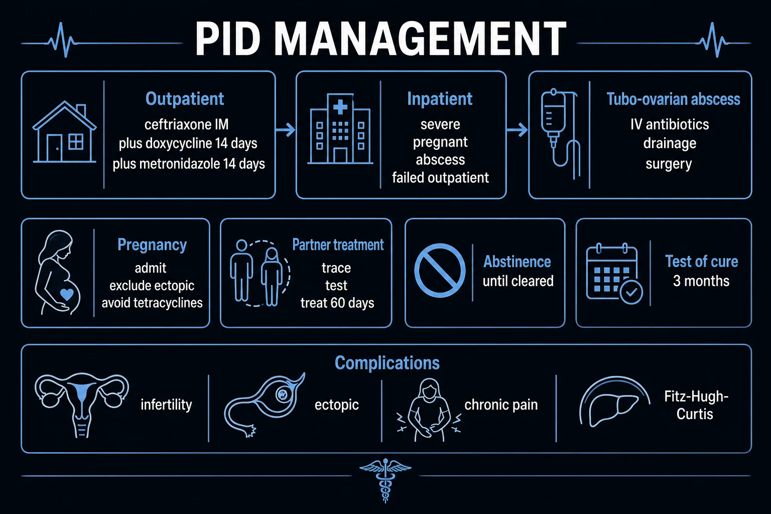
Definitive management — the antibiotic regimens
The definitive management is the antibiotic regimen, stratified by severity, pregnancy, the presence of a tubo-ovarian abscess, and the ability to tolerate and follow an oral course. The outpatient regimen for the woman with mild-to-moderate PID, no pregnancy, no abscess, who can tolerate oral therapy and will return for review, is ceftriaxone 500 mg intramuscularly as a single dose (1 g if she weighs over 150 kg), plus doxycycline 100 mg orally twice daily for 14 days, plus metronidazole 500 mg orally twice daily for 14 days. The metronidazole is now routine rather than optional, because it covers the anaerobes and bacterial-vaginosis organisms that complicate a polymicrobial infection. The European and United Kingdom convention often states metronidazole 400 mg twice daily — the same duration and intent, the regional dosing.[1][2]
The inpatient parenteral regimen is used for the pregnant woman, the severe or peritonitic case, the tubo-ovarian abscess, the failure of oral therapy, and the patient who cannot tolerate or follow an oral course. The pragmatic Australasian regimen is ceftriaxone 1 g intravenously daily plus doxycycline 100 mg intravenously or orally twice daily plus metronidazole 500 mg intravenously or orally twice to three times daily. The CDC parenteral options are the alternatives the candidate should name: cefotetan 2 g intravenously every 12 hours or cefoxitin 2 g intravenously every 6 hours plus doxycycline, or clindamycin 900 mg intravenously every 8 hours plus gentamicin (a 2 mg per kilogram loading dose then 1.5 mg per kilogram every 8 hours, or a once-daily 3 to 5 mg per kilogram). Parenteral therapy is converted to oral at clinical improvement and 24 to 48 hours of being afebrile, completing a total of 14 days.[1]
[1]The tubo-ovarian abscess
A tubo-ovarian abscess is the collection of pus within the ovary and tube, identified on ultrasound as a complex adnexal mass with internal debris and occasionally a fluid-debris level. These women are admitted for intravenous antibiotics and gynaecology-led care. Image-guided drainage (transvaginal ultrasound or CT-guided) or surgery is indicated when the abscess fails to respond to antibiotics, when it is large, or when rupture is suspected. The traditional size threshold taught for drainage is over 8 cm; recent evidence shows that abscesses from about 5 cm onwards are more likely to fail antibiotics and need drainage, so the threshold is shifting downward. A review at 48 to 72 hours is mandatory — persistent fever, pain or a rising inflammatory marker after appropriate intravenous therapy triggers drainage. A ruptured tubo-ovarian abscess is a surgical emergency presenting as peritonitis and sepsis.[6]
Partner management and follow-up
Treating the woman without treating her partners guarantees reinfection and recurrent PID. All sexual partners of the preceding 60 days are tested and treated, and expedited partner therapy is offered where the local jurisdiction permits. The woman abstains from intercourse until she and her partners have completed treatment and the symptoms resolve. A clinical review at 48 to 72 hours is arranged for every outpatient — no improvement at that point is defined as treatment failure and mandates admission, intravenous antibiotics, re-imaging for an unrecognised tubo-ovarian abscess, and reconsideration of appendicitis, ectopic pregnancy and torsion. A repeat NAAT at three months screens for reinfection, because the recurrence rate is high, and M. genitalium is treated per its macrolide-resistance profile (a macrolide first-line, moxifloxacin for resistant strains).[1][2][5]
Complications and prognosis
The real cost of PID is not the acute episode but its sequelae. Tubal factor infertility follows roughly 8 to 12 per cent of women after one episode, and the rate rises steeply with each subsequent episode and with the severity of the disease. Ectopic pregnancy risk increases six- to ten-fold because the scarred tube impairs ovum transit. Chronic pelvic pain affects around a third of women after PID, and recurrent PID is common, particularly in adolescents and when partners are untreated. The acute complications are the tubo-ovarian abscess, its rupture, peritonitis, and the rare but severe Fitz-Hugh–Curtis perihepatitis. The long-term prognosis is dominated by these reproductive sequelae, which is why fertility counselling is a mandatory part of the disposition.[3][4][5]
Special populations
Pregnancy transforms the management: the pregnant woman with suspected PID is admitted for parenteral antibiotics (tetracyclines such as doxycycline are avoided in pregnancy, and the regimen is adjusted accordingly), ectopic pregnancy is actively excluded by ultrasound, and the maternal and fetal risks are higher. The adolescent is at disproportionate risk of recurrence and sequelae — aggressive follow-up, partner treatment, contraception and re-screening at three months are essential. The HIV-positive woman tends to have more severe disease and a higher rate of tubo-ovarian abscess, and warrants inpatient management with broader anaerobic cover. The intrauterine device in situ is not removed routinely at diagnosis; the device is reassessed at the 72-hour review and removed only if the woman fails to improve. Postpartum and post-abortion PID are managed as severe disease with parenteral antibiotics.[1][5]
[1]ANZ practice note. The outpatient regimen follows the CDC 2021 standard (ceftriaxone 500 mg intramuscularly, doxycycline 100 mg twice daily for 14 days, metronidazole 500 mg twice daily for 14 days), with the Royal Australian and New Zealand College of Obstetricians and Gynaecologists and the Australasian Sexual Health Alliance guidance aligned to it. The metronidazole dosing is sometimes quoted as 400 mg twice daily in the United Kingdom and European convention — the same 14-day duration and the same intent. Mandatory reporting of gonorrhoea and chlamydia applies in most Australian states and in New Zealand, and contact tracing is offered through the sexual-health service. The 72-hour review is the ED safety-net. [1]
Evidence and regional guidelines
The contemporary evidence converges across the regions. The CDC Sexually Transmitted Infections Treatment Guidelines 2021 (Workowski, MMWR) is the global reference for the diagnostic criteria, the outpatient and inpatient antibiotic regimens, and the admission and partner-treatment recommendations. The 2017 European (IUSTI) guideline (Ross, International Journal of STD and AIDS) aligns with the CDC and adds explicit guidance on Mycoplasma genitalium and its macrolide resistance. The PEACH randomised trial (Ness, American Journal of Obstetrics and Gynecology 2002) established that outpatient and inpatient strategies are equivalent for mild-to-moderate PID, and its long-term follow-up (Ness, Obstetrics and Gynecology 2005) quantified the persistent reproductive sequelae. Trent (2011) documented the recurrent PID and adverse adolescent reproductive outcomes, and Jalloul (2022) defined the clinical predictors of failed medical treatment of the tubo-ovarian abscess, refining the drainage threshold.[1][2][3][4][6]
The microbiology in depth
PID is polymicrobial, and the Fellowship candidate must distinguish the sexually transmitted "primary" pathogens from the anaerobic and BV-associated "secondary" invaders that flourish once the cervical barrier is breached. The responsible organisms fall into three groups, and the antibiotic regimen is explicitly built to cover all three.[1][2]
Neisseria gonorrhoeae
- Gram-negative diplococcus; the classic PID pathogen, often the more acute and purulent presentation
- Ascends during menstruation and after instrumentation; produces a brisk endotoxin-mediated salpingitis
- Increasing cephalosporin minimum inhibitory concentrations reported globally; the 500 mg ceftriaxone IM dose reflects this
- NAAT from urine, self-taken vaginal swab or endocervix; co-infection with chlamydia is common, so treat for both
Chlamydia trachomatis
- Obligate intracellular bacterium; the commonest bacterial STI and the leading PID cause in many regions
- Often indolent or "silent" — the inflammation and tubal scarring proceed with minimal symptoms, which is why it drives much of the infertility
- Produces a cell-mediated immune response (the HSP-60 driven fibrosis) that scars the tube over weeks
- Doxycycline 100 mg twice daily for 14 days covers it; azithromycin 1 g single dose is inadequate for established PID
Mycoplasma genitalium
- Emerging, increasingly recognised PID pathogen with a discrete macrolide-resistance problem
- Lacks a cell wall — so beta-lactams (ceftriaxone) are inactive against it; covered by doxycycline but resistance is rising
- Macrolide resistance (the 23S rRNA A2058G mutation) is common; moxifloxacin is the second-line for resistant strains
- Test where available; resistance-guided therapy (azithromycin extended or moxifloxacin) outperforms empirical treatment
Anaerobes and BV organisms
- Prevotella, Peptostreptococcus, Gardnerella vaginalis and other BV-associated bacteria secondarily invade the upper tract
- Flourish in the dysbiosis of bacterial vaginosis, which itself doubles the PID risk
- Produce the pyosalpinx and the tubo-ovarian abscess; the polymicrobial pus
- Covered by metronidazole — the reason metronidazole is now routine, not optional, in every PID regimen
Enteric Gram-negatives
- Escherichia coli and related organisms, particularly after instrumentation, postpartum or post-abortion
- More prominent in non-STI PID and in the older or post-procedural patient
- Covered by ceftriaxone; broaden if a hospital-acquired or resistant organism is suspected
- A clue to the non-sexual PID pathway and to the need for broader initial cover in the post-procedural case
The ascending-infection pathophysiology
The cervical mucus is the physical and immunological barrier that confines vaginal organisms to the lower tract. PID begins when that barrier is breached, allowing organisms to ascend. The candidate should be able to walk the examiner through the sequence, because the tubal scarring that drives the long-term sequelae is a direct consequence of each stage.[1]
The ascending infection — from cervix to abscess
- The barrier breach. The cervical mucus plug fails at menstruation (the blood is a culture medium and raises vaginal pH), after instrumentation (IUD insertion, hysterosalpingography, termination), after pregnancy loss or childbirth, or in the dysbiosis of bacterial vaginosis. Organisms that were confined to the vagina now reach the endometrium.
- The endometritis. The organisms colonise the endometrium, producing an acute endometritis with submucosal neutrophil infiltration. This stage may be symptomatic (intermenstrual bleeding, discharge) or silent.
- The salpingitis. The organisms ascend through the tubal ostia into the fallopian tubes. The tubal mucosa becomes oedematous and inflamed, the ciliated epithelium is damaged, and a purulent exudate (the pyosalpinx) distends the tube lumen. This is the stage that matters for fertility.
- The oophoritis and parametritis. The inflamed tube adheres to the ovary and the broad ligament, producing a tubo-ovarian complex and a parametritis. The pelvic peritoneum may be involved, producing peritonism and a pelvic peritonitis.
- The tubo-ovarian abscess. If the fimbriated end of the tube seals, the purulent exudate cannot drain; the distended tube ruputures into the ovary or the peritoneal cavity, seeding a tubo-ovarian abscess — a collection of pus within the ovary and the occluded tube, detectable on ultrasound as a complex adnexal mass.
- The tubal scarring and the sequelae. The acute inflammation heals by fibrosis. The scarred, cilia-denuded, adhesion-laden tube impairs ovum transit — the substrate for tubal factor infertility, ectopic pregnancy, and chronic pelvic pain that may follow the patient for life.
Diagnostic criteria synthesised
The CDC frames PID as a syndrome diagnosed on clinical grounds, with no single sensitive and specific finding. The minimum criteria are deliberately inclusive — the trade-off is sensitivity (catch the disease, do not miss treatable tubal inflammation) over specificity. The additional criteria sharpen the specificity, and the elaborate criteria confirm it.[1]
Minimum criteria (treat)
- Cervical motion tenderness, OR uterine tenderness, OR adnexal tenderness on bimanual examination
- No other cause identified for the findings
- Empiric treatment is recommended when all of the above are met and no other cause is found — do not wait for the NAAT
Additional criteria (raise specificity)
- Oral temperature over 38.3°C
- Abnormal mucopurulent cervical or vaginal discharge
- Abundant white cells on saline wet-mount microscopy of vaginal fluid
- Raised ESR or raised CRP
Elaborate criteria (confirm)
- Positive NAAT for N. gonorrhoeae or C. trachomatis
- Pelvic ultrasound or MRI showing thickened fluid-filled tubes with or without free pelvic fluid or a tubo-ovarian complex
- Endometrial biopsy with histopathologic evidence of endometritis
- Laparoscopic findings consistent with PID (the gold standard)
Antibiotic regimens compared
The regimens all run for 14 days and all cover the three organism groups. The choice between outpatient (oral + a single IM dose) and inpatient (parenteral) is driven by severity, pregnancy, the presence of a tubo-ovarian abscess, and the ability to tolerate and follow an oral course. The PEACH trial established the equivalence of outpatient and inpatient strategies for mild-to-moderate disease.[1][3]
Outpatient (mild–moderate)
- Ceftriaxone 500 mg IM single dose (1 g if over 150 kg)
- PLUS doxycycline 100 mg PO twice daily for 14 days
- PLUS metronidazole 500 mg PO twice daily for 14 days (400 mg BD in UK convention)
- Use when: tolerates oral, will return at 72 hours, not pregnant, no abscess, no peritonitis or sepsis
Inpatient parenteral (ANZ pragmatic)
- Ceftriaxone 1 g IV daily
- PLUS doxycycline 100 mg IV/PO twice daily
- PLUS metronidazole 500 mg IV/PO two to three times daily
- Use when: pregnant, severe, peritonitis or sepsis, a tubo-ovarian abscess, failed oral therapy, or oral therapy not tolerable or follow-up not assured
Inpatient parenteral (CDC alternatives)
- Cefotetan 2 g IV 12-hourly OR cefoxitin 2 g IV 6-hourly, PLUS doxycycline; OR clindamycin 900 mg IV 8-hourly PLUS gentamicin (load then 8-hourly, or 3–5 mg/kg once daily)
- Convert to oral doxycycline (plus metronidazole for the clindamycin–gentamicin arm) at clinical improvement and 24–48 hours afebrile
- Complete a total of 14 days of therapy
- Cover the alternative regimens because examiners ask for them, and local resistance or allergy may force them
The PEACH randomised trial — inpatient versus outpatient treatment (Ness et al. 2002)
Multicentre randomised controlled trial with long-term follow-up
Population: Women with clinically suspected mild-to-moderate PID (n = 831)
Comparator: Outpatient ceftriaxone IM single dose plus doxycycline, with or without metronidazole
Key finding
Outpatient and inpatient strategies were equivalent for short-term outcomes in mild-to-moderate PID; long-term reproductive outcomes were also similar between groups
The tubo-ovarian abscess pathway
A tubo-ovarian abscess transforms PID from an outpatient disease into a gynaecology-led inpatient admission, and a ruptured abscess is a surgical emergency. The candidate must hold the diagnostic threshold, the antibiotic response window, and the drainage indications.[6]
The tubo-ovarian abscess — from diagnosis to drainage
- The recognition. A woman with PID and a palpable adnexal mass, severe unilateral pain, high fever, or sepsis is presumed to have a tubo-ovarian abscess until imaging excludes it. A transvaginal ultrasound is the first-line — it shows a complex adnexal mass with internal debris, sometimes a fluid–debris level or a dilated pyosalpinx.
- The admission. Admit for intravenous antibiotics (the inpatient regimen above) and analgesia. Consult gynaecology early. The IV antibiotics alone resolve a substantial proportion of smaller abscesses.
- The 48-to-72-hour review. Reassess at 48 to 72 hours. Persistent fever, worsening pain, or a rising inflammatory marker on appropriate IV therapy is failure — escalate to drainage.
- The drainage. Image-guided drainage (transvaginal ultrasound or CT-guided) is preferred to surgery in the stable patient — it preserves the tube and ovary where possible. The traditional size threshold is over 8 cm; recent evidence (Jalloul 2022) shows abscesses from about 5 cm are more likely to fail antibiotics, so the threshold is shifting downward.
- The surgery. Laparoscopy or laparotomy with drainage, salpingo-oophorectomy or hysterectomy is reserved for the ruptured abscess with peritonitis and sepsis, the failed drainage, the diagnostic uncertainty, or the older woman who has completed her family.
- The discharge and follow-up. Convert to oral at clinical improvement and 24 to 48 hours afebrile, completing 14 days total. Arrange partner treatment, contraception, a repeat NAAT at three months, and fertility counselling — the affected tube is at high risk of future infertility and ectopic pregnancy.
Clinical predictors of failed medical treatment in tubo-ovarian abscess (Jalloul et al. 2022)
External validation of a risk score; retrospective cohort
Population: Women with a tubo-ovarian abscess managed with IV antibiotics
Comparator: Observed rate of failed antibiotic monotherapy requiring drainage or surgery
Key finding
Larger abscess size (from approximately 5 cm), older age, diabetes, and higher inflammatory markers independently predicted failure of antibiotic monotherapy
Fitz-Hugh–Curtis perihepatitis
Fitz-Hugh–Curtis syndrome is the perihepatitis of PID — inflammation of the liver capsule and the adjacent peritoneum, classically (though not exclusively) from chlamydial or gonococcal spread up the right paracolic gutter. The candidate must recognise it because it presents with right upper quadrant pleuritic pain that mimics biliary or pulmonary disease and can divert the workup away from the pelvis.[1][2]
The presentation is a woman with PID symptoms (lower abdominal pain, discharge, cervical motion tenderness) who additionally has right upper quadrant pleuritic pain that worsens with inspiration, coughing or movement, sometimes referred to the right shoulder. The abdominal examination shows right upper quadrant tenderness with a normal liver and no Murphy sign, and the pelvic examination shows the PID findings. The liver function tests are typically normal or minimally deranged — this is a capsular inflammation, not a parenchymal hepatitis. The abdominal ultrasound or CT may show a small perihepatic fluid collection or "violin-string" adhesions between the liver and the anterior abdominal wall (the latter are best seen at laparoscopy). The management is the standard PID regimen — the perihepatitis resolves with the same antibiotics. The diagnostic trap is anchoring on the gallbladder or the lung and missing the pelvic source: in any young woman with unexplained right upper quadrant pleuritic pain, examine the pelvis and send the NAAT.[1]
[1]Long-term sequelae — the rates to quote
The exam questions on PID frequently probe the long-term sequelae, and the candidate should be able to quote the approximate rates and their relationship to the number of episodes and the severity of the disease.[3][4][5]
Tubal factor infertility
- Approximately 8–12 per cent after one episode of PID
- Rises to roughly 20 per cent after two episodes and 40 per cent after three or more
- Driven by the tubal scarring and ciliary loss from the acute salpingitis; chlamydial (often silent) PID is a major contributor
- Investigated by hysterosalpingography or laparoscopy with dye; managed by IVF for the bilateral tubal disease
Ectopic pregnancy
- Risk increases six- to ten-fold after PID
- The scarred, narrowed tube delays ovum transit and favours tubal implantation
- Any woman with a prior PID history and a positive beta-hCG is managed as ectopic-prone — early ultrasound and a low threshold for the ectopic pathway
- Represents the leading non-obstetric cause of first-trimester maternal death
Chronic pelvic pain
- Affects around one in three women after PID
- Driven by tubo-ovarian adhesions, a hydrosalpinx, and the neuropathic sensitisation of chronic inflammation
- Persists for months to years and is notoriously difficult to treat; multidisciplinary pain management is often required
- The prevention is the prompt and complete treatment of the acute episode and reinfection prevention
Tubo-ovarian abscess
- The acute severe complication; presents with a mass, high fever, sepsis
- Detected by transvaginal ultrasound; drained if it fails antibiotics or is large (over 8 cm classic, from 5 cm recent)
- A ruptured TOA is a surgical emergency with peritonitis and septic shock
- More common in older women, with diabetes, and in the HIV-positive patient
Fitz-Hugh–Curtis perihepatitis
- RUQ pleuritic pain from perihepatic capsular inflammation
- Classically chlamydial; treated with the standard PID regimen
- Diagnosed clinically with the pelvic findings; the LFTs are typically normal
- The trap is the biliary workup that misses the pelvic source
Mycoplasma genitalium and the resistance problem
Mycoplasma genitalium is the emerging PID pathogen that the contemporary guidelines address explicitly. It lacks a cell wall, so the beta-lactams (ceftriaxone, cefotetan, cefoxitin) are inactive against it; doxycycline has limited efficacy; and the macrolides (azithromycin) face a rising resistance problem driven by the 23S rRNA mutation. Resistance-guided therapy — testing for the mutation and choosing azithromycin extended course versus moxifloxacin accordingly — outperforms empirical treatment. The candidate should name the organism, its resistance mechanism, and its second-line agent (moxifloxacin).[2][8]
[1]The intrauterine device and PID
The IUD is a common source of diagnostic confusion. The evidence is clear: the IUD raises the PID risk only in the first three weeks after insertion, almost certainly because it introduces organisms at placement; after three weeks the risk returns to the background rate, and the IUD user has the same PID risk as the non-user. An IUD found in situ in a woman with PID is therefore not removed routinely at diagnosis — it is left in place and reassessed at the 72-hour review, and removed only if the woman fails to improve on appropriate therapy. The Actinomyces-like organisms occasionally seen on the cervical smear of a long-term IUD user are not, by themselves, an indication for removal in the absence of symptoms.[1][7]
[1]The partner treatment and the public-health arm
Treating the woman without treating her partners is the commonest reason for recurrence. Every sexual partner of the preceding 60 days is tested and treated, ideally with the same presumptive regimen, and expedited partner therapy is offered where the local jurisdiction permits. Abstinence from intercourse until both the woman and her partners have completed treatment and the symptoms resolve is counselled explicitly. Mandatory notification of gonorrhoea and chlamydia applies in most Australian states and in New Zealand, and the contact tracing is coordinated through the sexual-health service. The repeat NAAT at three months screens for reinfection, because the recurrence rate is high, particularly in the adolescent and in the woman whose partners were not treated.[1][5]
[1]SAQs
SAQ — Suspected PID in a young woman with lower abdominal pain
10 minutes · 10 marks
A 22-year-old nulliparous woman presents to the emergency department with three days of bilateral lower abdominal pain, a yellow-green vaginal discharge and dyspareunia. She is sexually active with two male partners in the last two months and uses no barrier contraception. Her last menstrual period was two weeks ago. On examination she is afebrile at 37.6 degrees C, HR 92, BP 116/72; bimanual examination reveals cervical motion tenderness, bilateral adnexal tenderness and a mucopurulent discharge. The urine beta-hCG is negative.
SAQ — Tubo-ovarian abscess complicating PID with sepsis
10 minutes · 10 marks
A 31-year-old woman presents to the emergency department with five days of worsening left-sided pelvic pain, fevers and rigors, and two days of vomiting. She has a three-year-old intrauterine device in situ. On arrival: T 39.1 degrees C, HR 128, BP 84/52 (MAP 62), RR 26, SpO2 95 per cent on room air, GCS 15. Bimanual examination is limited by pain but suggests a tender left adnexal mass. The urine beta-hCG is negative. WCC 19.2, CRP 240, lactate 3.6. The transvaginal ultrasound shows a 7 cm complex left adnexal mass with internal debris.
Exam pearls
- Beta-hCG first. Ectopic pregnancy is the mimic that kills; exclude it before you treat PID.
- The minimum CDC criteria are cervical motion, uterine, or adnexal tenderness — sharpened by fever over 38.3°C, mucopurulent discharge, raised ESR or CRP, vaginal white cells, or a positive NAAT.
- The outpatient regimen is ceftriaxone 500 mg intramuscularly plus doxycycline 100 mg twice daily for 14 days plus metronidazole 500 mg twice daily for 14 days. Metronidazole is not optional.
- Pregnant, or a tubo-ovarian abscess, or severe, or a surgical mimic: admit.
- Treat the partners of the preceding 60 days and offer expedited partner therapy; re-screen at three months.
- The 72-hour review is mandatory — no improvement is treatment failure and triggers admission and re-imaging.
- Fitz-Hugh–Curtis perihepatitis is the right upper quadrant pleuritic pain of PID; manage as PID.
- The sequelae — infertility, ectopic, chronic pelvic pain — are the exam's real question; quote the rates. [1]
Red flags
[1]References
- [1]Workowski KA, Bachmann LH, Chan PA, et al. Sexually Transmitted Infections Treatment Guidelines, 2021 MMWR Recomm Rep, 2021.PMID 34292926
- [2]Ross J, Jensen JS, Ison C, et al. 2017 European guideline for the management of pelvic inflammatory disease Int J STD AIDS, 2018.PMID 29198181
- [3]Ness RB, Soper DE, Holley RL, et al. Effectiveness of inpatient and outpatient treatment strategies for women with pelvic inflammatory disease: results from the Pelvic Inflammatory Disease Evaluation and Clinical Health (PEACH) Randomized Trial Am J Obstet Gynecol, 2002.PMID 12015517
- [4]Ness RB, Trautmann G, Richter HE, et al. Effectiveness of treatment strategies of some women with pelvic inflammatory disease: a randomized trial Obstet Gynecol, 2005.PMID 16135590
- [5]Trent M, Ellen JM, Walker A. Adverse adolescent reproductive health outcomes after pelvic inflammatory disease Arch Pediatr Adolesc Med, 2011.PMID 21199980
- [6]Jalloul RJ, Liu Y, Al-Hammoud A, et al. Clinical Predictors of Failed Medical Treatment in Patients with Tubo-ovarian Abscess: External Validation of a Recently Published Risk Score J Minim Invasive Gynecol, 2022.PMID 35051659
- [7]Workowski KA, Bolan GA. Sexually transmitted diseases treatment guidelines, 2015 MMWR Recomm Rep, 2015.PMID 26042815
- [8]Couldwell DL, Lewis DA. Mycoplasma genitalium infection: current treatment options, therapeutic failure, and resistance-associated mutations Infect Drug Resist, 2015.PMID 26060411