EM · Subdural and extradural haematoma
Subdural and extradural haematoma
Also known as Acute subdural haematoma · Chronic subdural haematoma · Extradural haematoma · Epidural haematoma
Subdural and extradural haematoma — the two extra-axial traumatic collections distinguished by anatomy, source, and tempo. Extradural (epidural) haematoma: arterial bleed from the middle meningeal artery after a temporal fracture, the lentiform biconvex shape on CT, the lucid interval, the young trauma patient, the urgent craniotomy. Acute subdural haematoma: venous bridging-vein or cortical bleed, the crescent shape crossing sutures, the elderly and anticoagulated patient, the craniotomy. Chronic subdural: the insidious cognitive decline in the atrophic elderly brain, the burr-hole evacuation. Management: ABCDE, ICP control (mannitol 0.5 g per kg, head up 30 degrees, hypertonic saline 3 per cent 250 mL), urgent neurosurgical referral, and early reversal of anticoagulation. Differential: subarachnoid, contusion, tumour. ACEM-primary, globally tagged.
On this page & tools
Your progress
Saved locally on this device.
Target exams
Red flags
Subdural and extradural haematoma are the two extra-axial traumatic collections that dominate emergency neurosurgery, and the Fellowship candidate must hold them side by side. The extradural (epidural) haematoma is the arterial bleed between the skull and the dura — classically from the middle meningeal artery torn by a temporal bone fracture, producing a lentiform collection that expands fast, often after a lucid interval, in a young patient with an otherwise uninjured brain. The subdural haematoma is the venous or cortical collection between the dura and the arachnoid — crescent-shaped, crossing the suture lines, common in the elderly, the alcoholic, and the anticoagulated patient, where the underlying brain is usually injured in the acute form. Both kill by the same mechanism — rising intracranial pressure and cerebral herniation — and both are managed by the same sequence: ABCDE, control the intracranial pressure, urgent neurosurgical referral, and early reversal of any anticoagulation. The examiner rewards the candidate who can distinguish the two on the CT in seconds and act on the one that is expanding.[1][2][3]
Definition and classification
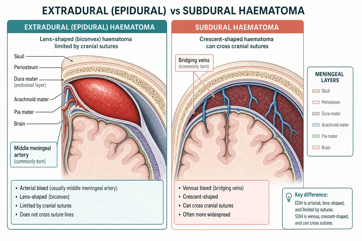
The two collections are defined by their anatomical compartment, and the compartment dictates the source, the CT shape, and the urgency. [1]
[1]The extradural haematoma (EDH) is further classified by location — temporoparietal (the commonest, from the middle meningeal artery), frontal, occipital, and posterior fossa (rare but rapidly fatal). The acute subdural haematoma (aSDH) appears within 72 hours of the injury as a hyperdense crescent; the subacute subdural (3 days to 3 weeks) is isodense and easy to miss; the chronic subdural (cSDH) presents beyond 3 weeks as a hypodense collection, often in the elderly patient who cannot recall the injury. The acute and chronic subdurals are clinically distinct enough that the examiner treats them as separate entities.[1][2]
Epidemiology and risk
The two lesions target different populations, and recognising the population is part of the diagnosis. The extradural haematoma occurs most often in the young male trauma patient — the cyclist, the assault victim, the fall — and accounts for 2 to 8 per cent of head-injury admissions; a temporal or parietal skull fracture is the commonest precursor. The acute subdural complicates severe traumatic brain injury (up to a third of severe TBI) and carries a mortality of 40 to 60 per cent because the underlying brain parenchyma is contused and oedematous. The chronic subdural is the disease of the elderly, the anticoagulated, the alcoholic, and the shunt-dependent patient — the atrophic brain stretches the bridging veins, and a trivial, often-forgotten injury (a stumble, a head bump) tears one; it recurs after evacuation in 10 to 15 per cent. Anticoagulant and antiplatelet therapy multiplies the risk of every one of these lesions and is the modifiable factor the emergency physician must address first.[2][4]
Pathophysiology — arterial, venous, and the Monro-Kelly ceiling
A single haemodynamic principle governs both: the CPP = MAP − ICP relationship. As the collection expands inside the rigid skull, the intracranial pressure rises, the cerebral perfusion pressure falls, and once the compensatory reserve (CSF displaced, venous blood compressed) is exhausted, a small further expansion produces a steep, dangerous rise in pressure and then herniation. The difference between the two lesions is the rate at which this happens. [1]
The extradural is arterial. A fracture across the pterion — where the middle meningeal artery grooves the inner table — lacerates the artery, and the high-pressure bleed strips the dura from the skull, forming the biconvex collection that can expand over hours. The venous sinuses and the diploic veins are less common sources. Because the underlying brain is often uninjured, the prognosis is excellent IF the collection is evacuated before herniation — this is the lesion where minutes matter and where a normal early CT can become a catastrophe hours later. [1]
The subdural is most often venous — the bridging veins that cross from the cortical surface to the dural sinuses tear under the shearing or the atrophic stretch — but in the acute traumatic form a cortical laceration or contusion is frequently the source. The venous tempo is slower, which is why the chronic subdural accumulates over weeks; but the acute subdural, layered on a contused brain, kills through the combined mass of the clot, the contusion, and the oedema.[1][2]
Clinical presentation
The extradural classically follows the lucid interval: the patient is concussed and briefly unconscious, wakes to a normal or near-normal GCS, and then — as the arterial collection expands — develops a worsening headache, vomiting, confusion, and a progressive fall in GCS, culminating in an ipsilateral fixed dilated pupil (uncal herniation from the third-nerve compression) and contralateral hemiparesis. The lucid interval is the textbook feature, but it occurs in only about a third of cases — its absence does NOT exclude the diagnosis, and the candidate who waits for it will lose the patient. A skull fracture, the temporal bruising, or a blow to the pterion heightens the suspicion. [1]
The acute subdural presents with a depressed conscious level from the impact — there is usually no true lucid interval because the underlying brain is injured. A rapid deterioration, an abnormal pupil, and a hemiparesis signal the rising pressure. The chronic subdural is the insidious presentation that masquerades as dementia, depression, or simply "off legs" in the elderly: a fluctuating cognitive decline, a gait disturbance, a headache, a mild hemiparesis, and sometimes a seizure, developing over weeks after a trivial or unrecalled head injury. The Cushing triad — hypertension, bradycardia, irregular respiration — is a late, pre-terminal sign in any of these and means herniation is imminent.[1][2]
Differential diagnosis — the intracranial mass after trauma or in the elderly
The reduced GCS, the focal deficit, or the headache-plus-confusion presentation demands the CT, but the candidate must hold the differential before the image loads. [1]
Extradural haematoma
- Young trauma; temporal blow; the lucid interval then the rapid drop
- CT: lentiform (biconvex), does NOT cross sutures, often with a skull fracture
- Urgent craniotomy; excellent prognosis if evacuated before herniation
- Arterial (middle meningeal); expands over hours
Subdural haematoma
- Acute: severe TBI, depressed GCS, no lucid interval; chronic: elderly, insidious decline
- CT: crescent-shaped, crosses sutures, does NOT cross the midline falx
- Craniotomy (acute) or burr-hole (chronic); underlying brain often injured
- Venous (bridging veins) or cortical; acute mortality 40 to 60 per cent
Subarachnoid haemorrhage
- Thunderclap headache maximal in seconds; neck stiffness, photophobia
- CT: blood in the basilar cisterns and the sulci, not a focal collection
- Nimodipine 60 mg every 4 hours; BP under 160 mmHg; coiling or clipping
- Aneurysmal; the sentinel bleed precedes the catastrophic rupture
Contusion / intracerebral haematoma
- Coup and contrecoup pattern; the temporal and frontal poles
- CT: patchy hyperdense blood within the parenchyma, surrounding oedema
- Manage the ICP; surgery only if the mass effect is severe
- "Blossoms" over 24 to 72 hours as the oedema and the delayed bleed evolve
The brain tumour and the metastasis are the non-traumatic masses that present subacutely with progressive headache, focal deficit, and seizures; a CT with surrounding vasogenic oedema and ring enhancement distinguishes them, and the chronic subdural is the commonest mimic of a tumour in the elderly patient. Meningitis adds fever and neck stiffness but can coexist or confuse after a head injury — image first if any focal sign or reduced GCS is present.[2]
Bedside assessment
Assess the airway, breathing, and circulation first — hypoxia and hypotension are themselves causes of secondary brain injury and demand immediate correction. Perform a focused neurological examination and reassess it serially: the Glasgow Coma Score (recorded as its eye, verbal, and motor components; the best motor response the most informative single element), because a drop of two or more points is the single most reliable bedside sign of an expanding haematoma and mandates an urgent repeat CT; the pupils — a unilateral dilated unresponsive pupil is uncal herniation; the vital signs for the Cushing triad; the motor examination for a hemiparesis or abnormal posturing; and the head for a fracture, a Battle sign, raccoon eyes, or haemotympanum (basal skull fracture). Take a specific anticoagulant, antiplatelet, and bleeding-disorder history on every patient, and check the bedside glucose to exclude hypoglycaemia. The mechanism of injury, the time from injury, and any co-ingestion of alcohol are recorded because they bear on the CT threshold and on the interpretation of the GCS.[3]
Investigations — CT first, never the lumbar puncture
The non-contrast CT head is the definitive investigation and is performed as soon as the patient is stabilised. The examiner expects the candidate to read the two shapes: [1]
[1]The midline shift is measured — over 5 mm is significant and a surgical trigger. The effacement of the basal cisterns signals the raised intracranial pressure. A skull fracture, if not seen on the soft-tissue windows, is sought on the bone window. The CT indications for a head injury (the NICE criteria, used in ANZ and UK practice) include a GCS below 13 at 2 hours, a suspected open or depressed skull fracture, any sign of a basal skull fracture, a post-traumatic seizure, a focal neurological deficit, more than one episode of vomiting, a coagulopathy, and a dangerous mechanism — any of which earns an immediate CT. The lumbar puncture is NEVER performed when an extra-axial collection is possible: it releases the spinal-compartment pressure below a supra-tentorial gradient and precipitates tonsillar herniation. Blood tests include the full blood count, the coagulation (the anticoagulated patient is reversed early), the group and save or crossmatch, the electrolytes, and the bedside glucose.[1][2][3]
Immediate management and resuscitation
Resuscitation follows the ABCDE framework with the brain as the priority organ and the protection of the cerebral perfusion pressure as the explicit goal. [1]
[1]The resuscitation targets and the osmotherapy doses
Definitive management — the surgical thresholds and the anticoagulation reversal
The definitive treatment is surgical, and the emergency physician's role is to deliver the patient to the neurosurgeon with the brain perfused, the pressure controlled, and the coagulopathy reversed. The surgical thresholds, drawn from the Brain Trauma Foundation guidelines, are reproduced because the examiner asks for them. [1]
Extradural haematoma
- Volume over 30 mL, or any midline shift, or a neurological deficit — evacuate
- Urgent CRANIOTOMY regardless of the GCS if the volume is over 30 mL
- A GCS below 9 with a pupil abnormality is the emergency — operate within hours
- Excellent prognosis if evacuated before the herniation
Acute subdural haematoma
- Thickness over 10 mm OR a midline shift over 5 mm — evacuate by craniotomy
- If under 10 mm and shift under 5 mm: operate if the GCS drops by 2 or more, or the pupils are asymmetric, or the ICP is over 20 mmHg
- Craniotomy within 2 hours of the deterioration for the best outcome
- Underlying contusion and oedema often warrant a decompressive craniectomy
Chronic subdural haematoma
- Symptomatic collection over 10 mm or with a midline shift — BURR-HOLE evacuation
- Burr-hole craniostomy with a subdural or subgaleal drain is the standard
- Asymptomatic or mildly symptomatic: observe with the serial imaging
- Recurrence 10 to 15 per cent; the middle meningeal artery embolisation is an emerging adjunct
The reversal of anticoagulation is started as soon as the coagulopathy is identified, because the expanding haematoma is the danger in the anticoagulated patient. For warfarin, give prothrombin complex concentrate (PCC) 25 to 50 IU per kg intravenously plus vitamin K 10 mg IV (the PCC is faster and more complete than the fresh-frozen plasma, and the vitamin K sustains the reversal over days). For dabigatran, give idarucizumab 5 g IV (two 2.5 g vials). For apixaban or rivaroxaban, give andexanet alfa (or 4-factor PCC 50 IU per kg if andexanet is unavailable). For the antiplatelet agents, the platelet transfusion is generally NOT recommended in the spontaneous intracerebral haemorrhage (the PATCH trial), but the surgical lesion and the neurosurgical procedure may alter the local practice — discuss with the neurosurgeon. Desmopressin 0.4 microgram per kg is considered for the antiplatelet-related surgical bleeding. The reversal is delivered in parallel with the resuscitation and the neurosurgical referral, never as a sequential step that delays the transfer.[3][4]
Subtypes and special scenarios
The posterior fossa extradural is rare but rapidly fatal — it compresses the brainstem directly and may present with ataxia, a decreased conscious level, and an obstructive hydrocephalus; it is evacuated urgently. The bilateral chronic subdural and the interhemispheric subdural (in the shaken-impact or the non-accidental injury) are the variants the paediatric and the elderly examiners probe. The spontaneous (non-traumatic) extradural, from an arteriovenous malformation or a coagulopathy, is uncommon but occurs in the anticoagulated patient and the skull-base vascular lesion. The subdural empyema is the infected subdural collection that complicates the sinusitis, the mastoiditis, or the skull-base fracture — it presents with fever, headache, and a rapid neurological decline, and it is a neurosurgical and an infectious-disease emergency treated with the evacuation and the empirical antibiotics (ceftriaxone plus metronidazole).[2]
Complications and pitfalls
The complications are the cerebral herniation (the commonest cause of death), the rebleeding and the recurrence (especially the chronic subdural, at 10 to 15 per cent), the post-traumatic seizures (the anticonvulsant prophylaxis is given for the high-risk lesion — the intracranial haematoma, the depressed fracture, the penetrating injury), the infection (the osteomyelitis, the meningitis, the subdural empyema from an open or a basal fracture), and the secondary brain injury from the hypoxia, the hypotension, and the uncontrolled intracranial pressure. The recurring pitfalls are the inverse of the management: waiting for the lucid interval in the extradural instead of acting on the falling GCS; attributing a reduced GCS to the alcohol, the dementia, or the age without the CT; missing the isodense subacute subdural (look for the effaced sulci and the midline shift, not just the hyperdense crescent); not reversing the anticoagulation early; performing a lumbar puncture in a mass lesion; and delaying the neurosurgical referral for a lesion that meets the surgical threshold. The co-ingestion of alcohol is the classic confounder — the CT excludes the mass, always.[1][3]
Prognosis and disposition
The prognosis hinges on the lesion and the speed of the decompression. The extradural haematoma evacuated before herniation has a mortality under 10 per cent and a good neurological recovery, because the underlying brain is often uninjured — this is the lesion where the emergency physician's speed is most directly rewarded. The acute subdural carries a mortality of 40 to 60 per cent even with the evacuation, because the underlying contusion and oedema dominate the outcome; the modifiable determinant is the secondary injury (the hypoxia, the hypotension, the delayed surgery). The chronic subdural has a good functional outcome after the burr-hole evacuation, with a recurrence rate of 10 to 15 per cent and a small procedural-morbidity. Every patient with an acute extra-axial haematoma is admitted to a neurosurgical or an intensive-care bed, intubated and ventilated if comatose, with an intracranial-pressure monitor in the severe case. The patient with a small, asymptomatic chronic subdural may be managed conservatively with the serial imaging and the safety-net advice. The disposition decision is made with the neurosurgical team, never unilaterally.[1][2][3]
Special populations
The elderly patient has the cerebral atrophy that stretches the bridging veins and enlarges the subdural space, allowing a larger collection to accumulate before the symptoms — this is why the chronic subdural is the disease of the elderly, and why the insidious cognitive decline, the gait change, or the "off legs" presentation warrants a CT. The lower physiological reserve and the comorbidity raise the surgical risk and lengthen the recovery. The anticoagulated patient (warfarin or a DOAC) has a several-fold higher risk of an expanding intracranial haematoma and is reversed early and completely — the reversal is delivered in parallel with the resuscitation, and the neurosurgical decision may be held until the coagulopathy is corrected. The paediatric TBI uses the paediatric GCS and the different thresholds; the open sutures and the fontanelle in the infant allow some pressure dissipation but also mask the signs; the non-accidental injury is considered in the inconsistent-history case, particularly the subdural, the retinal haemorrhage, and the posterior interhemispheric collection. The intoxicated patient has a GCS masked by the alcohol or the drug — the CT excludes the mass, and the reduced GCS is never attributed to the alcohol alone.[1][2][4]
Evidence and regional guidelines
The contemporary framework is the Brain Trauma Foundation 2020 update of the severe-TBI guidelines (PMID 32761068), which sets the intracranial-pressure treatment threshold (above 22 mmHg), the cerebral-perfusion-pressure target (60 to 70 mmHg), the SBP threshold (110 or above), the normocapnia target, the osmotherapy recommendations, and the surgical thresholds for the extra-axial haematoma. The NICE head-injury guideline (UK, adopted widely across the ANZ and the UK practice) defines the CT indications and the observation-or-discharge rule for the minor head injury. The contemporary reviews of the epidural and the subdural haematoma (PMID 30085524 and 30422565) consolidate the anatomy, the clinical presentation, the imaging, and the management. The reversal of the direct oral anticoagulants (PMID 34521332) and the anticoagulated-trauma literature underpin the early-reversal principle. The airway, the breathing, and the circulation targets, the no-LP-before-CT rule, and the no-corticosteroids-in-trauma rule are global; the neurosurgical pathway and the CT indications follow the local trauma and the neurosurgical protocol.[1][2][3][4]
ANZ practice note. The subdural and extradural haematoma are managed by the Brain Trauma Foundation 2020 framework via the state-wide neurosurgical referral. The non-negotiable targets are the intubation at GCS 8 or below, the SBP at or above 110 mmHg, the normocapnia (PaCO₂ 35 to 40 mmHg), the head elevated to 30 degrees, and the mannitol 0.5 g per kg or the hypertonic saline 3 per cent 250 mL for the signs of herniation. The anticoagulation is reversed early (the PCC and the vitamin K for the warfarin; the idarucizumab for the dabigatran; the andexanet or the PCC for the apixaban or the rivaroxaban). The CT indications follow the NICE head-injury criteria, and the surgical thresholds (the extradural over 30 mL, the acute subdural over 10 mm or the shift over 5 mm, the chronic subdural by the burr-hole) drive the neurosurgical referral. [1]
SAQ — The extradural haematoma and the lucid interval
10 minutes · 10 marks
A 19-year-old man is brought to the emergency department after being struck on the side of the head during an assault. He was briefly unconscious, then walked to the ambulance talking, but in the department an hour later he is increasingly confused, vomits twice, and his Glasgow Coma Score has fallen from 15 to 12. The right pupil is becoming dilated.
SAQ — The anticoagulated elderly patient with a chronic subdural haematoma
10 minutes · 10 marks
A 78-year-old woman who takes apixaban for atrial fibrillation is brought by her family with three weeks of a progressive headache, a fluctuating confusion and an unsteady gait, after a trivial fall at home six weeks earlier that she did not think worth mentioning. The Glasgow Coma Score is 14, the pupils are equal and reactive, and there is a mild left hemiparesis.
Exam pearls
- Lentiform (biconvex) and does NOT cross the sutures — the extradural; crescent and crosses the sutures but not the midline falx — the subdural.
- The lucid interval is the extradural — but only a third of cases show it; its absence does NOT exclude the diagnosis.
- The extradural is arterial (the middle meningeal artery); the subdural is venous (the bridging veins) or cortical — the tempo and the prognosis follow the source.
- A drop of 2 or more in the GCS is the most reliable sign of an expanding haematoma — the urgent repeat CT and the neurosurgical referral.
- The acute subdural mortality is 40 to 60 per cent — the underlying brain is injured; the extradural mortality is under 10 per cent — the brain is often spared.
- The chronic subdural is the insidious mimic of the dementia, the depression, or the "off legs" in the elderly — the CT excludes it.
- The isodense subacute subdural is missed on a casual CT read — look for the effaced sulci and the midline shift.
- Reverse the anticoagulation early — the PCC and the vitamin K (warfarin), the idarucizumab (dabigatran), the andexanet or the PCC (apixaban or rivaroxaban).
- Never attribute a reduced GCS to the alcohol, the dementia, or the age alone — the CT excludes the mass.
- The burr-hole for the chronic subdural; the craniotomy for the acute subdural and the extradural; the decompressive craniectomy for the refractory intracranial hypertension. [1]
Red flags
[1]References
- [1]Khairat A, Waseem M. Epidural Hematoma 2026.PMID 30085524
- [2]Pierre L, M Das J. Subdural Hematoma 2026.PMID 30422565
- [3]Hawryluk GWJ, Aguilera S, Buki A, et al. Guidelines for the Management of Severe Traumatic Brain Injury: 2020 Update of the Decompressive Craniectomy Recommendations Neurosurgery, 2020.PMID 32761068
- [4]Karcioglu O, Adedoyin A. Direct (New) Oral Anticoagulants (DOACs): Drawbacks, Bleeding and Reversal Cardiovasc Hematol Agents Med Chem, 2022.PMID 34521332