EM · Intracerebral haemorrhage
Intracerebral haemorrhage
Also known as Intraparenchymal haemorrhage · Spontaneous intracerebral haemorrhage · Hypertensive haemorrhage · Lobar haemorrhage
Intracerebral haemorrhage is the spontaneous, non-traumatic bleeding into the brain parenchyma, most often from hypertensive small-vessel disease in the deep structures (the putamen, the thalamus, the pons, the cerebellum) or cerebral amyloid angiopathy in the elderly (the lobar haemorrhage). It presents with a sudden focal deficit, headache, vomiting, a depressed conscious level and marked hypertension. The non-contrast CT shows a hyperdense mass with mass effect, midline shift and intraventricular extension, graded by the Graeb score and the ICH score. Management is blood-pressure control to a systolic target of 140 mmHg (labetalol 10 to 20 mg IV or nicardipine 5 to 15 mg/h), urgent reversal of anticoagulation (prothrombin complex concentrate 25 to 50 IU/kg plus vitamin K 10 mg for warfarin; idarucizumab 5 g for dabigatran; andexanet alfa for apixaban or rivaroxaban), intracranial-pressure control (head elevation 30 degrees, mannitol 0.5 g/kg or hypertonic saline 3 per cent 250 mL), and neurosurgical evacuation for a cerebellar haematoma over 3 cm or a deteriorating lobar clot. ACEM-primary, globally tagged.
On this page & tools
Your progress
Saved locally on this device.
Target exams
Red flags
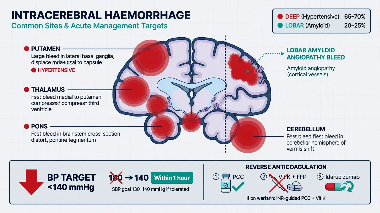
Intracerebral haemorrhage is the spontaneous, non-traumatic bleeding into the brain parenchyma, and it accounts for roughly 10 to 15 per cent of all strokes across Australia, New Zealand, the United Kingdom and North America. It is the most destructive stroke subtype: the 30-day mortality is around 40 per cent, and approximately half of all deaths occur within the first 48 hours. Unlike ischaemic stroke, where the task is reperfusion, the task here is haemostasis and the prevention of haematoma expansion — continued bleeding in the first day enlarges the clot in 20 to 30 per cent of patients and is the dominant driver of death and disability. Two mechanisms dominate the aetiology: hypertensive small-vessel disease, which ruptures the deep penetrating arteries and produces the putaminal, thalamic, pontine and cerebellar bleeds; and cerebral amyloid angiopathy, which deposits beta-amyloid in the leptomeningeal and cortical vessels of the elderly and produces the lobar haemorrhage. The Fellowship candidate must hold the whole pathway in mind: recognise the deficit, image with non-contrast CT to separate haemorrhage from ischaemia, control the blood pressure to a systolic target of 140 mmHg, reverse any anticoagulation immediately, control the intracranial pressure, and refer the surgically salvageable lesion — above all the cerebellar haematoma — without delay.[1][2][3]
Definition and classification
Intracerebral (intraparenchymal) haemorrhage is bleeding directly into the brain substance rather than into the subarachnoid, subdural, epidural or intraventricular space in isolation. In practice the term refers to the spontaneous, non-traumatic parenchymal bleed, and the location classifies it. Deep (hypertensive) haemorrhages arise from the rupture of the small penetrating arteries — the lenticulostriate, the thalamogeniculate and the paramedian pontine branches — that are exquisitely vulnerable to the lipohyalinosis and Charcot-Bouchard microaneurysms of chronic hypertension. They sit in the putamen (the commonest site, around half of all cases), the thalamus, the internal capsule, the pons and the cerebellum. Lobar haemorrhages lie in the cortex and subcortical white matter at the junction of grey and white matter, follow the distribution of the cerebral lobes (frontal, parietal, temporal, occipital), and are the signature of cerebral amyloid angiopathy in the elderly. A small proportion arise from an arteriovenous malformation, a cavernous haemangioma, an intracranial aneurysm that has burst into the parenchyma, a haemorrhagic tumour (melanoma, renal cell, lung, glioblastoma), coagulopathy, sympathomimetic drug use (amphetamine, cocaine), or haemorrhagic transformation of an infarct. The location is the first clue to the cause, and it dictates both the surgical decision and the secondary-prevention workup. [1]
Epidemiology and risk factors
Intracerebral haemorrhage is half as common as ischaemic stroke but twice as lethal. Its incidence rises steeply with age and is higher in people of Asian and Black ancestry, in whom hypertensive small-vessel disease is more prevalent and tends to present earlier. The single most important modifiable risk factor is chronic hypertension, which underlies roughly 60 to 70 per cent of deep bleeds; the others are advancing age, anticoagulation (the risk is increased roughly tenfold and rising as direct oral anticoagulant use grows), heavy alcohol intake, smoking, a low low-density-lipoprotein cholesterol (the paradoxical relationship seen in the SPARCL and TREND analyses), cerebral amyloid angiopathy, and sympathomimetic drug use. The anticoagulated patient deserves special emphasis: warfarin and the direct oral anticoagulants both enlarge the haematoma, prolong the window of expansion, and roughly double the mortality, which is why reversal begins in parallel with the first blood-pressure reading and never after it.[3]
Pathophysiology — why haematoma expansion is the enemy
When a small penetrating artery or an amyloid-laden cortical vessel ruptures, blood dissecting into the parenchyma both crushes and shears the surrounding tissue and, within minutes, forms a space-occupying mass. The bleeding is rarely a single event: in the first 24 hours the haematoma enlarges by a third or more in 20 to 30 per cent of patients, driven by continued leakage from the primary vessel and by secondary rebleeding at the margin. Every millilitre of expansion directly raises intracranial pressure, displaces midline structures, and converts a survivable deficit into a fatal one.[1][3] The mass effect provokes perihhaematomal oedema, which peaks at 48 to 72 hours and adds a second wave of pressure on top of the clot itself. When the bleed communicates with the ventricular system the blood obstructs cerebrospinal fluid drainage and produces an acute hydrocephalus, and blood in the ventricles is itself toxic to the ependyma. These three pressures — clot mass, oedema, and hydrocephalus — are the mechanisms of the secondary brain injury, and each is a target of the emergency management.
[1]Clinical presentation
Intracerebral haemorrhage presents as the sudden onset of a focal neurological deficit that maps to the site of bleeding, frequently accompanied by headache, vomiting and a depressed level of consciousness, and almost always with a markedly elevated blood pressure. These four accompaniments — headache, vomiting, coma and hypertension — are the clinical clues that distinguish haemorrhage from ischaemia at the bedside, though none is reliable enough to avoid CT. A putaminal bleed produces a contralateral hemiparesis and hemisensory loss with a conjugate gaze deviation towards the side of the lesion, and may cause aphasia when dominant. A thalamic bleed produces a contralateral sensory loss that dominates over weakness, often with a vertical-gaze palsy and a drowsy, apathetic state. A pontine bleed is catastrophic: a rapid coma, pinpoint pupils, quadriparesis and decerebrate posturing, with a poor prognosis when the haematoma is large. A cerebellar bleed produces vertigo, vomiting, ataxia and a headache that may be misread as a benign vestibular event, yet it is the one lesion in which an emergency operation saves a life. Lobar bleeds present with a deficit confined to a single cortical territory — a homonymous hemianopia, a fluent or non-fluent aphasia, or a hemiparesis — together with a prominent headache and sometimes a seizure, reflecting cortical irritation. Seizures are commoner in lobar than in deep haemorrhage, and a first-time seizure with a focal deficit and a raised blood pressure should prompt CT rather than an antiepileptic load. [1]
Differential diagnosis
The mimics of intracerebral haemorrhage overlap with the mimics of stroke in general, because a focal deficit with vomiting and a falling conscious level has the same first question — is this bleeding or ischaemia? — and only the non-contrast CT answers it. [1]
Intracerebral haemorrhage
- Sudden focal deficit with headache, vomiting, depressed GCS and marked hypertension
- Non-contrast CT: hyperdense mass, mass effect, possible intraventricular blood or midline shift
- Blood-pressure control to systolic 140 mmHg, reverse anticoagulation, consider surgery
- Cerebellar clot over 3 cm is the surgical emergency
Acute ischaemic stroke
- Sudden focal deficit maximal at onset, usually no headache or vomiting, GCS often preserved
- CT may be normal early; CTA may show large-vessel occlusion
- Thrombolysis or thrombectomy if within window
- Must be distinguished from ICH on CT before any lytic — giving lytic to a haemorrhage is fatal
Subarachnoid haemorrhage
- Thunderclap headache maximal in seconds, neck stiffness, photophobia, brief loss of consciousness
- CT: hyperdense blood in the basilar cisterns and Sylvian fissure, not a parenchymal mass
- Nimodipine 60 mg every 4 hours, BP control, urgent coiling or clipping
- Focal deficit is less prominent than in ICH; headache is the dominant feature
Hypoglycaemia
- Focal deficit reproduced with complete fidelity; confusion or coma possible
- Diabetic on insulin or sulfonylurea; finger-prick glucose typically below 2.5 mmol/L
- Reversed by intravenous glucose within minutes
- Check the glucose first in every suspected stroke — the one bedside test that changes the plan immediately
Hypertensive encephalopathy
- Diffuse encephalopathy rather than a clean focal deficit; markedly elevated BP, headache, visual disturbance, seizures
- CT may show posterior white-matter oedema (PRES); no hyperdense parenchymal mass
- Lower BP gradually with intravenous antihypertensive; treat seizures
- Gradual rather than truly sudden onset; the deficit does not map to a single vascular territory
Haemorrhage into a tumour
- Deficit with an irregular, heterogeneous mass on CT; may have a fluid-blood level
- Known primary (melanoma, renal cell, lung, choriocarcinoma) or a prior neurological symptom
- Contrast CT or MRI defines the underlying lesion
- Requires oncology and neurosurgical input; secondary prevention differs from a hypertensive bleed
The discriminating moves at the bedside are to check the finger-prick glucose, to ask about anticoagulant use and the last dose, to confirm the time of onset and any preceding headache or seizure, and to image every patient in whom stroke is a possibility — because the focal deficit of a small deep haemorrhage and of an ischaemic stroke are otherwise indistinguishable, and a haemorrhage given thrombolysis is a catastrophe. [1]
Investigations
The investigations run in parallel with resuscitation. A finger-prick glucose is checked first to exclude hypoglycaemia. A 12-lead ECG screens for atrial fibrillation (the cause and a guide to secondary prevention) and for the ischaemic changes that may coexist. Bloods include a full blood count, a coagulation screen (the INR is decisive in the anticoagulated patient), urea and electrolytes, and a group-and-save; a pregnancy test is sent in any woman of reproductive age. The coagulation result and the anticoagulant history drive the reversal pathway, so they are taken and chased immediately rather than batched with the routine panel. [1]
The pivotal imaging is non-contrast CT of the brain, and its purpose is to separate haemorrhage from ischaemia — the single decision that decides the entire plan. Acute blood appears as a hyperdense (50 to 70 Hounsfield unit) mass within the parenchyma, well defined and rounded or oval, with surrounding low-density oedema. The site, the size, the mass effect (effacement of the sulci or the ventricles), the midline shift (measured at the septum pellucidum or the pineal gland), the intraventricular extension, and any hydrocephalus are all documented, because each feeds into the surgical decision and the prognostic scores. A fluid-blood level within the clot suggests ongoing bleeding or a coagulopathy and warrants a coagulation screen and a contrast study. CT angiography is obtained in patients under 65, in lobar bleeds, in those without a clear hypertensive cause, and whenever an underlying vascular lesion is suspected; the spot sign — contrast extravasation within the haematoma — marks active bleeding and predicts haematoma expansion, a higher mortality, and a target for any haemostatic therapy under trial. MRI with susceptibility-weighted imaging is more sensitive to cerebral microbleeds and helps distinguish hypertensive small-vessel disease from amyloid angiopathy, but it is not the first-line emergency study. [1]
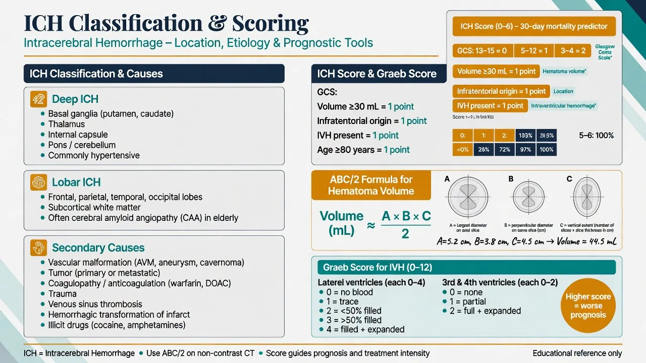
Two scores must be reproduced and documented on every intracerebral haemorrhage. The Graeb score grades the intraventricular extension, and the ICH score predicts the 30-day mortality. [1]
The Graeb score — intraventricular haemorrhage (maximum 12)
The ICH score sums five bedside variables to a maximum of 6. The Glasgow Coma Scale (GCS) — the sum of the eye response (1 to 4, from no opening to spontaneous), the verbal response (1 to 5, from none to orientated) and the motor response (1 to 6, from none to obeying commands), a total from 3 to 15 — contributes 0 for a GCS of 13 to 15, 1 for 5 to 12, and 2 for 3 to 4. The haematoma volume contributes 0 for under 30 mL and 1 for 30 mL or more. An infratentorial origin adds 1, any intraventricular extension adds 1, and an age of 80 or over adds 1. The 30-day mortality rises steeply with the total: a score of 0 carries near-zero mortality, 1 around 13 per cent, 2 around 26 per cent, 3 around 72 per cent, and 4 or more over 95 per cent. Recording the ICH score, the volume and the Graeb score at the first CT organises the case for the neurosurgical referral and for the family conversation that follows. [1]
Immediate management and resuscitation
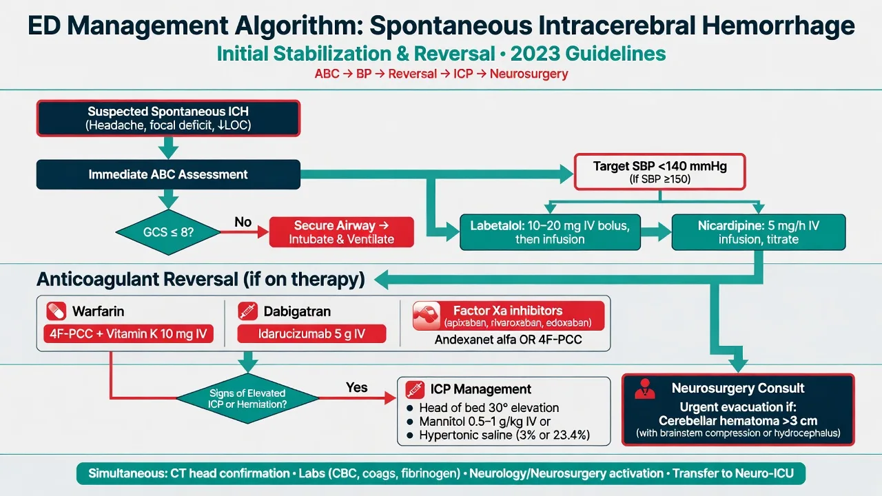
Resuscitation and the specific therapy run together and begin at the moment of suspicion. The airway is secured in the patient with a GCS of 8 or less, and oxygen is given only if the saturation falls below 94 per cent, because hyperoxia is harmful. Intravenous access is secured, the patient is kept nil by mouth pending a swallow screen, and glucose is treated only if low. Two manoeuvres run in parallel with the airway and oxygen and must not wait for the CT report: blood-pressure control and anticoagulation reversal, because both target haematoma expansion and every minute of delay is a minute in which the clot can grow. The neurosurgical team is notified early — particularly for a cerebellar or a large lobar bleed — and a hostile or deteriorating patient is moved to a resuscitation bay with continuous monitoring. [1]
[1]Blood-pressure management
Blood-pressure control is the resuscitation manoeuvre most often examined, and the evidence is precise. [1]
Management of the anticoagulated patient
The anticoagulated patient is the highest-risk subset, and reversal begins in parallel with the first blood-pressure reading. The aim is to restore normal haemostasis as fast as possible to arrest haematoma expansion, and the agent chosen depends on the drug. Warfarin is reversed with prothrombin complex concentrate at 25 to 50 IU per kilogram intravenously (it restores the factors within minutes and is superior to fresh-frozen plasma) together with vitamin K 10 mg intravenously to sustain the reversal over the following days; the INR is rechecked at 30 minutes and at intervals thereafter. Dabigatran, a direct thrombin inhibitor, is reversed with idarucizumab 5 g intravenously (given as two 2.5 g vials in succession), a specific monoclonal antibody fragment that binds dabigatran with high affinity. Apixaban and rivaroxaban, the factor Xa inhibitors, are reversed with andexanet alfa — a decoy factor Xa — dosed according to the drug, the dose and the timing (a low-dose regimen of a 400 mg bolus plus 480 mg infusion, or a high-dose regimen of an 800 mg bolus plus 960 mg infusion); where andexanet is unavailable, prothrombin complex concentrate at 50 IU per kilogram is a reasonable alternative. Heparin is reversed with protamine 1 mg per 100 units of heparin. The antiplatelet drugs are not reversed routinely; the PATCH trial showed that platelet transfusion after an intracerebral haemorrhage on antiplatelets worsened outcome and is not recommended, though it may be considered before an urgent neurosurgical procedure. [1]
Intracranial-pressure control and neuroprotection
Raised intracranial pressure develops as the clot mass, the perilesional oedema and any hydrocephalus combine, and it is the mechanism by which a deep or cerebellar bleed kills. The general measures come first: nurse the patient with the head of the bed elevated to 30 degrees and midline (to aid venous drainage), maintain normocapnia, normoglycaemia, normothermia and normoxia, and avoid hyperhydration and hypo-osmolar fluids. Mannitol 0.5 to 1 g per kilogram intravenously as a bolus (repeated 0.25 g per kilogram every 4 to 6 hours, checked against the serum osmolality and held if it exceeds 320 mOsm per kilogram) is the classic osmotic agent; hypertonic saline — 3 per cent at 250 mL as a bolus, or 23.4 per cent in 30 mL aliquots through a central line — is an equivalent or superior alternative that also supports the blood pressure. An external ventricular drain is placed for an obstructive hydrocephalus from intraventricular blood and allows both drainage and the monitoring of the intracranial pressure. Sedation and analgesia are titrated with short-acting agents (propofol, fentanyl) that do not accumulate, and seizure prophylaxis (levetiracetam 500 mg intravenously twice daily) is given if a seizure has occurred but not routinely prophylactically. [1]
Neurosurgical evacuation — who, and when
The surgical decision splits cleanly along the site of the bleed. A cerebellar haematoma over 3 cm in diameter, or any cerebellar bleed with brainstem compression, hydrocephalus or a deteriorating Glasgow Coma Scale, is a neurosurgical emergency and is evacuated urgently — a posterior fossa craniectomy that relieves the pressure on the brainstem and can convert a dying patient into a survivor.[3] A supratentorial lobar haematoma within 1 cm of the cortical surface with a deteriorating conscious level is considered for craniotomy and evacuation, though the STICH trials showed no clear overall benefit and the decision is individualised. Deep haemorrhages (putaminal, thalamic) are generally managed medically because surgery through the deep white matter causes as much damage as it relieves; a decompressive hemicraniectomy may be offered to a younger patient with a large clot and refractory intracranial pressure as a last resort. Minimally invasive surgery (stereotactic or endoscopic clot aspiration, with or without thrombolysis through a catheter) is under active investigation (MISTIE, ENRICH) and is not yet a standard Fellowship answer. The bottom line the examiner wants: the cerebellar clot over 3 cm is evacuated tonight; the deep clot is not.
Complications and pitfalls
The complications are the direct consequences of the mass and the blood. Haematoma expansion in the first 24 hours is the commonest cause of early deterioration and is targeted by the blood-pressure and reversal strategies above. Perilesional oedema peaks at 48 to 72 hours and may precipitate a transtentorial or tonsillar herniation. Obstructive hydrocephalus from intraventricular blood requires an external ventricular drain. Seizures are commonest with cortical (lobar) bleeds and present as a new neurological deterioration. Aspiration pneumonia (from dysphagia and a depressed conscious level), deep vein thrombosis (prophylaxis started after 48 hours once the bleed is stable), pressure injury, and fever (which worsens outcome and is treated actively) complete the early set. The commonest pitfalls are a delayed CT that allows expansion to proceed unseen, an under-treated blood pressure that misses the 140 mmHg target, a forgotten or slow anticoagulation reversal, a cerebellar clot dismissed as vertigo, and the reflex thrombolysis given to a haemorrhage misread as an ischaemic stroke — the one error that is always fatal and that the CT exists to prevent. [1]
Prognosis and disposition
Outcome tracks the size and the site of the clot, the presenting Glasgow Coma Scale, the presence and amount of intraventricular blood (the Graeb score), the patient's age and comorbidity, and — above all — whether the haematoma expanded. The ICH score at the first CT frames the prognosis: a low score predicts a good recovery, a score of 4 or more predicts death or severe disability. Every patient is admitted to a stroke or neurocritical-care unit with continuous monitoring, blood-pressure control, and early multidisciplinary rehabilitation. Secondary prevention is begun once the bleed is stable: rigorous blood-pressure control (the single most effective measure against recurrence), smoking cessation, and — in the patient whose haemorrhage was not caused by amyloid angiopathy and who has a strong indication such as atrial fibrillation — a careful, individualised decision about restarting anticoagulation, typically after a few weeks and often with a direct oral anticoagulant in preference to warfarin. [1]
Special populations
The elderly lobar haemorrhage patient often has cerebral amyloid angiopathy, is prone to recurrence, and may show multiple microbleeds and superficial siderosis on MRI; anticoagulation is restarted only with reluctance, if at all. The anticoagulated patient has been discussed in detail and is the subset in whom reversal is time-critical. The pregnant patient is managed jointly with obstetrics and neurosurgery; the blood-pressure target is unchanged, warfarin is avoided throughout pregnancy, and delivery planning follows the neurological status. The young patient with an unexplained bleed demands a search for an arteriovenous malformation, a cavernoma, a vasculitis, a thrombophilia, or drug use — the cause changes the prevention and the prognosis. The cerebellar haemorrhage is its own population: the only deep-site bleed in which an emergency operation routinely saves a life, and the one in which a delayed referral is the recurring, avoidable disaster. [1]
Evidence and regional guidelines
The evidence base for acute blood-pressure management rests on INTERACT2[1] and ATACH-2,[2] which together defined the systolic target of 140 mmHg as the safe and effective floor — effective enough to improve functional outcome, but no longer pushed below 130 mmHg after ATACH-2 showed harm. The 2022 AHA/ASA Guideline for the Management of Patients with Spontaneous Intracerebral Hemorrhage[3] is the contemporary global standard and synthesises the blood-pressure, reversal, intracranial-pressure and surgical recommendations cited throughout this topic. The STICH trials informed the surgical approach to supratentorial clots, and MISTIE and ENRICH continue to define the role of minimally invasive evacuation. ANZ practice follows the Stroke Foundation Clinical Guidelines for Stroke Management and the local state-wide neurosurgical retrieval service; the United Kingdom follows the NICE and Royal College of Physicians stroke pathways; Europe follows the ESO guideline. The targets and the agents are global; the local stroke and neurosurgical pathways govern the retrieval logistics and the exact reversal protocol.
ANZ practice note. Every Australian and New Zealand emergency department runs a stroke triage pathway that fast-tracks a suspected intracerebral haemorrhage to a non-contrast CT within minutes of arrival. Blood pressure is lowered to a systolic target of 140 mmHg with intravenous labetalol or nicardipine; anticoagulation is reversed immediately according to the agent; and the patient is referred to the state-wide neurosurgical service, where a cerebellar haematoma over 3 cm, or any clot with brainstem compression or a deteriorating GCS, is evacuated urgently. Prothrombin complex concentrate is preferred over fresh-frozen plasma for warfarin reversal; idarucizumab and andexanet alfa are available for the direct oral anticoagulants. [1]
Exam practice
SAQ — Hypertensive putaminal intracerebral haemorrhage with acute blood-pressure management
10 minutes · 10 marks
A 62-year-old man is brought to the emergency department one hour after the sudden onset of a left hemiparesis, headache and two episodes of vomiting. His blood pressure is 198/112 mmHg, GCS 13 (E3V4M6), with a dense left-sided pyramidal weakness, a left homonymous hemianopia and slurred speech. Finger-prick glucose is 7.1 mmol/L. The non-contrast CT shows a 25 mL right putaminal haemorrhage with mild perilesional oedema, no midline shift and no intraventricular extension; CT angiography demonstrates no spot sign. He takes perindopril, amlodipine and aspirin 100 mg daily and has no history of anticoagulation.
SAQ — Cerebellar haemorrhage with neurological deterioration
10 minutes · 10 marks
A 58-year-old woman is brought to the emergency department three hours after the sudden onset of an occipital headache, vomiting and progressive drowsiness. Over the preceding 30 minutes her GCS has fallen from 13 to 11 (E2V4M5) and her blood pressure is 184/98 mmHg. She has a left gaze palsy, a left lower motor neurone facial weakness and an ataxic left arm. The non-contrast CT shows a 3.6 cm right cerebellar haemorrhage with effacement of the fourth ventricle, early obstructive hydrocephalus and compression of the brainstem, with no supratentorial shift.
Exam pearls
- A focal deficit with vomiting, a depressed conscious level and marked hypertension is an intracerebral haemorrhage until the non-contrast CT proves otherwise — never thrombolyse before CT.
- Haematoma expansion in the first 24 hours drives mortality — the systolic target of 140 mmHg, the rapid anticoagulation reversal, and the spot sign on CTA are the three things that limit it.
- The systolic blood-pressure target is 140 mmHg — INTERACT2 showed benefit, ATACH-2 showed no further benefit and more renal harm below 130, and the 2022 AHA/ASA guideline synthesises the two.
- Reverse the anticoagulant by its class — prothrombin complex concentrate plus vitamin K for warfarin, idarucizumab for dabigatran, andexanet alfa for the factor Xa inhibitors, protamine for heparin.
- A cerebellar haematoma over 3 cm, or with brainstem compression, hydrocephalus or a falling GCS, is a neurosurgical emergency evacuated tonight; a deep supratentorial bleed is not.
- Estimate the volume with the ABC/2 formula and grade the intraventricular blood with the Graeb score — a volume of 30 mL is the ICH-score threshold and the surgical-decision pivot.
- Do not transfuse platelets for an antiplatelet-associated bleed — the PATCH trial showed harm; reserve transfusion for the urgent neurosurgical procedure. [1]
Red flags
[1]References
- [1]Anderson CS, Heeley E, Huang Y, et al. Rapid blood-pressure lowering in patients with acute intracerebral hemorrhage N Engl J Med, 2013.PMID 23713578
- [2]Qureshi AI, Palesch YY, Barsan WG, et al. Intensive Blood-Pressure Lowering in Patients with Acute Cerebral Hemorrhage N Engl J Med, 2016.PMID 27276234
- [3]Greenberg SM, Ziai WC, Cordonnier C, et al. 2022 Guideline for the Management of Patients With Spontaneous Intracerebral Hemorrhage: A Guideline From the American Heart Association/American Stroke Association Stroke, 2022.PMID 35579034