EM · Testicular torsion
Testicular torsion
Also known as Spermatic cord torsion · Acute scrotum · Torsion of the testis
The testicular torsion — the twisting of the spermatic cord on its longitudinal axis that obstructs the venous return and then the arterial supply, producing the testicular ischaemia and necrosis; the quintessential time-critical surgical emergency of the acute scrotum with the salvage window of six hours. The clinical picture (the sudden severe unilateral testicular pain, the swelling, the high-riding testis, the absent cremasteric reflex, the horizontal lie, the nausea and vomiting), the diagnosis (the clinical diagnosis, the Doppler ultrasound if uncertain but never to delay surgery, the manual detorsion), the management (the urgent surgical exploration within six hours, the bilateral orchiopexy, the orchidectomy if necrosis). The differential (the epididymo-orchitis, the appendix testis torsion, the incarcerated hernia, the renal colic). ACEM-primary, globally tagged.
On this page & tools
Your progress
Saved locally on this device.
Target exams
Red flags
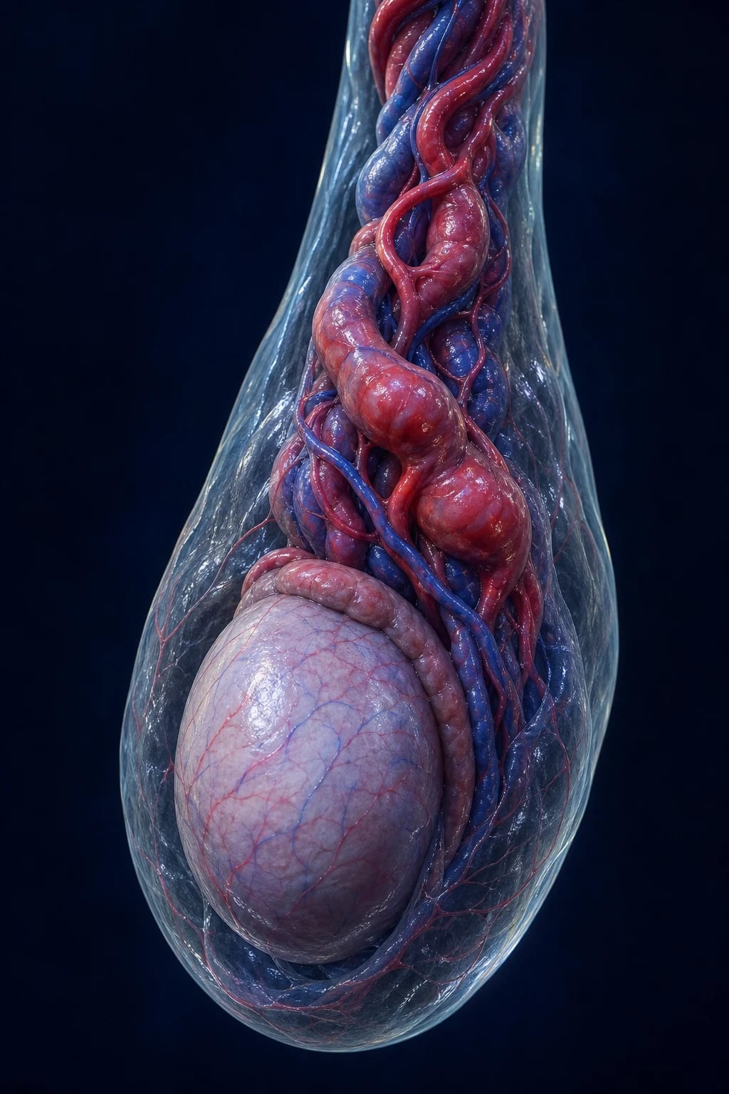
The testicular torsion is the twisting of the spermatic cord on its longitudinal axis that first obstructs the venous and lymphatic return and then, as the oedema mounts, the arterial inflow, producing testicular ischaemia and — if unrelieved — irreversible necrosis. It is the diagnosis that defines the acute scrotum: any male with the sudden testicular pain has torsion until proven otherwise, and the salvage of the testis is governed by the time from the onset of pain to the surgical detorsion. The Fellowship candidate must hold the six-hour clock in mind from the moment the triage note reads "scrotal pain", must make the diagnosis clinically, must not let the Doppler ultrasound delay the theatre in the clear-cut case, and must understand the bilateral orchiopexy, the manual detorsion, and the orchidectomy for the necrotic gland.[1][2]
Definition and epidemiology
The testicular torsion is the rotation of the testis on the spermatic cord axis that compromises the blood supply. Two anatomical types are recognised. Intravaginal torsion, by far the commoner, occurs within the tunica vaginalis and is enabled by the bell-clapper deformity — an abnormally high reflection of the tunica onto the spermatic cord rather than onto the testicle, which leaves the gland suspended and free to rotate. The deformity is bilateral in most cases, which is the anatomical basis for the bilateral orchiopexy. Extravaginal torsion occurs outside the tunica vaginalis, is almost exclusively a neonatal (prenatal or perinatal) event, and produces a non-viable testis at birth in most cases. The epidemiology is bimodal: a small neonatal peak and a large pubertal peak between 12 and 18 years, although torsion occurs at any age. The lifetime incidence is approximately 1 in 4000 males under 25 years. The left side is affected slightly more often than the right. The strongest risk factor is a prior episode — the patient with intermittent torsion is at high risk of a complete twist, and the patient with a bell-clapper deformity risks the contralateral side. Undescended testis carries a markedly elevated torsion risk.[1]
Pathophysiology
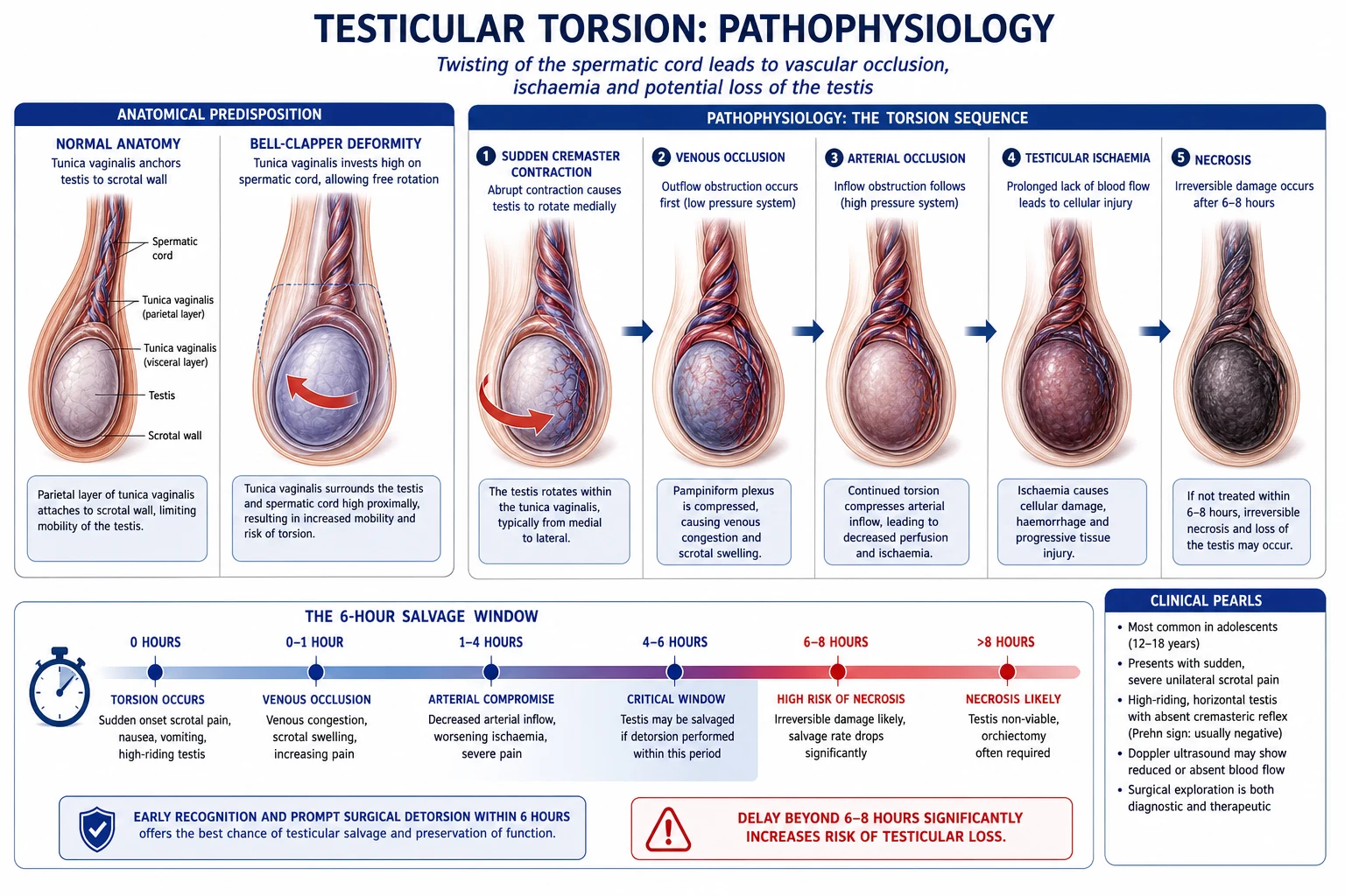
The torsion is fundamentally a vascular phenomenon governed by a pressure sequence. The twist of the cord first compresses the thin-walled venous and lymphatic channels, which have the lowest intraluminal pressure. The venous obstruction produces congestion and oedema of the testis; the rising tissue pressure within the inelastic tunica albuginea then collapses the arterial inflow, converting a congested but perfused testis into an ischaemic one. The arterial occlusion is the event that begins the countdown to necrosis — and it is why the salvage curve is so steep. The degree of twist (typically 180 to 1080 degrees) and the duration together determine viability: a high twist may occlude the artery within minutes, while a partial twist may permit some perfusion for hours. The intermittent torsion detorses spontaneously before infarction, only to recur; each episode is an ischaemic insult that may culminate in atrophy even without a complete infarct.[2]
The clinical presentation
The classic presentation is the sudden, severe, unilateral scrotal pain often waking the patient from sleep, frequently with nausea and vomiting (a sympathetic response to the visceral pain of the ischaemic testis that is itself a discriminator from the inflammatory causes). The pain may radiate to the lower abdomen or the groin, and the patient may report a preceding episode that resolved spontaneously — the signature of intermittent torsion. The physical examination signs are the syllables of the diagnosis. The high-riding testis sits abnormally high in the scrotum and may lie horizontally (the bell-clapper lie) because the twisted cord has shortened. The absent cremasteric reflex — stroking the inner thigh normally elevates the ipsilateral testis — is the most sensitive single sign of torsion. The testis is tender, swollen, and firm, and may be retained in an abnormal, high position that does not drop when the patient stands. The Prehn sign (relief of pain on elevation of the testis) is negative in torsion and positive in epididymitis, but it is not reliable enough to exclude torsion. The blue-dot sign — a small bluish discoloration at the upper pole — indicates torsion of the appendix testis, a mimic. Atypical presentations include pain referred wholly to the abdomen with a normal-looking scrotum, and the neonate with an asymptomatic, hard, non-transilluminating scrotal mass.[1]
Differential diagnosis
Testicular torsion
- Sudden severe pain, nausea and vomiting; the high-riding, horizontal testis
- The absent cremasteric reflex; the TWIST score 5 to 7
- The urgent surgical exploration within six hours; no imaging delay
- Salvage 90 to 100 per cent under 6 hours; the time-critical emergency
Epididymo-orchitis
- Gradual onset over hours to days; the fever, dysuria, discharge
- The Prehn sign positive (pain relieved by elevation); the cremasteric reflex present
- The urinalysis positive; the Doppler US shows increased flow
- The ceftriaxone 500 mg IM plus azithromycin 1 g PO (STI) or ofloxacin 400 mg BD (enteric)
Appendix testis torsion
- The blue-dot sign at the upper pole; the gradual pain
- The cremasteric reflex typically preserved; the normal testicular lie
- The Doppler US shows normal flow with an enlarged appendix
- The conservative management with analgesia; self-limiting
Incarcerated hernia
- The irreducible groin or scrotal mass; the bowel obstruction features
- The history of a reducible hernia; the cough impulse absent when incarcerated
- The tender, tense mass extending to the external ring
- The urgent surgical repair; the strangulation risk
Renal colic
- The flank to groin pain radiating to the testicle; the normal scrotal examination
- The haematuria; the CT KUB shows the stone
- The cremasteric reflex present; the testis normal
- The NSAIDs (diclofenac 75 mg IM) and the analgesia
The other differentials to hold in mind include the idiopathic scrotal oedema (a paediatric, painless, bilateral scrotal swelling that pits on pressure), the Henoch-Schönlein purpura (the palpable purpura with scrotal involvement), the scrotal trauma (the history and the haematocoele), and the very rare testicular tumour presenting with acute pain from a haemorrhage into the mass. The Fellowship rule stands: in the acute scrotum, the torsion is excluded only by surgical exploration or by a Doppler ultrasound that confirms the flow — never by the clinical impression alone in the indeterminate case.[5]
The bedside assessment and the clinical scores
The bedside assessment is clinical and rapid. The history establishes the sudden onset, the duration (which sets the salvage clock), the prior episodes (intermittent torsion), and the pubertal age. The examination tests the cremasteric reflex, the testicular lie and position, the swelling and tenderness, and the Prehn sign, then palpates the contralateral side (the bell-clapper is bilateral) and excludes a hernia. The structured risk tool is the TWIST score (the Testicular Workup for Ischemia and Suspected Torsion, Barbosa and colleagues), which stratifies the pre-test probability without replacing the surgical judgement. [1]
| TWIST criterion | Points |
|---|---|
| Hard testis | 2 |
| Absent cremasteric reflex | 1 |
| Nausea and vomiting | 1 |
| High-riding testis | 1 |
| Scrotal swelling | 2 |
The TWIST score interpretation
A high TWIST score is itself an indication for the operating theatre without imaging — the score formalises the rule that the diagnosis is clinical.[1]
The investigations
The investigations support but never delay the surgery. The diagnosis of torsion is clinical — the patient with the high-probability picture goes straight to the theatre. The colour Doppler ultrasound is reserved for the indeterminate case in which the clinical probability is low or moderate and the surgical team is not proceeding: it demonstrates the absent or markedly reduced intratesticular arterial flow on the affected side. The Doppler has a sensitivity of approximately 85 to 95 per cent and a specificity of 95 per cent, but a normal Doppler never fully excludes torsion because the partial or intermittent twist may preserve some flow, and the window to false reassurance is the window to the lost testis. The point-of-care ultrasound performed by the emergency clinician can demonstrate the absent flow at the bedside and accelerate the surgical referral.[3] The urinalysis screens for the infective causes (the pyuria of the epididymitis), the full blood count and CRP are non-specific, and the beta-hCG (where the scrotum is examined in a person of reproductive potential) is the standing safeguard. No blood test diagnoses torsion; the bloods are drawn for the anaesthetic and the theatre.[3]
Management — the time-critical approach
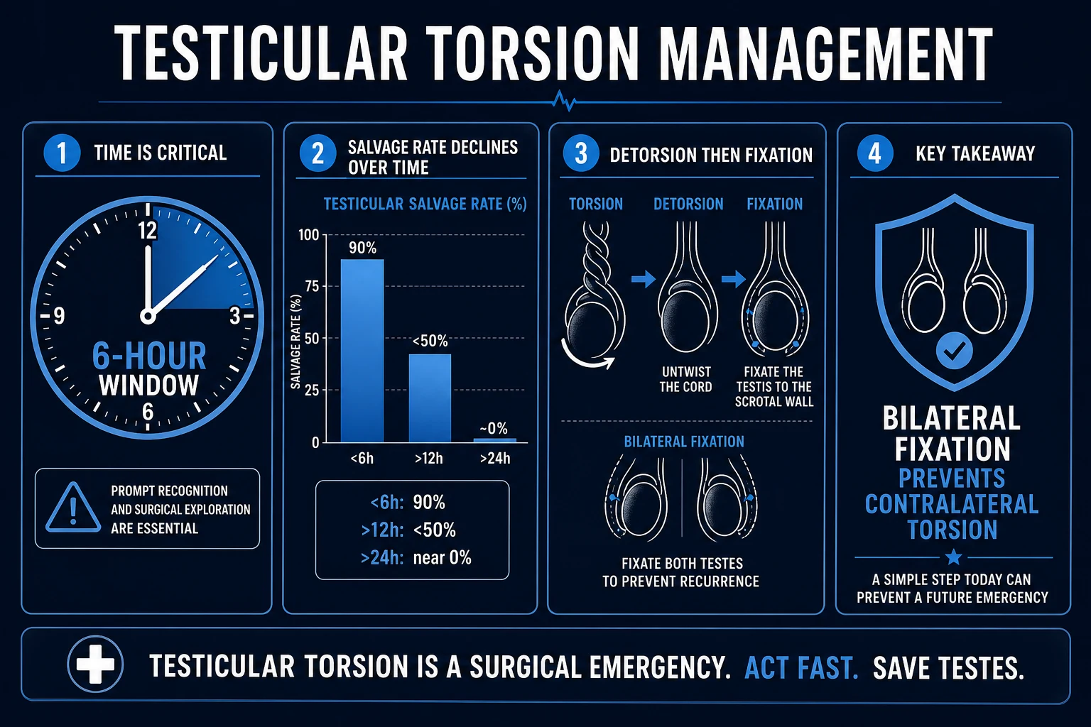
The management is the resuscitation, the analgesia, the surgical referral, and the theatre — in parallel, not in sequence. The intravenous access is established, the nil-by-mouth status is confirmed for the anaesthetic, and the surgical team is notified at the moment torsion is suspected, before any imaging. The analgesia is generous and does not obscure the diagnosis: the morphine 0.1 mg per kilogram intravenously (up to 10 mg) or the fentanyl 1 to 2 micrograms per kilogram intravenously, titrated to the pain, with the paracetamol 1 g intravenously (15 mg per kilogram in the child) as the baseline. The ondansetron 0.15 mg per kilogram intravenously (4 mg in the adult, up to 8 mg) treats the nausea and vomiting and aids the fasting. The pre-operative antibiotic, typically the cefazolin 25 mg per kilogram intravenously (up to 2 g), is given on the way to the theatre. The disposition is the operating theatre within six hours of the onset — every minute of delay beyond six hours trades salvage for orchiectomy.[2][5]
[1]The manual detorsion
The manual detorsion is the bedside untwisting of the cord, performed as a temporising bridge to the surgery in the patient with the high-probability torsion and the long transfer or theatre delay. The testis is gently manipulated medial to lateral — the "opening the book" manoeuvre — because most torsions twist inward (lateral to medial) and so detorse outward. The endpoint is the relief of pain and the return of the testis to a lower, vertical lie; the return of the flow on the Doppler confirms the success. The manual detorsion is successful in over 80 per cent of attempts, but it is never the definitive treatment — even a fully detorsed testis still requires the urgent bilateral orchiopexy because the recurrence is otherwise inevitable. The procedural analgesia or sedation may be used: the morphine 0.1 mg per kilogram intravenously for analgesia, or a procedural sedation with the ketamine 1 to 1.5 mg per kilogram intravenously (or 4 mg per kilogram intramuscularly) with full monitoring, or the midazolam 0.05 to 0.1 mg per kilogram intravenously for anxiolysis. The manual detorsion that fails to relieve the pain is attempted in the opposite direction (a minority twist the other way) and, if still unsuccessful, mandates the immediate surgery.[4]
The surgical management
The scrotal exploration is the definitive management. Through a scrotal incision (or occasionally an inguinal incision for the suspected tumour), the testis is delivered, the cord is detorsed, and the gland is assessed for the viability — the return of the pink colour and the bleed at the incision of the tunica albuginea. The viable testis is fixed in the scrotum with the orchiopexy (the non-absorbable sutures through the tunica albuginea to the dartos or the scrotal wall). The bilateral orchiopexy — fixing the contralateral testis at the same operation — is mandatory because the bell-clapper deformity is bilateral and the contralateral torsion would otherwise claim the second gland. The non-viable testis (the black, necrotic gland with no bleeding) is removed by the orchidectomy. The decision to retain a marginally viable testis balances the small chance of recovery against the risk of the anti-sperm antibody-mediated damage to the contralateral testis and the subsequent infertility. The window dressing, the post-operative analgesia, and the follow-up for the atrophy complete the surgical care.[5]
The high-probability features of torsion
TWIST
Complications and prognosis
The dominant complication is the loss of the testis (the orchidectomy), the rate of which climbs steeply with the duration of the torsion — the salvage is 90 to 100 per cent under six hours, 50 per cent from six to twelve hours, and under 10 per cent beyond twenty-four hours. The testicular atrophy may follow even a salvaged testis, particularly after the prolonged or repeated torsion. The infertility is a long-term consequence: the anti-sperm antibodies that develop after a necrotic or atrophied testis may impair the function of the contralateral gland, and the bilateral orchiopexy does not protect against this immunological injury. The recurrence on the contralateral side is prevented by the bilateral fixation but is otherwise a real risk. The psychological impact — the body-image and the fertility anxiety — is significant in the adolescent and warrants the explicit counselling. The prognosis is otherwise excellent for the promptly detorsed and fixed testis, and the patient is counselled that the future fertility depends on the viability of the retained testis and the absence of the immunological damage.[2]
The paediatric and adolescent population
The paediatric and the adolescent male is the core population of the torsion. The pubertal peak (12 to 18 years) reflects the rapid testicular growth on the bell-clapper deformity, and the new testicular pain in this age group is torsion until proven otherwise. The younger child may not localise the pain and may present with the abdominal pain, the limping gait, or the non-specific malaise; the scrotum must be examined in any boy with the unexplained abdominal pain. The neonatal torsion is typically extravaginal and prenatal, presenting at birth with a firm, painless, non-transilluminating scrotal mass and a dark scrotum; the salvage rate is very low, but the contralateral testis must be examined and, in the postnatal torsion, urgently explored. The undescended testis carries a markedly elevated torsion risk and presents with the empty hemiscrotum and the high, tender groin mass. The communication with the child and the parents — the clear explanation of the salvage clock, the consent for the orchiopexy and the possible orchidectomy, and the candour about the fertility — is itself an examined competency.[1][2]
ANZ practice note. The testicular torsion is managed as the surgical emergency under the transfer and the retrieval framework: the patient with the suspected torsion in the rural or the peripheral site is transferred urgently to the surgical centre, and the manual detorsion is the bridge during the retrieval. The ACEM and the urological guidance emphasise the clinical diagnosis, the immediate surgical referral, the avoidance of the imaging delay in the high-probability case, and the Doppler ultrasound only for the indeterminate case. The pre-operative analgesia, the nil-by-mouth status, and the antibiotic on the way to the theatre are the ED standard. [1]
Evidence and regional guidelines
The contemporary evidence is consistent across the regions. The European Association of Urology (EAU) paediatric urology guideline and the American Urological Association (AUA) guidance converge on the clinical diagnosis, the urgent exploration within six hours, the bilateral orchiopexy, and the manual detorsion as the bridge. The Doppler ultrasound is endorsed only for the indeterminate case. The transfer-status data show that the inter-hospital transfer adds hours to the time-to-theatre and is a major driver of the orchiectomy rate — the argument for the local manual detorsion and the early surgical referral in the rural setting.[2]
The bell-clapper deformity — the anatomical basis
The bell-clapper deformity is the structural lesion that makes the intravaginal torsion mechanically possible, and understanding it is to understand why the orchiopexy is bilateral. In the normal anatomy, the tunica vaginalis — the peritoneal derivative that forms the serous sac around the testis — reflects onto the testicle itself, investing the gland so that the posterior border (the bare area, where the epididymis and the vessels enter) is anchored to the scrotal wall. The testis thus fixed cannot spin freely. In the bell-clapper deformity, the tunica reflects abnormally high onto the spermatic cord rather than onto the testicle, so the entire gland — including its epididymal attachment — is invested by the visceral tunica and suspended within the tunical sac like the clapper of a bell. With no posterior fixation, the testis is free to rotate on its cord axis within the fluid-filled space. [1]
The deformity is a bilateral congenital variant in the large majority of affected individuals: both testes hang on their long mesenteric-like pedicles, and either may twist. This bilateralism is the single most important anatomical fact for the surgeon — it is the justification for the contralateral orchiopexy at the same operation, the fixation of the unaffected testis with the non-absorbable sutures that prevent the second, devastating torsion. The Fellowship candidate who explores one side and leaves the other unfixed has failed the patient and the examination. The prevalence of the bell-clapper deformity in the general population is estimated at 12 per cent, but only a fraction ever torse — the trigger is often the rapid testicular growth at puberty, the cold exposure (the cremasteric contraction), the trauma, or the nocturnal erection that rotates the gland on its slack pedicle.[7]
[1] [1]The time-critical salvage physiology
The salvage of the torsed testis is governed by a steep, time-dependent viability curve that the Fellowship candidate must reproduce from memory. The mechanism is the orderly, pressure-driven vascular collapse described above: the venous and lymphatic obstruction first, then the arterial occlusion, then the ischaemia, then the necrosis. The degree of twist matters — a complete 720-degree rotation may occlude the artery within minutes, while a partial 180-degree twist may preserve trickle-flow for hours — but the duration of the complete occlusion is the dominant variable. The salvage rates, drawn from the large cohorts and the systematic reviews, are as follows.[9]
The salvage rate as a function of the duration of symptoms
The implication for the ED is absolute: the surgical clock starts at the onset of the pain, not at the triage time. The patient who arrives four hours after the onset has two hours to reach the operating table within the salvage window; the patient who arrives at eight hours is already in the diminishing-returns zone. The time-to-theatre is the single most modifiable determinant of the orchiectomy rate, and the inter-hospital transfer is its greatest enemy — the rural patient who is not detorsed manually and not transferred with urgency loses the testis to the road. The manual detorsion, the early surgical notification, and the parallel (not sequential) workup are the ED's contribution to the salvage.[2]
The ED time-critical pathway — the parallel workup
Recognise: the sudden severe scrotal pain, the high-riding horizontal testis, the absent cremasteric reflex — the clock starts at the onset of pain, recorded to the minute
Resuscitate in parallel: the intravenous access, the nil-by-mouth, the baseline bloods for the anaesthetic (group and hold, FBC, U&E), the analgesia (morphine 0.1 mg/kg IV) and the antiemetic (ondansetron 0.15 mg/kg IV)
Refer immediately: the surgical or urological team is called the moment the torsion is suspected — before any imaging; the consent for the exploration, the orchiopexy, and the possible orchidectomy
Stratify: the TWIST score; if high (5 to 7), no imaging — straight to theatre; if intermediate or low, the Doppler ultrasound to confirm or exclude
Bridge if needed: the manual detorsion (medial-to-lateral, "opening the book") during the transfer or theatre delay; the documented return of the flow; the immediate bilateral orchiopexy still required
Theatre: the scrotal exploration, the detorsion, the viability assessment, the orchiopexy (viable) or the orchidectomy (necrotic), and the mandatory contralateral orchiopexy
Follow-up: the analgesia, the scrotal support, the counselling on the fertility and the body image, the review for the atrophy at six to twelve weeks
Intermittent torsion — the chameleon
The intermittent torsion is the under-recognised precursor of the complete infarct and the trap of the acute scrotum. The patient — typically an adolescent — describes the repeated episodes of acute testicular pain that resolve spontaneously within minutes to hours, often with the position change or the manual self-manipulation that unknowingly detorses the cord. Between the episodes, the examination may be entirely normal, and the Doppler ultrasound (if obtained in the pain-free window) shows the normal flow, falsely reassuring. Each episode, however, is an ischaemic insult: the cumulative damage may produce the testicular atrophy even without a single complete infarct, and any episode may fail to detorse, converting the intermittent pattern into the surgical emergency. [1]
The diagnosis of the intermittent torsion is clinical and historical — the recurrent, stereotyped, self-resolving acute scrotal pain in the pubertal male is the signature, and the normal examination between the attacks does not exclude it. The management is the elective bilateral orchiopexy to prevent the progression to the complete torsion. The Fellowship message: the patient with the history of the recurrent self-resolving testicular pain is not reassured and sent home — he is referred for the elective orchiopexy, because the next episode may be the one that does not resolve.[7]
[1]The imaging — when, and when not
The imaging rule is the most examined and the most violated principle of the torsion, and the Fellowship candidate must state it without hedging: the high-probability torsion goes to the theatre without the imaging. The colour Doppler ultrasound is reserved for the indeterminate case — the clinical probability that is neither high enough to justify the exploration nor low enough to dismiss the torsion — and it is obtained only when the surgical team is genuinely undecided and the imaging will not materially delay the operation. [1]
High suspicion (TWIST 5–7)
- No imaging — straight to the operating theatre
- The Doppler delay converts the salvageable testis into the necrotic one
- The manual detorsion as the bridge during the theatre delay
- The bilateral orchiopexy at the exploration
Intermediate (TWIST 3–4)
- The urgent Doppler ultrasound if the surgical team is undecided
- The scan obtained in parallel with the surgical preparation — never serially
- A normal Doppler does not fully exclude the partial or intermittent torsion
- The threshold to explore if the scan is equivocal or unavailable
Low suspicion (TWIST 0–2)
- The Doppler ultrasound to confirm the alternative diagnosis (the epididymitis, the appendix torsion)
- The urinalysis and the STI screen
- The clear safety-net advice and the early return for the recurrent or worsening pain
- The documented plan and the review
The colour Doppler ultrasound demonstrates the absent or the markedly reduced intratesticular arterial flow on the affected side, often with the increased peri-testicular flow (the reactive hyperaemia around the dead gland). The sensitivity is approximately 85 to 95 per cent and the specificity approximately 95 per cent, but the false negative — the partial twist, the intermittent torsion, the early presentation with the preserved trickle-flow, and the operator-dependent scan — is the reason that a normal Doppler never fully excludes the torsion. The point-of-care ultrasound by the trained emergency clinician, demonstrating the absent intratesticular flow at the bedside, accelerates the surgical referral and is increasingly part of the ED competency.[3]
The TWIST score — systematic review and meta-analysis
PMID 35238603
Population: 14 studies, 2878 patients with the acute scrotum, across the paediatric and the adult populations
Comparator: The surgical confirmation of the torsion as the reference standard
Key finding
The high-score group (TWIST 5 to 7) had a torsion rate of over 80 per cent; the low-score group (0 to 2) a rate under 10 per cent
The systematic review and the meta-analysis consolidated the TWIST score as the most validated clinical stratification tool for the acute scrotum, with the high score carrying the torsion in the large majority and the low score effectively excluding it in most cohorts. The score does not replace the surgical judgement — the intermediate group remains the zone of the individual decision — but it formalises the rule that the diagnosis of the high-probability torsion is clinical and does not require the imaging.[6][8]
[1] [1] [1] [1] [1] [1] [1] [1] [1] [1] [1] [1]Exam practice
SAQ — Acute scrotum: high-probability testicular torsion
10 minutes · 10 marks
A 16-year-old boy presents to the emergency department with two hours of sudden, severe left testicular pain that woke him from sleep, with one episode of vomiting. He had a similar but shorter episode one week ago that resolved spontaneously. On examination he is distressed with pain. The left testis is high-riding, lying horizontally, exquisitely tender, with an absent cremasteric reflex on the left. The right testis is normal. HR 102, BP 124/72.
SAQ — Intermittent torsion and the equivocal acute scrotum
10 minutes · 10 marks
A 14-year-old boy presents with eight hours of gradually worsening right testicular pain after football. He has had three similar episodes over the past two months, each resolving completely within an hour. On examination the right testis is uncomfortable but not high-riding, the cremasteric reflex is present bilaterally, and there is mild swelling. TWIST score is 3.
Exam pearls
- The salvage clock starts at the onset of pain, not at the ED arrival. Ask the patient exactly when the pain began and record the duration.
- The absent cremasteric reflex is the most sensitive single sign of torsion; its presence does not exclude torsion, but its absence heightens the suspicion enormously.
- The Doppler ultrasound is for the indeterminate case only. In the high-probability torsion, the imaging delays the theatre and converts the salvageable testis into the necrotic one.
- The bell-clapper deformity is bilateral — the contralateral testis must be fixed at the same operation (the bilateral orchiopexy) to prevent the second torsion.
- The manual detorsion is medial-to-lateral ("opening the book"), is the bridge not the cure, and still requires the urgent bilateral orchiopexy.
- The intermittent torsion is the chameleon — the repeated self-resolving episodes are the warning that the complete twist is coming.
- The neonatal torsion is extravaginal, prenatal, and usually non-viable at birth — but the postnatal torsion is the surgical emergency.
- Examine the scrotum in any boy with the unexplained abdominal pain — the referred pain is the trap. [1]
Red flags
[1]References
- [1]Zouari M, et al. Time-Critical Diagnosis of Pediatric Testicular Torsion in a Tertiary Pediatric Emergency Setting: Integrating Clinical Predictors With Selective Doppler Ultrasound J Emerg Med, 2026.PMID 42379130
- [2]Erpenbeck SP, et al. Impact of transfer status, time to operation, and orchiectomy rates in pediatric testicular torsion: A retrospective cohort analysis J Pediatr Urol, 2026.PMID 41895038
- [3]Taha K, et al. Rapid testicular salvage in the emergency department using point of care ultrasound [POCUS] Int J Emerg Med, 2025.PMID 40247183
- [4]Ribeiro S, et al. Lessons learned after 10 years of manual detorsion in testicular torsion J Pediatr Urol, 2026.PMID 41412855
- [5]Mensah JE, et al. Scrotal exploration for suspected testicular torsion: Lessons from 61 consecutive cases presenting at an adult emergency room Trop Doct, 2026.PMID 42060586
- [6]Barbosa JA, Tiseo BC, Barayan GA, et al. Development and initial validation of a scoring system to diagnose testicular torsion in children J Urol, 2013.PMID 23103800
- [7]Visser AJ, Heyns CF. Testicular function after torsion of the spermatic cord BJU Int, 2003.PMID 12887467
- [8]Qin K, Qu L, et al. Diagnosing with a TWIST: Systematic Review and Meta-Analysis of a Testicular Torsion Risk Score J Urol, 2022.PMID 35238603
- [9]MacDonald C, Kronfli R, Carachi R, et al. A systematic review and meta-analysis revealing realistic outcomes following paediatric torsion of testes J Pediatr Urol, 2018.PMID 30404723