EM · Paediatric abdominal emergencies
Paediatric abdominal emergencies — intussusception, volvulus, appendicitis and pyloric stenosis
Also known as Intussusception · Midgut volvulus · Malrotation · Paediatric appendicitis · Infantile hypertrophic pyloric stenosis · The acute abdomen in children
Paediatric abdominal emergencies — the four surgical conditions an ACEM Fellow must distinguish at the bedside: intussusception (3 months to 2 years, intermittent colicky pain with drawing up of the legs, a palpable sausage-shaped RUQ mass and redcurrant-jelly stool as a late sign, ultrasound target/donut sign, pneumatic or ultrasound-guided enema reduction with 80 to 90 per cent success and approximately 10 per cent recurrence), midgut volvulus from malrotation (bilious vomiting in the newborn and infant, never normal, upper GI contrast or ultrasound whirlpool sign, the time-critical Ladd procedure), atypical paediatric appendicitis (diffuse pain and diarrhoea in the under-5, over 50 per cent perforation at presentation, the Samuel Pediatric Appendicitis Score reproduced, ultrasound first), and infantile hypertrophic pyloric stenosis (projectile non-bilious vomiting at 3 to 6 weeks, palpable olive, hypochloraemic hypokalaemic metabolic alkalosis with paradoxical aciduria, correct chemistry before Ramstedt pyloromyotomy). Common management thread — weight-based 0.9 per cent saline plus KCl, nasogastric decompression, surgical referral — distinguished from the medical mimics of constipation, UTI and mesenteric adenitis. ACEM-primary, globally tagged.
On this page & tools
Your progress
Saved locally on this device.
Target exams
Red flags
Related topics
- Acute appendicitis
- Acute abdominal pain — the emergency department approach
- The sick child and paediatric resuscitation
- Paediatric sepsis and septic shock (the septic child in the emergency department)
- Paediatric fever and serious bacterial illness (the febrile child in the emergency department)
- Neonatal emergencies (the sick neonate in the emergency department)
- Bowel obstruction
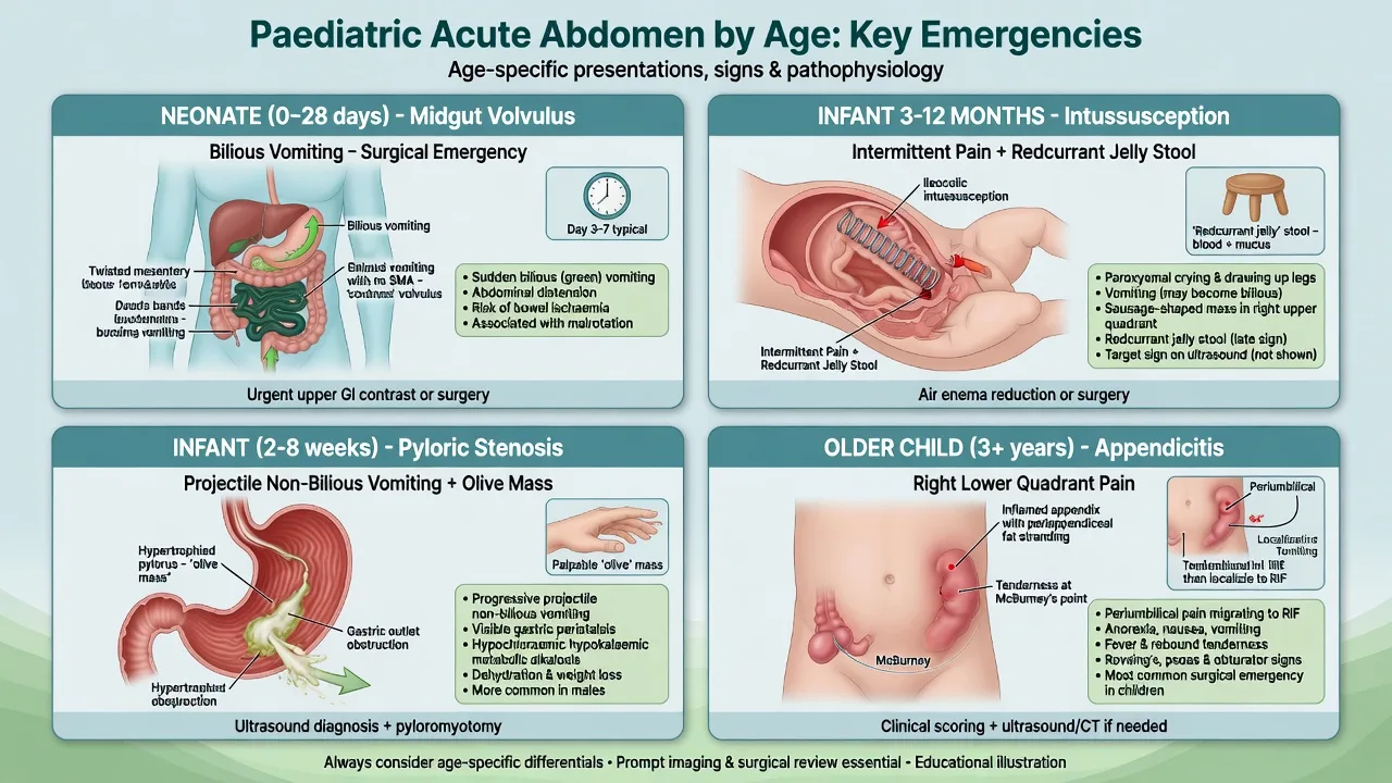
The paediatric acute abdomen is governed by age. Four surgical conditions dominate the emergency department and each has a characteristic window: intussusception in the infant aged three months to two years, midgut volvulus in the newborn and young infant with bilious vomiting, infantile hypertrophic pyloric stenosis at three to six weeks with projectile vomiting, and acute appendicitis as the most common surgical emergency in the child over two but a deceptive, frequently perforated diagnosis in the child under five. The Fellowship candidate must hold all four in mind simultaneously, recognise that the history (intermittent colicky screaming, bilious vomit, projectile non-bilious vomit, refusal to walk) drives the diagnosis more than the examination, and resuscitate with weight-based fluid, decompress with a nasogastric tube, and refer to surgery in parallel with investigation. The medical mimics — constipation, urinary tract infection, mesenteric adenitis and gastroenteritis — are the default for most children, and the art is to prove the dangerous surgical diagnosis rather than to assume the common medical one.[1][3]
An age-based framework
The single most useful triage device is the age of the child, because each surgical emergency clusters into a narrow age band. The candidate who maps the age to the likely diagnosis is rarely surprised at the bedside. [1]
The age window for each surgical diagnosis
The age band not only flags the four cardinal surgical diagnoses but also the broader differential that an ACEM Fellow must hold when the picture does not fit the classic presentation. Each developmental window carries its own cluster of surgical and medical possibilities, and a child outside the expected window for a diagnosis (an intussusception in a ten-year-old, a volvulus in an adolescent) should prompt a deliberate search for a pathological lead point, a structural anomaly, or a diagnosis from an adjacent age band. The table below expands the framework into a working differential by age.[6][10]
Infant (0–1 yr)
- Pyloric stenosis — 2 to 8 weeks, projectile non-bilious vomiting, palpable olive, hypochloraemic hypokalaemic metabolic alkalosis
- Intussusception — 6 months to 2 years, intermittent colicky pain, drawing up legs, sausage-shaped RUQ mass, redcurrant-jelly stool is late
- Malrotation with midgut volvulus — bilious vomiting in the newborn is never normal; upper GI contrast or ultrasound whirlpool sign; Ladd procedure
- Incarcerated inguinal hernia — irreducible groin mass, vomiting, irritability; the leading cause of bowel obstruction in the first year; reduce gently or operate
- Hirschsprung-associated enterocolitis — delayed passage of meconium, abdominal distension, foul explosive stool on rectal exam; a surgical and infectious emergency
Toddler (1–4 yr)
- Meckel diverticulum — the rule of twos; painless brick-red rectal bleeding from ectopic gastric mucosa; confirm with a 99mTc pertechnetate (Meckel) scan
- Intussusception still possible through to 2 years, and any atypical ileoileal intussusception should prompt the search for a pathological lead point
- Appendicitis begins to appear — still atypical, frequently perforated, often diffuse pain with diarrhoea and a limp
- Constipation is now the commonest cause of recurrent pain; exclude obstruction and urinary pathology before attributing
- Urinary tract infection — non-specific in the toddler (vomiting, fever, abdominal pain); always dipstick and culture
Child (5–11 yr)
- Appendicitis — now the dominant surgical diagnosis; the migratory periumbilical-to-RIF pain becomes recognisable; ultrasound first
- Mesenteric adenitis — the commonest mimic; recent or concurrent viral illness, less localised pain, enlarged nodes without an inflamed appendix on ultrasound
- Constipation — faecal loading, palpable left iliac fossa mass, normal inflammatory markers
- Urinary tract infection — dysuria, frequency, foul urine; a right-sided pyelonephritis can masquerade as appendicitis
- Lower-lobe pneumonia and diabetic ketoacidosis may present as abdominal pain — examine the chest, check the glucose
Adolescent (12+ yr)
- Ovarian torsion — sudden severe lower abdominal pain, nausea, vomiting, an enlarged adnexal mass on ultrasound with absent flow; a time-critical surgical diagnosis
- Ectopic pregnancy — every adolescent girl with abdominal pain has a pregnancy test; rupture presents with shock and shoulder-tip pain
- Testicular torsion — acute scrotal pain referred to the abdomen; the testis is high-riding with an absent cremasteric reflex; the 6-hour window
- Biliary disease — gallstones (haemolytic, obesity, female), biliary colic and acute cholecystitis; Murphy sign
- Pancreatitis — epigastric pain radiating to the back, raised lipase; consider trauma, gallstones, drugs and alcohol
The cardinal feature to elicit at each age band
Intussusception
Intussusception is the invagination of a segment of proximal bowel (the intussusceptum) into the lumen of the distal adjacent bowel (the intussuscipiens), dragging the attached mesentery with it. It is the most common cause of intestinal obstruction in infants, with a peak incidence between three months and two years (the classical peak is five to nine months) and a slight male predominance. Approximately 90 per cent of infantile cases are idiopathic and ileocolic, the lead point being hypertrophied Peyer patches of the terminal ileum, often following a viral enteritis (rotavirus, adenovirus). In the child over three, or in any atypical location such as ileoileal intussusception, suspect a pathological lead point — a Meckel diverticulum, a juvenile polyp, an intestinal duplication cyst, a lymphoma, or in Henoch-Schönlein purpura an intramural haematoma.[1]
The pathophysiology is mechanical and progressive. The mesentery drawn into the intussuscipiens becomes oedematous and then venously obstructed; the venous obstruction progresses to arterial compromise, ischaemia, mucosal sloughing (the bloody, mucus stool) and eventual perforation. The diagnosis is made clinically and confirmed by ultrasound. [1]
Clinical presentation
The classic triad is intermittent colicky abdominal pain, vomiting and redcurrant-jelly stool. The pain is dramatic: the child, previously well, suddenly screams, draws the knees up to the abdomen, becomes pale and diaphoretic, then settles and appears normal between episodes, only to repeat the cycle every 15 to 30 minutes. Vomiting follows. Between attacks the child may be remarkably lethargic, which is itself a recognised presentation and can mislead the clinician toward a neurological or infective diagnosis. [1]
Redcurrant-jelly stool is a late sign, present in only about a third of cases at presentation, and reflects mucosal ischaemia and sloughing — its absence does not exclude intussusception, and waiting for it is a fatal error. The palpable sausage-shaped mass is best felt in the right upper quadrant or epigastrium, with an emptiness in the right iliac fossa (Dance sign). The full triad of pain, vomiting and bloody stool is present in fewer than half at presentation.[1]
Investigations
Ultrasound is the first-line and gold-standard imaging modality, with sensitivity above 95 per cent. The transverse view shows the target or donut sign (alternating hypoechoic and hyperechoic rings of oedematous bowel wall and mesenteric fat, typically around 5 cm), and the longitudinal view shows the pseudo-kidney sign. Free fluid, trapped mesenteric lymph nodes or a pathological lead point may be visible. Abdominal X-ray is insensitive early but may show a paucity of bowel gas in the right upper quadrant, a soft-tissue mass, or free air if perforation has occurred. A blood gas and electrolytes assess dehydration and perfusion; a raised lactate signals ischaemia.[1]
Management of intussusception
Resuscitation comes first — the dehydrated, shocked infant needs weight-based 0.9 per cent saline or Hartmann's solution boluses of 10 mL per kilogram, a nasogastric tube for decompression, broad-spectrum antibiotics, and analgesia. The definitive reduction is by image-guided enema — ultrasound-guided saline or pneumatic, or fluoroscopic air enema — which is successful in 80 to 90 per cent of cases. The recurrence rate is approximately 10 per cent, most often within 48 hours, and a single recurrence can be re-reduced; multiple recurrences or a pathological lead point favour surgery. Absolute contraindications to enema reduction are peritonitis, free intraperitoneal air (perforation), severe dehydration or shock, and ascites — these mandate immediate laparotomy with manual reduction or resection of non-viable bowel. [1]
The ED workup of suspected intussusception — the order of operations
- Resuscitate in parallel. Estimate the weight, gain intravenous access, give 10 mL per kilogram boluses of 0.9 per cent saline or Hartmann to perfusion, insert a large-bore nasogastric tube for decompression, give morphine 0.1 mg per kilogram and ondansetron 0.15 mg per kilogram, and take a venous gas and electrolytes for perfusion and dehydration.
- Confirm the diagnosis with ultrasound. The transverse target/donut sign and the longitudinal pseudo-kidney sign confirm the intussusception; look for free fluid, a trapped mesenteric lymph node and a pathological lead point.
- Screen for the contraindications to enema reduction. Peritonitis, free intraperitoneal air (perforation), shock or severe dehydration, and ascites are absolute contraindications — these children go straight to theatre.
- Pneumatic (air) enema reduction under fluoroscopy. With the surgical team on standby, insufflate air per rectum to a controlled pressure (typically 80 to 120 mmHg, held for up to three minutes and repeated up to three attempts). Ultrasound-guided saline reduction is the alternative where air is unavailable. Success is signalled by free reflux of gas into the small bowel and resolution of the mass.
- Observe after reduction. Admit for 24 to 48 hours; the recurrence rate is about 10 per cent, most often within 48 hours, and a single recurrence can be re-reduced.
- Surgical referral for failure, recurrence or a pathological lead point. A failed reduction, peritonitis, recurrent intussusception beyond a single episode, or a child over three (where a Meckel diverticulum or polyp is the lead point) goes to laparotomy for manual reduction or resection of non-viable bowel.
Malrotation and midgut volvulus
Midgut malrotation is an abnormal fixation of the midgut during the tenth week of embryological rotation, leaving a narrow mesenteric base and peritoneal (Ladd) bands that cross and can obstruct the duodenum. The lethal complication is midgut volvulus — a clockwise twist of the small bowel around the superior mesenteric artery, producing acute obstruction and strangulation of the entire midgut. It is a surgical emergency measured in hours: delay converts to bowel necrosis and lifelong short-gut syndrome.[4][5]
Malrotation affects approximately 1 in 500 live births. Around 75 per cent of symptomatic patients present in the first month of life and most within the first week, but a minority present later with intermittent bilious vomiting, chronic abdominal pain, or malabsorption from episodes of partial volvulus. [1]
Clinical presentation
The cardinal sign is bilious vomiting. Bile-stained (green) vomit in a newborn or young infant indicates obstruction below the ampulla of Vater and is malrotation with volvulus until proven otherwise — it is never normal. The child may initially appear surprisingly well between episodes, then deteriorate rapidly as the bowel ischaemia progresses, passing to bloody stools, peritonitis and shock. Less acute presentations include failure to thrive, intermittent colicky pain and recurrent bilious vomiting. [1]
[1]Investigations
An upper gastrointestinal contrast study is the diagnostic gold standard, demonstrating the duodenojejunal flexure to the right of the vertebral body or at a lower level than the pylorus, and in frank volvulus the corkscrew or bird's-beak appearance of the twisted duodenum. Ultrasound is increasingly first-line, showing the whirlpool sign (the superior mesenteric vein wrapping around the superior mesenteric artery) and inversion of the normal SMA–SMV relationship, with high accuracy in experienced hands.[4] The abdominal X-ray is frequently normal early — a normal film does not exclude volvulus — and may show a double-bubble of duodenal obstruction only later.
Management of volvulus
This is a time-critical surgical emergency. Resuscitate in parallel with referral: a large-bore nasogastric tube for decompression, aggressive weight-based fluid resuscitation (repeated 10 mL per kilogram boluses of 0.9 per cent saline or Hartmann's) for the shocked, ischaemic child, broad-spectrum antibiotics (ceftriaxone plus metronidazole), and immediate surgical referral for a Ladd procedure — untwisting of the volvulus, division of the Ladd bands, widening of the mesenteric base, resection of any non-viable bowel, and an incidental appendicectomy, leaving the bowel in a position of non-rotation.[5]
Infantile hypertrophic pyloric stenosis
Infantile hypertrophic pyloric stenosis is the progressive hypertrophy of the circular muscle of the pylorus, producing a gastric outlet obstruction. It presents at three to six weeks of life, is four to five times commoner in males, and carries a familial tendency. The progressive obstruction generates a stereotyped biochemical picture that the Fellowship examiner tests relentlessly.[3]
Clinical presentation
The vomiting is non-bilious and projectile — the cardinal feature — beginning as effortless possetting and escalating over days to forceful ejection across the room. Characteristically the infant is hungry immediately after the vomit and takes the feed again. Without treatment the child dehydrates, loses weight and becomes alkalotic. On examination, visible gastric peristalsis crosses the upper abdomen, and the hypertrophied pylorus is palpable as a firm mobile olive in the epigastrium or right upper quadrant, best felt during or just after a feed when the abdominal wall is relaxed; the test is operator-dependent and may be missed. [1]
The metabolic derangement — hypochloraemic hypokalaemic metabolic alkalosis with paradoxical aciduria
Persistent loss of gastric hydrochloric acid generates a hypochloraemic, hypokalaemic metabolic alkalosis. The alkalosis is the textbook blood gas answer: a high pH, high bicarbonate, high pCO2 from respiratory compensation, low chloride and low potassium. The hypokalaemia is multifactorial — secondary hyperaldosteronism from volume depletion, intracellular shift, and some gastric potassium loss. The urinary sodium is driven low by aldosterone, and in the volume-depleted, hypokalaemic state the distal tubule excretes hydrogen ions in preference to potassium to reabsorb sodium and conserve volume, producing paradoxical aciduria — an acidic urine despite a systemic alkalosis.[3]
[1]Investigations
Ultrasound is the diagnostic gold standard: pyloric muscle wall thickness more than 4 mm and pyloric channel length more than 16 mm confirm the diagnosis. A blood gas and urea and electrolytes define the metabolic derangement and guide correction; the chloride, potassium and bicarbonate are followed to readiness for surgery.[3]
Management of pyloric stenosis — correct the chemistry, then operate
Pyloric stenosis is not a surgical emergency — it is a metabolic emergency. Operating on an alkalotic, hypokalaemic infant risks postoperative apnoea and arrhythmia. Resuscitation precedes surgery: a nasogastric tube (large-bore) for decompression, and intravenous fluid to correct the deficit, typically 0.9 per cent saline with potassium chloride (KCl) supplemented once urine output is established, with the chloride, potassium and bicarbonate trended to a chloride above 100 mmol per litre and bicarbonate below 30 mmol per litre. Only then is a Ramstedt pyloromyotomy performed — a longitudinal incision through the hypertrophied pyloric muscle down to the mucosa, leaving the mucosa intact.[3]
Pyloric stenosis — correct the chemistry, then the Ramstedt pyloromyotomy
- Make the infant nil by mouth and decompress. Insert a large-bore nasogastric tube and leave it on free drainage with intermittent suction; this halts the ongoing gastric loss and the progressive alkalosis.
- Resuscitate and correct the chemistry — the metabolic, not the surgical, emergency. Give intravenous 0.9 per cent saline to re-expand the volume; once urine output is established, add potassium chloride (10 to 20 mmol per litre of fluid) to replace the deficit. The goals are a chloride over 100 mmol per litre, a potassium over 3.5 mmol per litre and a bicarbonate below 30 mmol per litre — the markers of a safe anaesthetic.
- Watch for paradoxical aciduria to resolve. As the volume and the chloride are restored, the distal tubule stops exchanging sodium for hydrogen and resumes excreting bicarbonate; the urine becomes alkaline, signalling correction.
- Confirm the diagnosis if not already done. Ultrasound — pyloric muscle wall over 4 mm and channel over 16 mm — confirms the diagnosis; a contrast study is not needed.
- Perform the Ramstedt pyloromyotomy once the chemistry is corrected. A longitudinal incision through the hypertrophied pyloric muscle down to the mucosa, leaving the mucosa intact; performed open or laparoscopically. Feeds are advanced within hours of surgery.
- Do not delay surgery for the chemistry — but do not operate until it is corrected. The correction typically takes 24 to 72 hours; the only indication for urgent operation is a perforation, which does not occur in uncomplicated pyloric stenosis.
Bilious (green) vomit
- Obstruction BELOW the ampulla of Vater — bile is reaching the stomach and cannot pass
- Surgical emergency until proven otherwise: malrotation with midgut volvulus is the feared cause in the newborn
- Also: duodenal atresia (the double-bubble), annular pancreas, Hirschsprung disease, a high small-bowel obstruction
- Act immediately — upper GI contrast or ultrasound whirlpool sign, nasogastric decompression, fluid resuscitation, surgical referral
Non-bilious vomit
- Obstruction ABOVE the ampulla of Vater, or a functional gastric-outlet problem
- Pyloric stenosis is the classic — projectile non-bilious vomiting at 2 to 8 weeks with a palpable olive
- Also: gastro-oesophageal reflux, overfeeding, gastritis, a gastric web
- Less immediately surgical, but pyloric stenosis is a metabolic emergency — correct the alkalosis before the Ramstedt pyloromyotomy
Acute appendicitis in children — the atypical presentation
Appendicitis is the most common surgical emergency in children over two years of age, but it is uncommon under two — and the younger the child, the more atypical the presentation and the higher the perforation rate. In the under-five age group over 50 per cent have perforated at presentation, because the diagnosis is delayed by the atypical features and the child's inability to localise pain. [1]
The classic adult sequence of migratory periumbilical pain to the right iliac fossa with anorexia and low-grade fever is often absent in the young child. Instead the child may present with diffuse abdominal pain, prominent vomiting, diarrhoea (from a pelvic appendix irritating the rectum), irritability, refusal to walk or a limp, and a high fever — and the diagnosis is missed as gastroenteritis. Tenderness on coughing, hopping or percussion is a useful equivalent of rebound in the cooperative child. [1]
The Pediatric Appendicitis Score
The Pediatric Appendicitis Score (Samuel) is the bedside risk-stratification tool validated for the child with equivocal right iliac fossa pain, summarised below. A score of 6 or above supports appendicitis (high sensitivity, moderate specificity) and warrants imaging and surgical review; a low score does not exclude appendicitis, and clinical judgement governs.[2]
| Component | Points |
|---|---|
| Nausea or vomiting | 1 |
| Migration of pain to the right iliac fossa | 1 |
| Tenderness in the right iliac fossa | 2 |
| Pain elicited by cough, percussion or hopping | 2 |
| Anorexia | 1 |
| Leukocytosis (white cell count over 10 × 10⁹ per litre) | 1 |
| Left shift (neutrophilia) | 1 |
| Total | 9 |
Interpretation of the Pediatric Appendicitis Score
Management of paediatric appendicitis
Ultrasound is the first-line imaging to spare the child ionising radiation, with CT reserved for the equivocal, complicated or obese child. Management is analgesia (morphine 0.1 mg per kilogram intravenously), antiemetic (ondansetron 0.15 mg per kilogram), fluid resuscitation, empirical antibiotics (ceftriaxone 50 mg per kilogram plus metronidazole), and surgical referral for appendicectomy, with an appendiceal mass or abscess managed conservatively by drainage and interval appendicectomy. [1]
Meckel diverticulum — the rule of twos and painless rectal bleeding
Meckel diverticulum is the most common congenital anomaly of the gastrointestinal tract, a true diverticulum (containing all bowel-wall layers) arising from failure of obliteration of the vitelline (omphalomesenteric) duct. It is the surgical maxim of the rule of twos: present in about 2 per cent of the population, located within 2 feet (60 cm) of the ileocaecal valve, about 2 inches (5 cm) long, twice as common in males, and most commonly symptomatic before the age of 2 years. The complications — bleeding, obstruction, intussusception (as a pathological lead point), and diverticulitis — overlap with the surgical abdomen at every age.[6]
The classic presentation in the toddler is painless, brisk, brick-red (maroon) lower gastrointestinal bleeding, the result of acid secretion by ectopic gastric mucosa lining the diverticulum causing a contiguous ileal mucosal ulcer. Unlike an upper-GI bleed the blood is bright or maroon (not melaena), and unlike infectious colitis there is no diarrhoea, fever or tenesmus — the child is typically pain-free between bleeds. Obstruction (from an associated fibrous band, volvulus around the diverticulum, or intussusception with the diverticulum as the lead point) and Meckel diverticulitis (mimicking appendicitis, but the pain is periumbilical or lower) are the other modes of presentation. [1]
The diagnostic test is the 99mTc pertechnetate scan (the Meckel scan), which is taken up by the ectopic gastric mucosa and lights up the diverticulum. It is sensitive in the actively bleeding child with gastric mucosa but can be negative in the non-bleeding presentation or when no gastric mucosa is present; a nasogastric aspirate positive for blood and a tagged red-cell scan are adjuncts for the brisk bleed. Management of the symptomatic diverticulum is surgical — a segmental small-bowel resection with primary anastomosis or a diverticulectomy.[6]
[1] [1]Incarcerated inguinal hernia
An incarcerated inguinal hernia is the leading cause of intestinal obstruction in the first year of life and a surgical emergency that the Fellowship candidate must not overlook. The infant or child presents with an irreducible groin mass, irritability, vomiting (initially non-bilious, becoming bilious with bowel obstruction), and signs of obstruction; the great risk is strangulation — ischaemia and necrosis of the incarcerated bowel, visible as a tense, tender, discoloured mass with signs of peritonitis and systemic toxicity.[11]
The hernia is reduced by gentle, sustained taxis after analgesia and sedation, with the child in the Trendelenburg position, and is successful in the majority if attempted early — within the first few hours and before strangulation. Contraindications to manual reduction are the signs of strangulation (a tense, discoloured, tender mass, peritonitis, or systemic toxicity), which mandate immediate operative reduction and bowel assessment. A successfully reduced hernia is repaired electively within days to prevent recurrence; an irreducible or strangulated hernia goes to theatre. The dictum is: reduce if early and uncomplicated, operate if strangulated or irreducible.[11]
[1]The adolescent acute abdomen — torsion, ectopic, biliary and pancreatitis
As the child moves into adolescence the differential expands to incorporate the reproductive tract and the adult-type hepatobiliary and pancreatic pathologies. The cardinal rules for the adolescent with abdominal pain are three: examine the scrotum in every boy (testicular torsion refers pain to the abdomen), do a pregnancy test in every girl (a ruptured ectopic kills), and measure a lipase for the epigastric pain radiating to the back. Each of the following is a time-critical surgical diagnosis that the emergency physician must not miss. [1]
Ovarian torsion
Ovarian torsion is the twisting of the ovary (and often the tube) on its vascular pedicle, producing venous then arterial occlusion and ischaemic necrosis of the adnexa. It presents with sudden, severe, unilateral lower abdominal pain with nausea and vomiting, often with a prior history of similar self-limiting episodes (intermittent torsion). The pain may radiate to the groin, flank or back. Ultrasound with Doppler is the first-line investigation: an enlarged, oedematous ovary with a peripheral follicular pattern and reduced or absent venous and arterial flow is characteristic, but the presence of flow does not exclude torsion (intermittent detorsion), and the diagnosis ultimately rests on the clinical picture and the surgical evaluation. Management is urgent diagnostic laparoscopy with detorsion and oophoropexy, with ovarian preservation if viable.[8]
Ectopic pregnancy
Every adolescent girl with abdominal pain, vaginal bleeding, syncope or atypical gastrointestinal symptoms has a urine beta-hCG. A positive pregnancy test with abdominal pain is an ectopic until a proven intrauterine pregnancy is demonstrated on transvaginal ultrasound. Rupture presents with haemodynamic collapse, shoulder-tip pain (from diaphragmatic irritation by the haemoperitoneum) and cervical motion tenderness; the treatment of the ruptured ectopic is simultaneous resuscitation and theatre, while the unruptured stable ectopic may be managed surgically or with methotrexate. [1]
Testicular torsion
Testicular torsion is the twisting of the spermatic cord, producing testicular ischaemia that becomes irreversible within six to eight hours. It presents with acute unilateral scrotal pain (often referred to the abdomen or groin, which is why every boy with abdominal pain has a scrotal examination), nausea and vomiting, a high-riding testis with a horizontal lie and an absent cremasteric reflex. The cremasteric reflex is the single most sensitive sign. Colour Doppler ultrasound demonstrates reduced or absent flow, but the diagnosis is clinical and a suspected torsion in a boy within the salvage window goes straight to theatre without waiting for imaging. The TWIST score (Testicular Workup for Ischaemia and Suspected Torsion) — hard testis, absent cremasteric reflex, nausea or vomiting, high-riding testis, and acute scrotal pain — is a validated bedside risk tool; a high score mandates immediate surgical exploration.[7]
[1]Biliary disease
Gallstones in the adolescent are associated with haemolytic disease (sickle cell, hereditary spherocytosis), obesity, female sex, and a family history. Biliary colic presents with post-prandial right upper quadrant pain; acute cholecystitis adds fever, a positive Murphy sign and a raised white cell count. Ultrasound is the first-line imaging (gallstones, wall thickening, a positive sonographic Murphy sign). Management is analgesia, antibiotics and early surgical referral for cholecystectomy. A complication specific to the child is gallstone pancreatitis and gallstone ileus, both of which extend the workup. [1]
Acute pancreatitis
Acute pancreatitis in the child presents with epigastric pain radiating to the back, nausea and vomiting, and a serum lipase at least three times the upper limit of normal. The common causes differ from the adult (gallstones, alcohol): in children the leading causes are biliary disease, trauma (including non-accidental injury), drugs (valproate, L-asparaginase, corticosteroids, azathioprine), infections and idiopathic disease. The NASPGHAN criteria define the diagnosis by two of three features (the characteristic pain, a lipase three times the upper limit, or imaging consistent with pancreatitis). Management is aggressive intravenous fluid resuscitation, analgesia, early enteral nutrition (within 72 hours, which supersedes the historical nil-by-mouth approach), and treatment of the cause.[9]
Qin & Qu — the TWIST score meta-analysis (J Urol 2022)
Systematic review and meta-analysis of the Testicular Workup for Ischaemia and Suspected Torsion score
Population: Children and adolescents presenting with acute scrotal pain across the included studies
Key finding
The TWIST score had a high pooled sensitivity and specificity for testicular torsion; a score of 5 (high risk) carried a very high positive likelihood ratio supporting immediate surgical exploration without imaging, while a score of 0 to 2 (low risk) safely supported discharge with follow-up. The intermediate group (3 to 4) required urgent Doppler ultrasound.
Practice change
The TWIST score is a validated bedside risk-stratification tool for the acute scrotum; it does not replace clinical judgement but supports the decision to operate immediately in the high-scoring boy within the 6-hour salvage window.
Ovarian torsion
- Adolescent girl; sudden severe unilateral lower abdominal pain with nausea and vomiting
- Enlarged oedematous adnexa with reduced or absent flow on Doppler — but flow may be present in intermittent torsion
- Pregnancy test to exclude ectopic; urgent diagnostic laparoscopy with detorsion and oophoropexy
- A surgical emergency — the longer the delay, the lower the ovarian salvage rate
Ectopic pregnancy
- Adolescent girl with abdominal pain, vaginal bleeding or syncope; a positive beta-hCG is the trigger
- No intrauterine gestation on transvaginal ultrasound with a positive test; rupture gives shock and shoulder-tip pain
- Ruptured — simultaneous resuscitation and theatre; unruptured stable — surgery or methotrexate
- The leading cause of first-trimester maternal death; every adolescent girl with abdominal pain has a pregnancy test
Testicular torsion
- Adolescent boy; acute unilateral scrotal pain referred to the abdomen, nausea and vomiting
- High-riding horizontal testis, absent cremasteric reflex (the most sensitive sign); reduced flow on Doppler
- Within the 6-hour salvage window — straight to theatre, no imaging; the TWIST score guides the decision
- A urological emergency — the salvage rate falls steeply after 6 hours and is negligible after 24
Appendicitis
- Either sex; migratory periumbilical-to-RIF pain, anorexia, low-grade fever
- Non-compressible appendix over 6 mm on ultrasound; the PAS guides imaging
- Ultrasound first, analgesia, antibiotics, surgical referral for appendicectomy
- Still the commonest surgical emergency in the over-two — do not let the reproductive differential displace it
Differential diagnosis — the medical mimics
Most children with abdominal pain in the emergency department have a medical cause, and the four surgical conditions above must be distinguished from them. The common mimics are constipation, urinary tract infection and mesenteric adenitis, with gastroenteritis, Hirschsprung-associated enterocolitis, an incarcerated inguinal hernia, testicular torsion (referred to the abdomen) and diabetic ketoacidosis the dangerous or deceptive further considerations. Mesenteric adenitis is the commonest mimic of appendicitis in the school-age child — a recent or concurrent viral illness, less localised pain, and enlarged mesenteric nodes without an inflamed appendix — and is a diagnosis of exclusion made only after the appendix has been cleared.[10]
Constipation
- Infrequent, hard stools; a palpable faecal-loaded colon, often in the left iliac fossa; no systemic features
- Normal inflammatory markers and urinalysis; a normal or faecal-loaded abdomen on imaging
- Disimpaction and oral macrogol; no surgical role unless Hirschsprung suspected
- The commonest cause of abdominal pain in children, but exclude obstruction before attributing pain to it
Urinary tract infection
- Fever, dysuria, frequency, foul-smelling urine; in the infant, non-specific — irritability, poor feeding, vomiting
- Positive urinalysis (leucocytes and nitrites) and a significant culture; bacteraemia possible
- Antibiotics guided by age and severity; ceftriaxone for the toxic or young infant
- A right-sided pyelonephritis can masquerade as appendicitis; the pelvic appendix can cause dysuria in reverse
Mesenteric adenitis
- A recent or concurrent viral upper respiratory or gastrointestinal illness; pain that does not migrate and is less localised
- Mildly raised or normal white cell count; enlarged mesenteric nodes on ultrasound without an inflamed appendix
- Supportive care, analgesia and hydration; self-limiting
- The commonest mimic of appendicitis in the child; a diagnosis of exclusion after the appendix is excluded
Gastroenteritis
- Vomiting and diarrhoea predominate over pain; diffuse, cramping, non-localised; often a contact history
- Normal inflammatory markers; no focal peritonism; the pain is colicky and shifting
- Oral or intravenous rehydration; antiemetics; usually self-limiting
- Beware the child labelled gastroenteritis who has bilious vomiting or colicky screaming — intussusception and volvulus kill the child sent home as a stomach bug
Bedside assessment
Assessment follows ABCDE, with attention to the paediatric specifics. Estimate the weight before any drug — a Broselow tape or the formula (age plus four) times two gives the weight in kilograms for a child between one and ten years. The circulation is assessed for dehydration and shock (capillary refill, peripheral perfusion, tearfulness of the mucous membranes, reduced urine output). The abdomen is examined for distension, tenderness, guarding, masses and visible peristalsis. The scrotum is examined in every boy with abdominal or groin pain — testicular torsion refers pain to the abdomen — and the inguinal canals for an irreducible hernia. Rectal examination is rarely needed in the child. In the adolescent girl, consider a pregnancy test and ovarian pathology. [1]
Immediate management and resuscitation
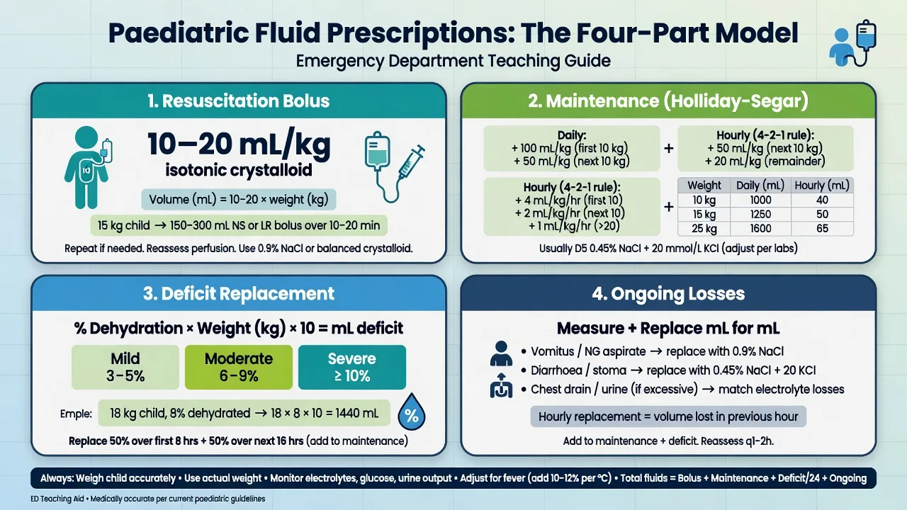
The four conditions share a common resuscitation thread. Establish intravenous access, give weight-based isotonic fluid — 0.9 per cent saline or Hartmann's solution in 10 mL per kilogram boluses titrated to perfusion, with potassium chloride added to the maintenance once urine output is confirmed — and place a nasogastric tube (large-bore for pyloric stenosis and volvulus) for decompression. Analgesia is generous and does not mask the diagnosis: morphine 0.1 mg per kilogram intravenously for surgical pain, with ondansetron 0.15 mg per kilogram for vomiting. Refer to the paediatric surgical team in parallel with the investigation, not after it. [1]
The weight-based doses for the paediatric acute abdomen
Imaging strategy
Ultrasound is the default first-line modality across all four conditions in children, avoiding ionising radiation and answering the specific question: target sign for intussusception, whirlpool sign and SMA–SMV relationship for malrotation, pyloric muscle thickness and channel length for pyloric stenosis, and a non-compressible appendix over 6 mm for appendicitis. Upper GI contrast remains the gold standard for malrotation when ultrasound is equivocal. CT is reserved for the equivocal, complicated or obese child in whom ultrasound is non-diagnostic, and for the adult-type presentation. [1]
ANZ practice note. The Royal Children's Hospital Melbourne and the Paediatric Research in Emergency Departments International Collaborative (PREDICT) networks endorse an ultrasound-first, radiation-sparing pathway for paediatric abdominal emergencies. Pneumatic or ultrasound-guided enema reduction of intussusception is standard where paediatric surgical and radiological expertise exists, with a surgical team on standby. The threshold for surgical referral is low and the senior decision-maker is the paediatric surgeon, not the emergency registrar. [1]
Complications and pitfalls
The complications are ischaemia, perforation and peritonitis for intussusception and volvulus; short-gut syndrome after extensive midgut infarction in delayed volvulus; the metabolic derangement and postoperative apnoea of uncorrected pyloric stenosis; and the perforation and intra-abdominal abscess of delayed appendicitis. The principal pitfalls are anchoring on gastroenteritis in the vomiting infant with intussusception or volvulus; relying on the presence of redcurrant-jelly stool to diagnose intussusception; trusting a normal abdominal X-ray to exclude volvulus; sending an alkalotic, hypokalaemic infant straight to theatre without correcting the chemistry; and missing the atypical appendicitis in the under-five who presents with diarrhoea or a limp. [1]
[1]Prognosis and disposition
All four conditions require admission under the surgical team. Pyloric stenosis corrected and pyloromyotomised has an excellent prognosis with a short hospital stay. Intussusception reduced by enema is observed for recurrence and discharged within 24 to 48 hours. Volvulus is time-critical — the prognosis depends on the viability of the midgut at laparotomy, and a delay of hours converts a healthy child into one dependent on long-term parenteral nutrition. Paediatric appendicitis treated promptly has a low mortality, but perforation extends the stay and the complication rate. The medical differentials — constipation, urinary tract infection, mesenteric adenitis — are discharged with a clear safety-net: return immediately with bilious or bloody vomiting, worsening or localising pain, fever, drowsiness, or refusal to drink. [1]
Special populations
The newborn with bilious vomiting is the highest-risk child — malrotation with volvulus is assumed until excluded, and a normal examination and X-ray do not wait. The infant under one presenting with vomiting or colicky pain has intussusception until proven otherwise. The child under five with appendicitis presents atypically and is frequently perforated at first contact. The adolescent girl with abdominal pain has a pregnancy test and ovarian pathology excluded. The immunocompromised or neutropenic child may present with muted signs and typhlitis (neutropenic enterocolitis) rather than the standard surgical diagnoses. [1]
Evidence and regional guidelines
The contemporary evidence base comprises the World Society of Emergency Surgery guidelines on intussusception, the PREDICT network pathways for paediatric abdominal emergencies, the American Pediatric Surgical Association Outcomes and Evidence Based Practice Committee guidance on appendicitis, and the Royal Children's Hospital Melbourne clinical practice guidelines. These converge on ultrasound-first imaging, weight-based resuscitation, nasogastric decompression, a low threshold for surgical referral, and the principle that the metabolic derangement of pyloric stenosis is corrected before the Ramstedt pyloromyotomy.[1][2][3]
Model answer — the ED workup of suspected intussusception
A nine-month-old with intermittent screaming, drawing up of the legs, vomiting and a palpable sausage-shaped right upper quadrant mass has intussusception. Estimate the weight (age plus four times two equals 10 kg). Resuscitate with 0.9 per cent saline 10 mL per kilogram boluses, insert a nasogastric tube, give morphine 0.1 mg per kilogram and ondansetron 0.15 mg per kilogram, and take a blood gas for perfusion. Confirm with ultrasound (target or donut sign). In the absence of peritonitis, shock or perforation, arrange image-guided enema reduction (success 80 to 90 per cent, recurrence about 10 per cent) with the surgical team on standby; a peritonitic or shocked child goes straight to theatre. Admit for observation. The pathological lead point is a hypertrophied Peyer patch in the infant.
SAQ — Atypical paediatric appendicitis in the under-five with a limp and diarrhoea
10 minutes · 10 marks
A 4-year-old boy is brought to the emergency department by his mother with two days of intermittent abdominal pain, four episodes of loose stool, and a one-day history of refusing to walk, preferring to be carried. He is miserable, clutches his abdomen intermittently, and points to his right thigh when asked where it hurts. On arrival: T 38.8 degrees Celsius, HR 132, BP 98/62, RR 26, SpO2 96 per cent on room air. The abdomen is mildly distended with diffuse tenderness greatest in the right iliac fossa and pelvis, voluntary guarding, and a positive cough test. He refuses to hop. Capillary refill is 3 seconds. Bedside glucose 5.8 mmol/L. The urinalysis is negative for nitrites and leucocytes.
SAQ — Intussusception in the infant: colicky screaming, a sausage mass, and the enema decision
10 minutes · 10 marks
A 9-month-old, previously well boy is brought to the emergency department with a 12-hour history of intermittent episodes of screaming, drawing his knees up to his chest, and going pale and diaphoretic, each lasting two to three minutes and recurring every 20 minutes. Between episodes he is listless. He has vomited twice, non-bilious. The mother reports one episode of stool mixed with blood and mucus on the nappy. On arrival between attacks: T 37.6 degrees Celsius, HR 118, BP 88/56, RR 32, capillary refill 2 seconds; the abdomen is soft with a palpable sausage-shaped mass in the right upper quadrant and a sense of emptiness in the right iliac fossa (Dance sign). Bedside glucose 5.2 mmol/L. Weight is estimated at 9 kg.
Exam pearls
- Bilious vomiting in a newborn is malrotation with midgut volvulus until proven otherwise — a green vomit is a surgical emergency, full stop.
- Redcurrant-jelly stool is a late sign of intussusception; its absence does not exclude the diagnosis.
- Pyloric stenosis is a metabolic, not a surgical, emergency — correct the hypochloraemic hypokalaemic alkalosis before the Ramstedt pyloromyotomy.
- Paradoxical aciduria in pyloric stenosis is a marker of established, severe volume and potassium depletion.
- The child under five with appendicitis presents atypically — diarrhoea, a limp, refusal to hop — and over half have perforated at presentation.
- A normal abdominal X-ray does not exclude volvulus; ultrasound (whirlpool sign) or upper GI contrast is required.
- Estimate the weight (Broselow or age plus four times two) before any drug in a child.
- Painless brick-red rectal bleeding in a well toddler is a Meckel diverticulum until the 99mTc pertechnetate scan says otherwise — do not dismiss it as gastroenteritis.
- The colour of the vomit is the most discriminating sign: bilious (green) means obstruction below the ampulla of Vater (volvulus); non-bilious projectile at 2 to 8 weeks is pyloric stenosis.
- Examine the scrotum in every boy with abdominal or groin pain — testicular torsion refers pain to the abdomen, and the cremasteric reflex is the most sensitive sign.
- A suspected testicular torsion within the six-hour salvage window goes straight to theatre without waiting for Doppler — the TWIST score (5 points) supports immediate exploration.
- Every adolescent girl with abdominal pain has a pregnancy test — a ruptured ectopic is the leading cause of first-trimester maternal death.
- An incarcerated inguinal hernia is the leading cause of bowel obstruction in the first year — reduce if early and uncomplicated, operate if strangulated or irreducible.
- Pneumatic (air) enema reduction of intussusception under fluoroscopy uses 80 to 120 mmHg for up to three attempts, with the surgical team on standby — it succeeds in 80 to 90 per cent.
- Paediatric pancreatitis is rarely alcoholic — think biliary disease, trauma (including non-accidental injury), and drugs (valproate, L-asparaginase); feed early.
- Doppler flow in an enlarged ovary does not exclude ovarian torsion — intermittent detorsion can restore flow, and the diagnosis is clinical and surgical. [1]
Red flags
[1]References
- [1]Hwang JY, Yoon H, Kim KM, Lee JS. Current diagnosis and image-guided reduction for intussusception in children Clin Exp Pediatr, 2023.PMID 35798026
- [2]Samuel M. Pediatric appendicitis score J Pediatr Surg, 2002.PMID 12037754
- [3]Rich BS, Dolgin SE. Hypertrophic Pyloric Stenosis Pediatr Rev, 2021.PMID 34599053
- [4]Nguyen HN, Navarro OM, Kahn E, et al. Diagnostic Performance of Ultrasound for Diagnosing Midgut Malrotation and Volvulus in Children: A Multiinstitutional Retrospective Review AJR Am J Roentgenol, 2025.PMID 40499019
- [5]Filion L, Puligandla PS, Baird R, et al. Infant malrotation with midgut volvulus: A retrospective review of clinical presentation and delays in care at a Canadian tertiary paediatric centre Paediatr Child Health, 2025.PMID 41049711
- [6]Brown RL, Azizkhan RG. Gastrointestinal bleeding in infants and children: Meckel's diverticulum and intestinal duplication Semin Pediatr Surg, 1999.PMID 10573430
- [7]Qin KR, Qu LG. Diagnosing with a TWIST: Systematic Review and Meta-Analysis of a Testicular Torsion Risk Score J Urol, 2022.PMID 35238603
- [8]Scheier E. Diagnosis and Management of Pediatric Ovarian Torsion in the Emergency Department: Current Insights Open Access Emerg Med, 2022.PMID 35770141
- [9]Abu-El-Haija M, Kumar S, Szabo F, et al. Classification of Acute Pancreatitis in the Pediatric Population: Clinical Report From the NASPGHAN Pancreas Committee J Pediatr Gastroenterol Nutr, 2017.PMID 28333771
- [10]Helbling R, Conficconi E, Wyttenbach M, et al. Acute Nonspecific Mesenteric Lymphadenitis: More Than No Need for Surgery Biomed Res Int, 2017.PMID 28261620
- [11]Abdulhai SA, Glenn IC, Ponsky TA. Incarcerated Pediatric Hernias Surg Clin North Am, 2017.PMID 27894423