Paeds · fetal-neonatal-and-perinatal
Gestational age assessment and preterm classification
Also known as Gestational age assessment · Preterm classification · Newborn maturity assessment · Gestational age scoring · Size for gestational age
Fellowship guide to assigning and classifying gestational age — first-trimester ultrasound dating, the New Ballard Score, the gestational-age and birthweight bands, size-for-gestational-age, and the corrected-age framework that flows from each label, with regional pathway differences and exam pearls.
On this page & tools
Your progress
Saved locally on this device.
Practise this topic
Target exams
Red flags
Life stages
Care settings
Clinical exam formats
Board mappings
Related topics
- Fetal assessment, prenatal screening and counselling
- Preterm infant: immediate and longitudinal care
- Extremely preterm infant: viability and periviable counselling
- Late preterm infant: risks and follow-up
- Fetal growth restriction and small-for-gestational-age infant
- Normal growth from fetal life through adolescence
Overview & Definition
Picture the delivery room at two in the morning. A baby is born weighing 2100 g, and the question that will shape every decision for the next two years is deceptively simple: how old, in weeks, is this baby? The answer is not the weight, and it is not always obvious from the appearance — it is the gestational age, the completed weeks from the first day of the mother's last menstrual period to the day of birth. [6]
Gestational age is the organising principle of neonatal medicine because it predicts, better than any other single variable, which organ systems are immature enough to fail. A 26-week infant and a 34-week infant are different patients — different lung, brain, gut and temperature risk — and the plan flows from the band they fall into. Establishing that number accurately, and reconciling antenatal with postnatal estimates when they disagree, is one of the first high-stakes tasks the paediatrician owns. [6] [1]
Three related ages travel with gestational age and the candidate must keep them separate. Postmenstrual age is the gestational age at birth plus the chronological age, used in the first weeks to time screening such as the first retinopathy examination around 31 to 34 weeks postmenstrual age. Chronological age is the time since birth. Corrected age is the chronological age minus the number of weeks the baby was born early, and it is the age you use to plot growth and interpret milestones for the first two years. [18] [19]
Classification
Classification by gestational age is the vocabulary the whole of neonatology is written in, and it is built on completed weeks. The preterm bands descend in step with organ maturation. Extremely preterm is less than 28 weeks. Very preterm is 28 to 31 weeks and six days. Moderate preterm is 32 to 33 weeks and six days. Late preterm is 34 to 36 weeks and six days. [10]
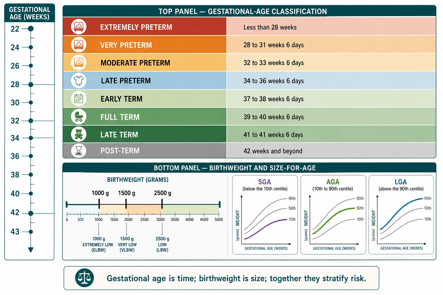
Term itself was once a single block from 37 to 42 weeks, but the Defining Term Pregnancy Workgroup split it because outcomes are not uniform across that range. Early term is 37 to 38 weeks and six days. Full term is 39 to 40 weeks and six days. Late term is 41 to 41 weeks and six days. Post-term is 42 weeks and beyond. The split matters because early-term and post-term infants each carry their own morbidity compared with full term. [10]
Birthweight categories describe size and travel alongside, but never replace, the gestational-age label. Low birthweight is under 2500 g. Very low birthweight is under 1500 g. Extremely low birthweight is under 1000 g. These thresholds are useful for population reporting and for triggering some screening, but a 2100 g term infant is not preterm. [4] [16]
Extremely preterm
< 28 weeks
- Highest respiratory, brain, gut and eye risk
- Tertiary intensive care from delivery room
- Cranial ultrasound and ROP screening expected
- Corrected-age follow-up to school age
Very preterm
28 – 31+6 weeks
- Often surfactant and caffeine pathway
- Special or intensive care
- Cranial ultrasound screening
- Structured neurodevelopmental follow-up
Late preterm
34 – 36+6 weeks
- Largest preterm group by number
- Feeding, temperature and glucose issues dominate
- Real readmission and morbidity risk
- Easily mistaken for 'almost term'
The third axis is size-for-gestational-age, and it is the one that most often catches candidates out. Small-for-gestational-age is a weight below the tenth centile for gestational age. Appropriate-for-gestational-age sits between the tenth and ninetieth centiles. Large-for-gestational-age is above the ninetieth centile. The point is that size-for-age is always read against gestational age, never against a fixed weight — the same 1800 g means very different things at 32 weeks and at 40 weeks. [4] [18]
[10] [16]Epidemiology & Risk Factors
The global preterm birth rate sits near 10 to 11 per cent of live births, with the majority of these infants born in South Asia and sub-Saharan Africa. High-income nations report lower rates, around 7 to 9 per cent, but no region is spared. Prematurity remains a leading cause of death in children under five, which is why accurate dating and classification are global health priorities, not just examination niceties. [16] [17]
Headline numbers for viva
Maternal risk factors for preterm delivery cluster into spontaneous and indicated pathways, and they matter because they change the antenatal optimisation the infant receives. Spontaneous preterm labour and preterm pre-labour rupture of membranes are driven by infection, cervical insufficiency, multiple gestation and short interpregnancy interval. Indicated delivery for pre-eclampsia, abruption or fetal growth restriction accounts for the remainder. [15] [17]
The late-preterm band is where epidemiology most often surprises candidates. These infants are the largest preterm group by absolute number, and because they look relatively mature they are easily underestimated. Their morbidity and readmission rates substantially exceed those of term infants, driven by feeding immaturity, temperature instability, hypoglycaemia and jaundice. [12] [13]
Inaccurate dating inflates apparent preterm rates and distorts every population estimate, which is why first-trimester ultrasound is the foundation of accurate epidemiology and of individual care. Where dating ultrasound access is limited — often in the very settings with the highest preterm burden — classification becomes less reliable and inequity compounds. [6] [16]
Pathophysiology
The reason a maturity score can estimate gestational age at all is that the body matures on a schedule. Each tissue that the New Ballard Score reads — skin, fat, cartilage, breast tissue, plantar surface, genitalia, neuromuscular tone — changes in a predictable way across trimesters, so the examiner can read maturity off the body. This section is about why those signs exist. [1]
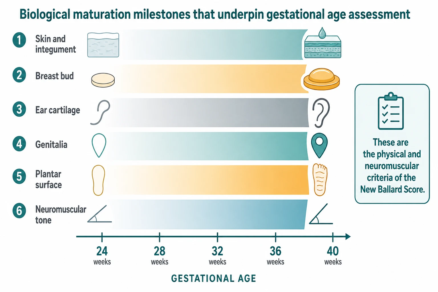
The skin tells a story because keratinisation and subcutaneous fat are late developers. An immature infant has thin, translucent skin through which veins are easily seen, abundant fine lanugo and little vernix. As gestation advances the skin thickens, the vernix accumulates and the lanugo thins and then mostly disappears. These are the physical criteria of the score, and they correlate with the skin's barrier function and transepidermal water loss, which is why they double as a guide to thermoregulation risk. [1]
Cartilage and glandular tissue mature in parallel. The pinna is soft and folds back on itself early, then its cartilage firms so it instantly springs back to shape. The breast bud grows from a flat areola to palpable tissue measured in millimetres, reflecting the maternal and fetal hormonal environment and overall nutritional maturity. The plantar surface develops creases that move from a single anterior crease toward creases covering the whole sole as the infant matures. [1] [2]
Genitalia and tone complete the picture. In the preterm infant the testes are high or in the inguinal canal and the scrotum is smooth, or the clitoris is prominent and the labia majora are flat; by term the testes are descended with rugose scrotum or the labia majora cover the labia minora. Neuromuscular tone stiffens with age — the square-window angle closes, the arm recoil becomes brisk, the popliteal angle tightens, and the heel-to-ear and scarf-sign movements become restricted. Read together, these signs estimate maturity to about plus or minus two weeks. [1]
The same schedule underlies the organ vulnerabilities that classification predicts. Surfactant production by type II pneumocytes ramps up in the third trimester, so respiratory distress risk falls steeply with each week gained. The germinal matrix overlying the caudate is highly vascular and fragile until around 32 weeks, and cerebral autoregulation is immature, which is why intraventricular haemorrhage risk is a function of gestational age. Gut barrier, brown fat and renal concentrating capacity all mature along the same curve. [15]
Clinical Presentation
The clinical appearance of an infant at the bedside is a maturity display, and reading it is part of confirming the dated gestational age. The extremely preterm infant has translucent, gelatinous skin, a flat nose, soft ears that fold back, no breast tissue, smooth soles, undescended testes or flat labia, and a hypotonic, extended posture. The very preterm infant shows partial maturation of these signs. The late preterm infant looks almost term but retains subtle clues. [1] [2]
The problems an infant presents with also group by band. The extremely and very preterm infant presents with surfactant-deficient respiratory distress, apnoea, temperature instability and feeding immaturity. The moderate and late preterm infant presents more often with feeding difficulty, hypoglycaemia, jaundice and temperature instability than with severe respiratory failure. The term infant presents along the standard newborn pathway. [11] [12]
Post-term infants present with their own signature. The dysmature, post-term infant may have dry, peeling, parchment-like skin, long nails, reduced vernix and meconium staining, and the hazards include meconium aspiration, macrosomia with birth injury such as shoulder dystocia, and oligohydramnios. Recognising the post-term phenotype matters because it shifts the differential and the early management. [10]
A useful bedside discipline is to compare the dated gestational age with the maturity impression. When they agree, the classification is straightforward. When they disagree by more than two weeks, the infant may be growth-restricted, dysmature, oedematous or have an anomaly that distorts the exam, and the discrepancy itself becomes the clinical finding to chase. [1] [4]
Differential Diagnosis
The classification step has its own differential, and the most dangerous error is conflating prematurity with smallness. A term infant who is small-for-gestational-age because of placental insufficiency is not preterm, and treating them as preterm triggers the wrong screening and the wrong follow-up. The correction is to assign gestational age first, then classify size-for-age against it. [4] [18]
Conditions that distort the maturity exam form the next layer of the differential. Congenital anomalies and chromosomal conditions can flatten the breast bud, alter the ear or the plantar creases, and change tone, so the score becomes unreliable. Congenital infection can cause growth restriction and hepatosplenomegaly that shift the clinical picture. In these infants, gestational-age estimation must lean on antenatal dating rather than the postnatal score. [1] [2]
Fluid shifts and treatments change the physical criteria. Oedema from resuscitation or fluid overload blurs skin assessment. Antenatal corticosteroids accelerate lung maturation and can shift the respiratory picture relative to the gestational age. A growth-restricted infant exposed to chronic stress may show advanced-appearing skin and creases that overstate maturity. The lesson is to treat the score as a confirmatory tool, not an oracle. [1]
Finally, dating error is itself a diagnosis to consider. An irregular cycle, recent cessation of hormonal contraception, post-implantation bleeding or an incorrectly remembered last menstrual period can all produce an antenatal estimate that disagrees with ultrasound and with the postnatal score. The ACOG framework gives explicit rules for reconciling these, and the candidate should know them. [6]
Clinical & Bedside Assessment
The assessment begins with the obstetric history and the dating record. Establish the first day of the last menstrual period, the cycle regularity, any recent contraception, and crucially the date and measurements of the first-trimester ultrasound. Reconcile the two according to the accepted rule: the ultrasound estimate is preferred when it differs from the menstrual estimate by more than about seven days in the first trimester. [6] [7]
The postnatal maturity exam then confirms or refines the estimate. The New Ballard Score combines six neuromuscular criteria — posture, square window, arm recoil, popliteal angle, scarf sign, heel-to-ear — with six physical criteria — skin, lanugo, plantar surface, breast, eye and ear, genitalia. Each is scored and the totals map to a gestational-age estimate that is reliable to about plus or minus two weeks in well infants. [1]
What are the neuromuscular criteria of the New Ballard Score?
The six neuromuscular criteria assess tone, which stiffens with maturity. Posture moves from an extended, limp posture to flexed hips and knees. The square-window angle of the wrist closes from more than 90 degrees toward zero. Arm recoil after extension becomes brisk. The popliteal angle tightens from flat to the abdomen toward a right angle. The scarf sign crosses the chest further from midline toward the opposite axillary line. The heel-to-ear moves from easy, touching the ear, to restricted. [1]
Older and alternative scores deserve a sentence. The Dubowitz exam was the original systematic maturity assessment, with more items, and the Capurro method is a simplified six-sign score still used in some regions. The New Ballard Score is the most widely adopted because it was validated and extended down to extremely preterm infants, where the earlier methods were less reliable. [2] [3]
Size-for-gestational-age is assessed by plotting weight, length and head circumference against the right centile chart. For preterm and early postnatal use the Fenton charts run to 50 weeks postmenstrual age, after which they align with the WHO and INTERGROWTH-21st standards that run from term through childhood. Plotting is not optional decoration — it is how a low birthweight is correctly attributed to prematurity, to growth restriction, or to both. [18] [19]
The limits of the postnatal exam must be named. Accuracy falls in sick, oedematous and extremely preterm infants, where the signs are blunted and the score can mislead by weeks. In these infants the antenatal ultrasound estimate is the anchor, and the postnatal score is confirmatory. The candidate who applies the score blindly to a deteriorating 25-week infant has misread the tool's range. [1] [9]
Investigations
Dating ultrasound is the reference investigation for gestational age. First-trimester crown-rump-length measurement, ideally between nine and fourteen weeks, estimates gestational age to within about five to seven days, which is more accurate than the last menstrual period. The crown-rump length is the measurement the candidate must be able to name and defend. [6] [7]
How dating accuracy changes with gestation
After the first trimester, dating relies on composite second-trimester biometry — biparietal diameter, head circumference, femur length and abdominal circumference — which is less accurate and yields an estimated fetal weight rather than a pure age. The ISUOG practice guidelines and the NICHD fetal imaging workshop frame how these parameters are used and when interval scanning distinguishes constitutional smallness from true growth restriction. [8] [9]
Growth and centile assessment is the other investigation stream. The INTERGROWTH-21st project established that fetal and newborn size are comparable across populations when nutritional, environmental and health needs are met, which is why international standards are used rather than single-population charts. Plotting weight, length and head circumference against gestational-age-specific centiles is how size-for-age is formally assigned. [4] [5]
There are no useful shotgun panels when the only question is gestational age. Routine admission blood tests answer other questions — glucose, infection, bilirubin — and should not be ordered in the name of dating. The investigation of gestational age is, almost entirely, the obstetric record plus the maturity exam and the growth chart. [6]
Management — Resuscitation
The acute threats at delivery are gated by gestational age, and the readiness posture is set before the baby arrives. The lower the gestational age, the more the team prepares for thermal failure, glucose instability and surfactant-need respiratory distress. Knowing the band in advance lets the right people, equipment and environment be in the room. [6] [11]
Warmth strategy is the clearest gestational-age-driven decision. For infants below 32 weeks a polyethylene wrap or bag at birth, plus a radiant warmer and a servo-controlled incubator, target a core temperature of 36.5 to 37.5 degrees, because hypothermia on admission independently worsens mortality. The starting oxygen strategy is also banded, with air or low oxygen preferred for the preterm infant and titration against the pre-ductal saturation curve. [11]
Anticipation of surfactant and of neonatal retrieval is the third band-driven decision. For an extremely or very preterm infant, a team capable of intubation, surfactant and stabilisation should attend, and the threshold for retrieval to a tertiary centre is set by the band. For the late preterm infant the focus shifts to feeding, glucose and temperature rather than advanced respiratory support. [11] [12]
Management — Definitive & Stepwise
Once gestational age is assigned, the care pathway is largely determined by the band. The level of care is the first decision: tertiary intensive care for the infant below 32 weeks, special care for 32 to 36 weeks and six days, and rooming-in or the well-baby pathway for the term infant whose weight is appropriate. Place of birth and the need for in-utero transfer follow the same thresholds. [11]
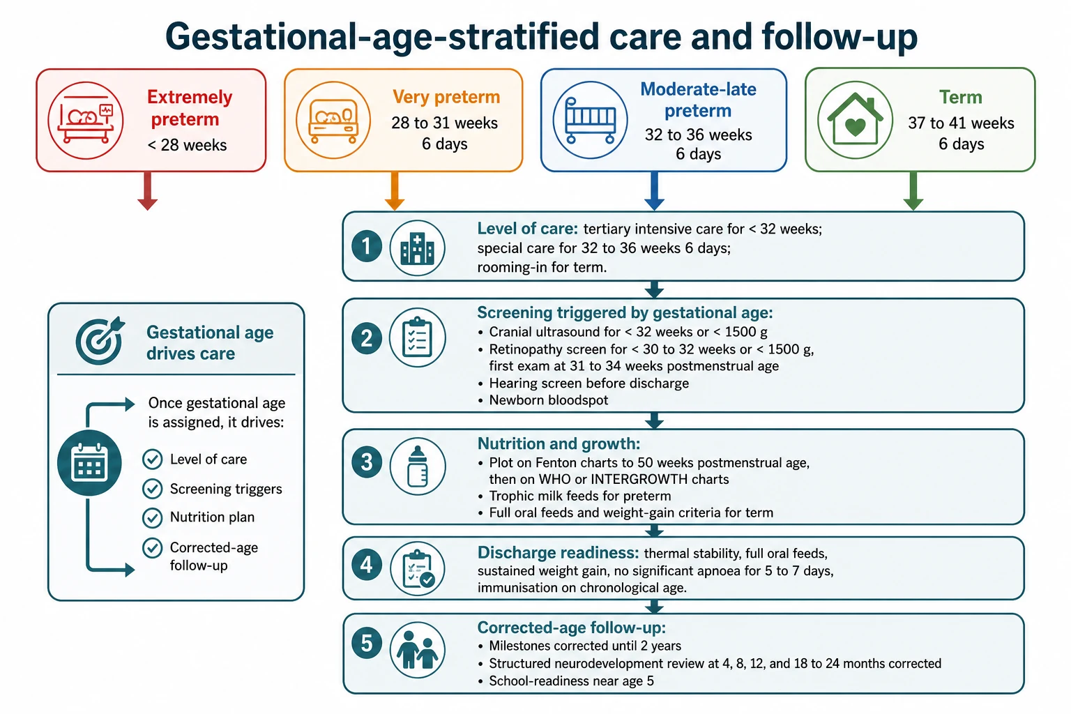
Gestational-age and birthweight thresholds then trigger the structured screening that every preterm graduate needs. Cranial ultrasound is performed for infants below 32 weeks or below 1500 g, with early and serial scans for intraventricular haemorrhage and periventricular leukomalacia. Retinopathy of prematurity screening is driven by gestational age and birthweight, typically below 30 to 32 weeks or below 1500 g, with the first examination around 31 to 34 weeks postmenstrual age. [9] [18]
DATE — what gestational age assignment unlocks
Dating — first-trimester ultrasound as the anchor, reconciled with the last menstrual period. Assessment — the New Ballard Score and the centile plot to confirm and classify. Thresholds — cranial ultrasound and retinopathy screening gated by gestational age and birthweight. Extrapolation — corrected age for growth and milestones, with structured follow-up to school age. [6] [1]
Growth plotting and developmental assessment use corrected age. Plot on Fenton charts to 50 weeks postmenstrual age, then transition to the WHO or INTERGROWTH-21st standards. Correct milestones for prematurity by subtracting the weeks born early, and keep correcting until about two years, after which most children align with their chronological peers. Forgetting to correct over-diagnoses delay; correcting for too long under-diagnoses it. [18] [19]
Discharge readiness is also gestational-age-aware but resolves into the same milestones for any preterm infant: thermal stability in an open cot, full oral feeds with sustained weight gain, no significant apnoea for five to seven days, and immunisation given on chronological age. Structured corrected-age follow-up at approximately 4, 8, 12 and 18 to 24 months, with a school-readiness assessment near age five, completes the longitudinal plan that flows from a single, well-assigned number. [11]
Specific Subtypes & Scenarios
The extremely preterm infant, below 28 weeks, sits at the top of every risk axis. Dating must be precise because a single week changes viability counselling and the level of active care offered. Expect tertiary intensive care from the first minute, surfactant and non-invasive respiratory support, cranial ultrasound surveillance and retinopathy screening, and a months-long admission with corrected-age follow-up to school age. [9] [15]
The very preterm infant, 28 to 31 weeks and six days, follows the continuous positive airway pressure and caffeine pathway, often with rescue surfactant, structured feeding advancement on human milk, and the same cranial and retinopathy surveillance. The discharge timeline is usually driven by feeding maturity and growth rather than by respiratory recovery. [11]
The moderate and late preterm infant, 32 to 36 weeks and six days, is the band that most often surprises. These infants look comparatively well but carry disproportionate morbidity — feeding immaturity, temperature instability, hypoglycaemia, jaundice and a real readmission risk. Discharge criteria must be met explicitly, and parental education and close follow-up are essential to prevent the morbidity that falls on this group. [12] [13]
The term infant, 37 to 41 weeks and six days, is where classification confirms the standard newborn pathway and routine screening applies. Within term, the early-term sub-band carries slightly higher morbidity than full term, which is one reason the workgroup split the category. The post-term infant, 42 weeks and beyond, brings meconium, macrosomia and dysmaturity into the differential. [10]
Two reconciliation scenarios complete the set. The growth-restricted but term infant weighs little but is mature, and must be classified as small-for-gestational-age rather than preterm. The discrepant-estimate infant, whose antenatal and postnatal ages disagree by more than two weeks, needs a documented reconciliation that weighs the quality of the dating evidence before a single best estimate is recorded and screening is triggered. [4] [6]
Complications & Pitfalls
The classification complications are mostly errors of conflation and omission, and examiners test them hard. Treating a low-birthweight term infant as preterm, or a growth-restricted infant as premature, misdirects every downstream decision. The correction is always to assign gestational age first, then classify size-for-age against it. [4] [18]
| Error | What goes wrong | Correction |
|---|---|---|
| Weight read as age | Growth-restricted term infant labelled preterm; wrong screening and follow-up | Assign gestational age from dating and the score, then plot size-for-age |
| Menstrual estimate trusted | Irregular cycle gives a systematically wrong age | Prefer first-trimester ultrasound when they disagree by 7–10 days |
| Over-reading the score in sick infants | Score off by weeks in oedematous or extremely preterm infants | Anchor to antenatal ultrasound; treat the score as confirmatory |
| Forgetting to correct | Milestones and growth over- or under-diagnosed as abnormal | Correct for prematurity until about 2 years; plot Fenton to 50 weeks PMA |
| Missing GA-triggered screens | Cranial ultrasound and ROP omitted | Record the single best estimate and trigger screening by band |
Over-reading the postnatal maturity score in a sick, oedematous or extremely preterm infant is a subtler trap. The score's reliability falls at the extremes and in illness, and leaning on it there produces a wrong age. Forgetting to correct for prematurity when plotting growth or interpreting milestones generates false abnormalities or false reassurance for up to two years. [1] [18]
Missing gestational-age-triggered screening turns a routine classification into a preventable harm. If the band is not recorded or is wrong, cranial ultrasound and retinopathy screening can be omitted, and the window for early intervention closes. Using different growth charts in sequence without aligning at 50 weeks postmenstrual age produces an artificial step in the centile trajectory that can be misread as growth failure. [9]
Prognosis & Disposition
Gestational age is the dominant predictor of survival and major morbidity, and it underpins every prognostic conversation. Survival improves steeply with each week gained across the preterm range, and survival without major morbidity becomes the harder endpoint below 26 to 28 weeks. The prognosis band is set the moment the age is assigned. [15] [17]
Size-for-gestational-age adds independent prognostic information on top of gestational age. Small-for-gestational-age infants, particularly those with placental insufficiency, carry additional risks of hypoglycaemia, hypothermia, polycythaemia and neurodevelopmental impairment. Large-for-gestational-age infants bring birth-injury and metabolic risks. Both axes must enter the prognostic conversation. [4]
Discharge and follow-up are banded but resolve into the same milestones for any preterm infant, and the plan should be stated in corrected-age terms so families understand the trajectory. Counsel families on probability, not certainty: outcomes have improved, most preterm graduates do well, but a real risk of developmental difference is watched for and supported with early intervention. [11] [18]
Special Populations
The extremely preterm infant near the threshold of viability is the population in whom a single day or a single gram changes classification and counselling. Antenatal counselling about anticipated outcomes and active versus comfort care must be individualised to gestational age, estimated weight, sex and antenatal steroid exposure. This leaf defers the counselling depth to the periviable leaf, but the classification decision flows directly from it. [9]
Growth-restricted infants with placental insufficiency are the population in whom size-for-age decouples from gestational age, and they are the population most at risk of being mislabelled preterm. They need careful catch-up nutrition and surveillance for the metabolic and cardiovascular consequences of fetal programming, and their gestational age must be anchored in dating, not weight. [4]
The late-preterm group is a special population in its own right because of the gap between appearance and risk. Multiple-gestation infants complicate dating, which is ultrasound-based, and growth discordance between twins makes size-for-age classification harder. For the smaller twin, the same cautions about conflating size with age apply in amplified form. [12] [14]
Rural, remote, Indigenous and migrant families face the equity dimension. Where dating ultrasound access is limited, classification is less reliable, and the conversation must be culturally safe and language-concordant. In the ANZ context, Te Tiriti-informed practice and attention to the inequities that drive preterm birth and worsen outcomes for Maori, Pacific and Aboriginal and Torres Strait Islander families are part of getting the classification and the care right. [16]
Evidence, Guidelines & Regional Differences
The INTERGROWTH-21st project reshaped the evidence base by showing that fetal and newborn size are comparable across populations when nutritional, environmental and health needs are met. Its fetal growth longitudinal study and newborn cross-sectional study provide the international standards against which size-for-gestational-age is now assessed, moving the field away from single-population charts. [5] [4]
The ACOG Committee Opinion 700 on Methods for Estimating the Due Date codified the dating hierarchy: first-trimester crown-rump-length is the reference standard, and explicit reconciliation rules apply when estimates disagree at each gestational range. The Hadlock crown-rump-length reevaluation and the ISUOG biometry and growth guidelines supply the technical measurement foundation. [6] [7] [8]
ANZ: Neonatal frameworks align dating with first-trimester ultrasound and tier care and screening by gestational-age and birthweight thresholds, with national outcome benchmarking. UK: NICE and BAPM frameworks for dating accuracy, classification and structured neonatal follow-up. US: AAP and ACOG guidance on dating, the Defining Term Pregnancy categories, and Medicaid-funded follow-up. Canada: Canadian Paediatric Society statements on growth-chart selection, classification and community follow-up networks. The principles converge; the operational details differ. [10] [14]
The maturity-score evidence spans half a century. Dubowitz built the first systematic exam, Capurro simplified it, and Ballard extended and validated the score down to extremely preterm infants, which is why the New Ballard Score is the most widely adopted. The Fenton preterm growth charts, built from a systematic review and meta-analysis, and the WHO Child Growth Standards complete the charting toolkit that bridges the preterm-to-childhood transition. [2] [3] [1] [18] [19]
Exam Pearls
Gestational age is time from the first day of the last menstrual period to the day of birth, measured in completed weeks — it is not weight, and the candidate who conflates the two fails the question before it starts. First-trimester crown-rump-length ultrasound is the reference standard at about five to seven days, and the last menstrual period yields to it when they disagree by more than seven to ten days. [6] [7]
The preterm bands are exam vocabulary: extremely preterm below 28 weeks, very preterm 28 to 31 and six, moderate 32 to 33 and six, late preterm 34 to 36 and six. Term splits into early, full and late term, and post-term is 42 weeks and beyond. Birthweight categories are low under 2500 g, very low under 1500 g, extremely low under 1000 g — and size-for-age is small below the tenth, appropriate tenth to ninetieth, large above the ninetieth centile for gestational age. [10] [4]
The New Ballard Score combines six neuromuscular and six physical criteria and is reliable to about plus or minus two weeks, but its accuracy falls in sick, oedematous and extremely preterm infants, where antenatal dating is the anchor. Postmenstrual age is gestational age plus chronological age; corrected age is chronological age minus the weeks born early; correct until about two years and use Fenton charts to 50 weeks postmenstrual age. [1] [18]
Screening is gestational-age-and-birthweight triggered, never symptom based: cranial ultrasound below 32 weeks or 1500 g, retinopathy below 30 to 32 weeks or 1500 g with the first exam at 31 to 34 weeks postmenstrual age. The late-preterm group is the largest preterm group and carries disproportionate morbidity, so never dismiss it as almost term. Always reconcile antenatal and postnatal estimates and record a single best estimate before classifying. [9] [12]
References
- [1]Ballard JL New Ballard Score, expanded to include extremely premature infants J Pediatr, 1991.PMID 1880657
- [2]Dubowitz LM Clinical assessment of gestational age in the newborn infant J Pediatr, 1970.PMID 5430794
- [3]Capurro H A simplified method for diagnosis of gestational age in the newborn infant J Pediatr, 1978.PMID 650322
- [4]Villar J International standards for newborn weight, length, and head circumference by gestational age and sex: the Newborn Cross-Sectional Study of the INTERGROWTH-21st Project Lancet, 2014.PMID 25209487
- [5]Papageorghiou AT International standards for fetal growth based on serial ultrasound measurements: the Fetal Growth Longitudinal Study of the INTERGROWTH-21st Project Lancet, 2014.PMID 25209488
- [6]American College of Obstetricians and Gynecologists Committee Opinion No 700: Methods for Estimating the Due Date Obstet Gynecol, 2017.PMID 28426621
- [7]Hadlock FP Fetal crown-rump length: reevaluation of relation to menstrual age (5-18 weeks) with high-resolution real-time US Radiology, 1992.PMID 1732970
- [8]Salomon LJ ISUOG Practice Guidelines: ultrasound assessment of fetal biometry and growth Ultrasound Obstet Gynecol, 2019.PMID 31169958
- [9]Reddy UM Fetal imaging: Executive summary of a Joint Eunice Kennedy Shriver National Institute of Child Health and Human Development, Society for Maternal-Fetal Medicine, American Institute of Ultrasound in Medicine, American College of Obstetricians and Gynecologists, American College of Radiology, Society for Pediatric Radiology, and Society of Radiologists in Ultrasound Fetal Imaging Workshop Am J Obstet Gynecol, 2014.PMID 24793721
- [10]Spong CY Defining term pregnancy: recommendations from the Defining Term Pregnancy Workgroup JAMA, 2013.PMID 23645117
- [11]Engle WA Late preterm infants, early term infants, and timing of elective deliveries Clin Perinatol, 2008.PMID 18456072
- [12]Engle WA Late-preterm infants: a population at risk Pediatrics, 2007.PMID 18055691
- [13]McIntire DD Neonatal mortality and morbidity rates in late preterm births compared with births at term Obstet Gynecol, 2008.PMID 18165390
- [14]Raju TNKR The Late Preterm Birth-Ten Years Later Pediatrics, 2017.PMID 28148728
- [15]Goldenberg RL Epidemiology and causes of preterm birth Lancet, 2008.PMID 18177778
- [16]Blencowe H National, regional, and worldwide estimates of preterm birth rates in the year 2010 with time trends since 1990 for selected countries: a systematic analysis and implications Lancet, 2012.PMID 22682464
- [17]Chawanpaiboon S Global, regional, and national estimates of levels of preterm birth in 2014: a systematic review and modelling analysis Lancet Glob Health, 2019.PMID 30389451
- [18]Fenton TR A systematic review and meta-analysis to revise the Fenton growth chart for preterm infants BMC Pediatr, 2013.PMID 23601190
- [19]WHO Multicentre Growth Reference Study Group WHO Child Growth Standards based on length/height, weight and age Acta Paediatr Suppl, 2006.PMID 16817681