Atopic Eczema (Adult)
Atopic eczema (atopic dermatitis, AD) is a chronic, relapsing inflammatory skin condition characterised by intense pruritus, xerosis, and eczematous lesions with age-dependent distribution patterns. It affects 15-20%...
What matters first
Atopic eczema (atopic dermatitis, AD) is a chronic, relapsing inflammatory skin condition characterised by intense pruritus, xerosis, and eczematous lesions with age-dependent distribution patterns. It affects 15-20%...
Eczema herpeticum (clusters of monomorphic vesicles, fever, systemic illness)
7 Jan 2026
Generated educational material; verify before clinical use.
Visible references section
See the concept before reading it
Study the key anatomy, imaging, and decision pathways as full teaching plates.
Clinical board
A visual summary of the highest-yield teaching signals on this page.
Urgent signals
Safety-critical features pulled from the topic metadata.
- Eczema herpeticum (clusters of monomorphic vesicles, fever, systemic illness)
- Erythroderma (less than 90% BSA involvement)
- Secondary bacterial infection (impetigo, cellulitis)
- Severe widespread eczema unresponsive to treatment
Linked comparisons
Differentials and adjacent topics worth opening next.
- Contact Dermatitis
- Seborrhoeic Dermatitis
Content status and exam context
This page is AI-generated educational content. It may contain errors or omissions and is not a substitute for current guidelines, local protocols, senior clinical judgement, or professional medical advice.
MedVellum does not claim an individual clinician reviewer, board certification, or professional credential for this page unless a future version names a real, verifiable reviewer.
Clinical explanation and evidence
Atopic Eczema (Adult)
1. Topic Overview
Summary
Atopic eczema (atopic dermatitis, AD) is a chronic, relapsing inflammatory skin condition characterised by intense pruritus, xerosis, and eczematous lesions with age-dependent distribution patterns. It affects 15-20% of children and 2-10% of adults globally, with increasing prevalence in developed countries over the past decades. [1,2] The disease represents a complex interplay between epidermal barrier dysfunction (notably filaggrin loss-of-function mutations), dysregulated type 2 immune responses (Th2/IL-4/IL-13 axis), environmental factors, and skin microbiome dysbiosis. [3]
Atopic dermatitis is the cutaneous manifestation of systemic atopy and frequently constitutes the first step in the "atopic march"—sequential development of allergic rhinitis and asthma. [4] Management is stepwise, centered on intensive emollient therapy (250-500g/week minimum), topical anti-inflammatory agents (corticosteroids, calcineurin inhibitors, PDE4 inhibitors, topical JAK inhibitors), and for moderate-to-severe disease, systemic therapies including dupilumab (IL-4Rα antagonist), JAK inhibitors (baricitinib, upadacitinib, abrocitinib), and phototherapy. [5,6]
The disease burden extends beyond visible skin lesions: patients experience profound sleep disruption, psychological distress, work/school absenteeism, and reduced quality of life scores comparable to other major chronic diseases. [7] Recognition of red flag complications—particularly eczema herpeticum (disseminated HSV superinfection) and erythroderma—is critical for timely intervention.
Key Facts
- Global Prevalence: 15-20% children; 2-10% adults (varies by geography)
- Age of Onset: 80% manifest before age 5; 60% in first year of life
- Genetic Risk: FLG loss-of-function mutations present in 20-30% of AD patients (European ancestry)
- Atopic March: 60-80% develop allergic rhinitis; 30-40% develop asthma
- First-Line Treatment: Emollients 250-500g/week minimum (often underprescribed)
- Topical Corticosteroids: Potency-matched to site and severity; backbone of flare management
- Biologic Therapy: Dupilumab (IL-4Rα inhibitor) for moderate-to-severe disease; 70-75% achieve EASI-75
- JAK Inhibitors: Oral baricitinib, upadacitinib, abrocitinib; rapid onset (2-4 weeks)
- Red Flag: Eczema herpeticum—clusters of monomorphic punched-out erosions/vesicles with fever
Clinical Pearls
"Emollients Are the Foundation, Not an Adjunct": Intensive emollient therapy (minimum 250g/week, often 500g+) is the cornerstone of AD management. The "soak and seal" method (bathing followed by immediate emollient application within 3 minutes) maximizes skin hydration. [5] Inadequate emollient use is the most common cause of treatment failure.
"Treat the Itch, Not Just Inflammation": AD is "the itch that rashes." Pruritus drives the itch-scratch cycle, causing lichenification and barrier disruption. IL-31 is the key pruritogenic cytokine. Breaking this cycle requires anti-pruritic strategies: sedating antihistamines at night (sleep), cool compresses, topical anti-inflammatory agents, and systemic therapies targeting IL-4/IL-13 or JAK-STAT pathways. [8]
"Age Determines Distribution":
- Infants (0-2y): Face, scalp, extensor surfaces
- Children (2-12y): Flexural areas (antecubital, popliteal fossae)
- Adults (> 12y): Hands, face, neck, flexures; lichenified plaques
"Eczema Herpeticum Is a Dermatological Emergency": Widespread vesicular eruption in AD patient = eczema herpeticum (Kaposi varicelliform eruption) until proven otherwise. Caused by HSV-1/2 dissemination through compromised skin barrier. Presents with clusters of monomorphic punched-out erosions, fever, malaise. Requires urgent IV aciclovir (10mg/kg TDS). [9]
"Potency Ladder for Topical Corticosteroids": Match potency to site and severity:
- Face/Genitals/Flexures: Mild (hydrocortisone 1%) or moderate (clobetasone 0.05%)
- Body/Limbs: Moderate (clobetasone) to potent (betamethasone valerate 0.1%)
- Palms/Soles: Very potent (clobetasol 0.05%)
- Apply once daily (as effective as twice daily); use "fingertip unit" (FTU) dosing
"New Era of Systemic Therapy": Dupilumab revolutionized severe AD management as first biologic approved for AD. Blocks IL-4 and IL-13 signaling (dual inhibitor of type 2 inflammation). SOLO 1 and 2 trials demonstrated 70-75% EASI-75 response. [10] JAK inhibitors (baricitinib, upadacitinib) offer even faster onset (2-4 weeks vs 8-12 weeks) with broader immune suppression. [11]
Why This Matters Clinically
Atopic dermatitis profoundly impacts quality of life across all age groups. Sleep disruption affects 60-80% of patients and families, leading to daytime fatigue, impaired work/school performance, and psychological sequelae including anxiety and depression. [7] The Dermatology Life Quality Index (DLQI) scores in moderate-to-severe AD average 12-18/30—comparable to coronary artery disease and diabetes. [12]
Economic burden is substantial: direct costs (medications, healthcare visits) and indirect costs (work loss, caregiver burden) exceed £5,000/patient/year in severe cases. [13] Early aggressive management and appropriate step-up to systemic therapies can prevent disease progression, reduce complication rates, and restore quality of life.
The atopic march phenomenon has public health implications: AD in infancy predicts subsequent development of asthma (30-40%) and allergic rhinitis (60-80%). [4] Early intervention targeting skin barrier restoration may potentially prevent or attenuate respiratory allergic disease, though this remains an area of active research.
2. Epidemiology
Incidence & Prevalence
| Measure | Value | Notes |
|---|---|---|
| Global Prevalence (Childhood) | 15-20% | Higher in developed countries (20-25%); lower in rural/developing regions (5-10%) [1] |
| Global Prevalence (Adult) | 2-10% | Varies by geography; 2-3% Europe/USA; up to 10% in some Asian populations [2] |
| Lifetime Cumulative Incidence | ~25% | Approximately 1 in 4 individuals will experience AD at some point |
| Trend | Increasing | 2-3 fold increase in prevalence over past 30 years in industrialized nations [1] |
| Point Prevalence (UK) | 5-20% children; 3-5% adults | Primary care consultation rate: 6-8% children annually |
Demographics
| Factor | Details |
|---|---|
| Age of Onset | 60% in first year of life; 80% before age 5; 10-15% adult-onset (> 18y) [2] |
| Sex | Slight female predominance in adults (1.3:1 F:M); equal in children |
| Geography | Highest in Northern Europe, Australia, urban USA; lowest in rural Africa, Eastern Europe, rural China |
| Socioeconomic Status | Higher prevalence in higher SES in developed countries (hygiene hypothesis); reverse in developing countries |
| Urbanization | 2-fold higher in urban vs rural populations (environmental exposures, pollution, reduced microbial diversity) [14] |
Exam Detail: Examination Focus: MRCP/Dermatology candidates should know:
- Prevalence difference between childhood (15-20%) vs adult (2-10%) AD
- Age-stratified onset pattern: 60% first year, 80% before age 5
- Geographic variation: highest in developed/urban areas (supports hygiene hypothesis)
- Increasing prevalence over decades (environmental factors > genetic drift)
- Association with atopic march: 30-40% develop asthma, 60-80% allergic rhinitis
Common Viva Question: "A patient asks why their child developed eczema despite no family history. What environmental factors contribute?" Model Answer: While genetic predisposition (especially FLG mutations) confers highest risk, environmental factors play major roles: (1) Hygiene hypothesis—reduced microbial exposure in early life leads to Th2-skewed immunity; (2) Urban environment—air pollution, allergen exposure; (3) Western diet—reduced omega-3 fatty acids; (4) Increased use of soaps/detergents disrupting skin barrier. Approximately 30% of AD cases have no family history of atopy, highlighting environmental contributions.
Risk Factors
Genetic Factors:
| Factor | Relative Risk | Mechanism |
|---|---|---|
| Family History | 2-3× if one parent; 3-5× if both parents atopic | Polygenic inheritance pattern |
| Filaggrin (FLG) Mutations | 3-5× risk; present in 20-30% AD patients (European) | Loss-of-function → impaired stratum corneum formation, increased TEWL [3] |
| Epidermal Differentiation Complex | Variable | Genes encoding loricrin, involucrin, small proline-rich proteins |
| HLA Associations | Modest | HLA-DR4, DQ8 (modest associations, not clinically useful) |
Environmental Risk Factors:
- Hygiene Hypothesis: Reduced microbial exposure in early life → inadequate Th1/regulatory T-cell development → Th2 skewing [14]
- Urban Environment: Air pollution (particulate matter, NO₂) disrupts skin barrier and promotes inflammation [14]
- Allergen Exposure: House dust mite, pet dander, pollens (particularly in extrinsic/IgE-mediated AD)
- Occupational Exposures: Wet work, irritants (hairdressers, healthcare workers, cleaners)
- Climate: Low humidity, cold dry winters worsen xerosis and barrier dysfunction
- Tobacco Smoke: Maternal smoking in utero increases offspring AD risk (1.5-2×)
Dietary Factors:
- Breastfeeding: Controversial; some studies suggest protective effect in first 3-6 months, others show no effect
- Food Allergens: Cow's milk protein, egg, wheat, soy, nuts (relevant in subset with IgE-mediated AD, especially infants)
- Vitamin D Deficiency: Observational associations with higher AD prevalence/severity (mechanistic role in skin immunity)
Microbiome:
- Skin Dysbiosis: Reduced bacterial diversity; overgrowth of Staphylococcus aureus (colonizes 70-90% of AD lesions) [15]
- Gut Microbiome: Altered gut microbiota in infancy may predispose to AD (lower Bifidobacterium, increased Clostridia)
3. Pathophysiology
Molecular Mechanisms
Atopic dermatitis pathogenesis involves a complex, bidirectional interaction between epidermal barrier dysfunction, type 2 immune dysregulation, neuroimmune signaling, and skin microbiome alterations. [3]
1. Epidermal Barrier Dysfunction
Filaggrin Loss-of-Function Mutations:
- FLG Gene: Encodes filaggrin, a key structural protein in the stratum corneum
- Function: Aggregates keratin filaments; degraded into natural moisturizing factors (NMF: amino acids, urocanic acid, pyrrolidone carboxylic acid)
- Loss-of-Function Mutations: R501X, 2282del4 (European); 3321delA (Asian)—present in 20-30% of AD patients (European ancestry) [3]
- Consequences:
- Impaired corneocyte formation → abnormal stratum corneum architecture
- Reduced NMF → xerosis (dry skin)
- Increased transepidermal water loss (TEWL)
- Enhanced allergen/irritant penetration → immune activation
- Altered skin pH (↑ alkaline) → ↑ protease activity → further barrier disruption
Beyond Filaggrin:
- Tight Junction Proteins: Claudin-1, claudin-4 downregulation → increased permeability
- Ceramides: Reduced ceramide 1, 3, 6 → impaired lipid lamellae in stratum corneum
- Antimicrobial Peptides (AMPs): Reduced β-defensins, cathelicidin (LL-37) → ↑ infection risk
Exam Detail: Key Examination Concept: Filaggrin mutations are necessary but not sufficient for AD. Only ~40% of individuals with FLG mutations develop AD, indicating requirement for additional factors (immune, environmental). Conversely, 70-80% of AD patients have normal FLG, demonstrating immune-driven pathway is equally important.
Viva Question: "Why do filaggrin mutations increase infection risk in AD?" Model Answer: Filaggrin degradation products (amino acids, urocanic acid) maintain acidic skin pH (4.5-5.5). Loss-of-function mutations → elevated pH (> 6) → increased serine protease activity → further barrier breakdown AND reduced activity of antimicrobial peptides (β-defensins, cathelicidin) which function optimally at acidic pH → enhanced colonization by S. aureus and HSV.
2. Type 2 Immune Dysregulation
Acute AD (Early Lesions):
-
Th2 Predominance: Activated CD4+ T-cells produce IL-4, IL-13, IL-31
- "IL-4 + IL-13: "
- Downregulate filaggrin, loricrin, involucrin gene expression → barrier impairment
- Induce TARC/CCL17 (recruits more Th2 cells)
- Stimulate B-cells → ↑ IgE production
- Inhibit antimicrobial peptide production
- "IL-31: Major pruritogenic cytokine → activates sensory neurons → itch [8]"
- "IL-5: Eosinophil recruitment (visible in acute lesions)"
- "IL-4 + IL-13: "
-
TSLP, IL-25, IL-33 (Epithelial Alarmins): Released from damaged keratinocytes → activate type 2 innate lymphoid cells (ILC2s) → amplify Th2 response
Chronic AD (Lichenified Lesions):
- Th1 Component: IFN-γ production increases in chronic lesions
- Th17 Component: IL-17, IL-22 contribute to epidermal hyperplasia and acanthosis
- Th22 Component: IL-22 further inhibits terminal differentiation genes
Type 2 Inflammation Dominates: Even in chronic AD, the Th2 axis (IL-4/IL-13) remains the central driver—this is why dupilumab (IL-4Rα antagonist) is highly effective. [10]
3. Itch-Scratch Cycle (Neuroimmune Loop)
- IL-31: Produced by Th2 cells; binds IL-31 receptor on sensory neurons → pruritus [8]
- Scratching: Mechanical trauma → keratinocyte damage → release of TSLP, IL-33 → further Th2 activation
- Neurogenic Inflammation: Sensory neuron activation → release of substance P, CGRP → mast cell degranulation → histamine (minor contributor to itch in AD, explaining poor antihistamine efficacy)
- Self-Perpetuating: Pruritus → scratch → barrier damage → inflammation → pruritus
Clinical Pearl: Why Antihistamines Don't Work Well in AD Itch: Unlike urticaria (histamine-driven itch), AD pruritus is mediated primarily by IL-31, IL-4, IL-13, and neuronal activation pathways independent of histamine. Antihistamines may help sleep via sedation (cetirizine, promethazine) but lack direct anti-pruritic efficacy in AD. Dupilumab and JAK inhibitors effectively reduce itch by blocking IL-4/IL-13 and downstream JAK-STAT signaling, respectively. [8]
4. Skin Microbiome Dysbiosis
-
Staphylococcus aureus Colonization: Present in 70-90% of AD lesions (vs 5-30% healthy skin) [15]
- Produces α-toxin (pore-forming toxin) → keratinocyte damage → barrier disruption
- Superantigens (SEA, SEB) → non-specific T-cell activation → Th2 amplification
- Biofilm formation → chronic colonization and infection risk
- "IgE to S. aureus: Present in 50-60% of AD patients; correlates with disease severity"
-
Reduced Bacterial Diversity: Dominance of Staphylococcus and Corynebacterium; reduced Streptococcus and commensal Cutibacterium acnes
-
Malassezia (Yeast): Colonizes scalp/upper trunk in AD; can trigger type 2 inflammation via fungal allergens
Classification Systems
1. Intrinsic vs Extrinsic Atopic Dermatitis
| Feature | Extrinsic AD (70-80%) | Intrinsic AD (20-30%) |
|---|---|---|
| IgE Levels | Elevated (> 200 IU/mL) | Normal (less than 200 IU/mL) |
| Allergen Sensitization | Positive (foods, aeroallergens) | Negative |
| Atopic Comorbidities | High (asthma, rhinitis common) | Lower |
| Eosinophilia | Often present | Usually absent |
| FLG Mutations | Less common | More common |
| Th2 Markers | Very high IL-4, IL-13 | Moderate elevation |
| Age Distribution | More common in children | More common in adults |
Clinical Relevance: Extrinsic AD may benefit from allergen avoidance (dust mite covers, dietary elimination if proven allergy). Intrinsic AD more likely driven by primary barrier defect (FLG mutations).
2. Severity Classification (Validated Scores)
EASI (Eczema Area and Severity Index): 0-72 scale
- Mild: 0-7
- Moderate: 7.1-21
- Severe: 21.1-50
- Very Severe: > 50
SCORAD (SCORing Atopic Dermatitis): 0-103 scale
- less than 25: Mild
- 25-50: Moderate
-
50: Severe
IGA (Investigator's Global Assessment): 0-4 scale
- 0: Clear
- 1: Almost clear
- 2: Mild
- 3: Moderate
- 4: Severe
Clinical Use: EASI and SCORAD used in clinical trials (dupilumab trials used EASI-75 as primary endpoint = 75% reduction in EASI from baseline). IGA used for treatment decisions (target IGA 0-1).
4. Clinical Presentation
Cardinal Features
Pruritus: The hallmark symptom—"the itch that rashes." Intensity often worse at night (circadian IL-31 variation), leading to sleep disruption in 60-80% of patients. [7]
Xerosis: Generalized dry, scaly skin even in non-eczematous areas (reflects widespread barrier dysfunction and reduced NMF).
Eczematous Morphology: Varies by disease phase:
- Acute: Erythematous papules and vesicles, oozing, crusting
- Subacute: Erythematous, excoriated, scaly plaques
- Chronic: Lichenified (thickened, leathery) plaques with exaggerated skin markings
Age-Dependent Distribution: See table below.
Clinical Presentation by Age
| Age Group | Distribution | Morphology | Key Features |
|---|---|---|---|
| Infants (0-2y) | Face (cheeks, forehead), scalp, extensor surfaces (knees, elbows) | Acute: erythema, vesicles, oozing, crusting | Spares diaper area (too moist for AD); drooling worsens perioral eczema |
| Children (2-12y) | Flexural areas: antecubital fossae, popliteal fossae, wrists, ankles | Subacute-to-chronic: excoriated plaques, early lichenification | Classic flexural distribution; periorbital involvement common |
| Adolescents/Adults (> 12y) | Hands (especially palmar), face, neck, upper trunk, flexures | Chronic: lichenified plaques; acute flares superimposed | Hand dermatitis (irritant/atopic overlap); head-neck predominant pattern in some adults |
Exam Detail: Examination Pearl: In OSCE/PACES stations, age-dependent distribution is a key diagnostic clue:
- Infant with eczema on cheeks and extensor surfaces → Think atopic dermatitis (NOT seborrhoeic dermatitis if pruritic and sparing diaper area)
- Child with lichenified plaques in antecubital/popliteal fossae → Classic atopic dermatitis
- Adult with chronic hand dermatitis + history of childhood eczema → Atopic dermatitis (irritant contact dermatitis overlap)
Viva Trap: "Why does infant AD spare the diaper area?" Answer: Diaper area is occluded, moist environment → hydrates stratum corneum → less xerosis/barrier dysfunction. AD preferentially affects dry, exposed areas. Conversely, seborrhoeic dermatitis favors moist/sebaceous areas (diaper area involved).
Symptoms
| Symptom | Frequency | Details |
|---|---|---|
| Pruritus | 100% | Defining feature; severe itch often worse at night; drives itch-scratch cycle |
| Sleep Disturbance | 60-80% | Patients and caregivers; average 1-2h sleep loss per night in moderate-severe AD [7] |
| Skin Pain | 40-60% | Fissures (especially hands, feet), excoriations, secondary infection |
| Burning/Stinging | 30-50% | Particularly with topical treatments, water exposure |
| Psychological Distress | 40-60% | Anxiety, depression, social embarrassment, low self-esteem |
Signs
General Inspection:
- Xerosis: Dry, scaly skin on non-eczematous areas (arms, legs)
- Pallor: Facial pallor (vasoconstriction in chronic inflammation)
- Infraorbital Darkening: "Allergic shiners"
- Dennie-Morgan Folds: Prominent infraorbital creases (atopic stigma)
- Keratosis Pilaris: Follicular keratotic papules on upper arms, thighs (common in atopics)
- Ichthyosis Vulgaris: Fine scaling on lower legs (associated with FLG mutations)
- Hyperlinear Palms: Exaggerated palmar creases (atopic hand sign)
Eczematous Lesions:
| Phase | Clinical Signs |
|---|---|
| Acute | Ill-defined erythematous plaques, vesicles (may be subtle), oozing, crusting, excoriations |
| Subacute | Erythematous scaly plaques, less oozing, more scaling, excoriation marks |
| Chronic | Lichenified plaques (thickened, leathery), exaggerated skin markings, hyperpigmentation (post-inflammatory), fissures |
Special Patterns:
- Nummular Eczema: Coin-shaped lesions (often on lower legs in adults)
- Pompholyx (Dyshidrotic Eczema): Vesicles on palms, soles, fingers (can coexist with AD)
- Lichen Simplex Chronicus: Isolated lichenified plaque from repeated scratching (nape, ankles, vulva)
Associated Features (Atopic Stigmata)
| Sign | Description | Clinical Relevance |
|---|---|---|
| Dennie-Morgan Folds | Infraorbital creases | Atopic marker (also seen in allergic rhinitis) |
| Allergic Shiners | Periorbital venous congestion → dark circles | Suggests concurrent allergic rhinitis |
| Hertoghe Sign | Loss of lateral third of eyebrows | Chronic rubbing in atopic patients |
| Pityriasis Alba | Hypopigmented patches on face (especially cheeks in children) | Post-inflammatory; resolves with moisturization |
| White Dermographism | White line after stroking skin (abnormal vascular response) | Seen in AD; opposite of normal red dermographism |
Red Flags (Complications Requiring Urgent Action)
[!CAUTION] Red Flags in Atopic Dermatitis
1. Eczema Herpeticum (Kaposi Varicelliform Eruption)
- Presentation: Widespread monomorphic vesicles (2-3mm) progressing to punched-out erosions, clusters on eczematous skin; fever, malaise, lymphadenopathy
- Pathogen: HSV-1 (most common) or HSV-2 dissemination through compromised barrier
- Risk Factors: Moderate-to-severe AD, FLG mutations, early-onset AD, head-neck predominant AD
- Diagnosis: Clinical + viral swab (PCR for HSV); Tzanck smear (multinucleated giant cells, less sensitive)
- Management: Urgent IV aciclovir 10mg/kg TDS (or oral 400mg 5× daily if mild); hospitalize moderate-severe cases [9]
- Complications: Keratitis (ocular HSV), viremia, secondary bacterial infection
Clinical Pearl: Any AD patient with sudden eruption of vesicles/erosions + fever should be treated empirically with aciclovir pending viral PCR. Do NOT wait for results. Mortality 1-9% if untreated.
2. Erythroderma (Exfoliative Dermatitis)
- Definition: > 90% body surface area involvement
- Presentation: Generalized erythema, scaling, possible exfoliation; systemic features (hypothermia, high-output cardiac failure, hypoalbuminemia)
- Management: Hospitalize; aggressive emollients, wet wraps, systemic therapy (dupilumab, cyclosporine), monitor fluid/electrolyte balance
- Differential: Exclude psoriatic erythroderma, drug reaction, CTCL
3. Secondary Bacterial Infection
- Clinical Signs: Golden crusting (impetiginized eczema), pustules, increased warmth/tenderness, rapidly worsening eczema
- Pathogen: S. aureus (90%)
- Management:
- "Localized: Topical fusidic acid or mupirocin"
- "Widespread: Oral flucloxacillin 500mg QDS × 7d (or clarithromycin if penicillin-allergic)"
- "MRSA: Doxycycline or co-trimoxazole"
- Adjunct: Dilute bleach baths (reduce S. aureus burden)
4. Failure to Thrive (Children)
- Causes: Protein/calorie malnutrition (especially if restrictive diet for presumed food allergy), chronic inflammation, sleep deprivation
- Assessment: Growth charts (weight, height, weight-for-height); assess dietary intake
- Management: Dietician referral; optimize eczema control; liberalize unnecessary dietary restrictions
5. Clinical Examination
Structured Approach (OSCE/PACES)
1. Introduction: Wash hands, introduce self, confirm patient identity, explain examination, obtain consent
2. General Inspection (from end of bed):
- Obvious eczematous lesions (distribution)
- Patient comfort level (scratching behavior)
- Atopic facies (allergic shiners, Dennie-Morgan folds)
- Growth/nutrition (particularly children)
3. Systematic Skin Examination:
Distribution Pattern:
- Note age-dependent pattern (infant: face/extensors; child: flexures; adult: hands/face/flexures)
- Assess body surface area involvement (palmar method: patient's palm = 1% BSA)
Morphology Assessment:
| Feature | Acute | Subacute | Chronic |
|---|---|---|---|
| Color | Bright red | Red-pink | Red-brown, hyperpigmented |
| Surface | Vesicles, oozing | Scaling, crusting | Lichenified, thickened |
| Border | Ill-defined | Ill-defined | More defined |
4. Specific Anatomical Sites:
- Face: Periorbital, perioral, cheeks
- Flexures: Antecubital fossae, popliteal fossae, wrists, ankles, neck
- Hands: Palms (pompholyx), dorsal hands (irritant exposure)
- Trunk: Upper chest, back (follicular pattern possible)
- Scalp: Scaling, alopecia (rare, from scratching)
5. Look for Complications:
- Infection: Crusting (golden = S. aureus; honey-colored = impetiginization), vesicles (HSV), pustules
- Lichenification: Chronic thickening from repeated scratching
- Excoriations: Linear erosions (itch-scratch cycle)
- Fissures: Deep cracks (especially hands, feet)
6. Atopic Stigmata:
- Dennie-Morgan folds, allergic shiners, hyperlinear palms, keratosis pilaris, ichthyosis vulgaris, pityriasis alba
7. Complete Examination:
- Ask to examine for asthma signs (wheeze on auscultation)
- Ask to review drug chart (topical treatments, systemic medications)
- Ask about family history (atopy in parents/siblings)
Exam Detail: Common OSCE/PACES Stem: "This 25-year-old has had itchy skin since childhood. Please examine the skin and present your findings."
Model Presentation: "This is a comfortable young adult with evidence of atopic dermatitis. On general inspection, I note allergic shiners and Dennie-Morgan folds. The distribution is predominantly flexural, with lichenified erythematous plaques in the antecubital and popliteal fossae bilaterally. There are excoriation marks and xerosis on the extensor surfaces of the limbs. No evidence of secondary infection or eczema herpeticum. Additional atopic stigmata include hyperlinear palms and keratosis pilaris on the upper arms. I estimate body surface area involvement at approximately 10-15%, consistent with moderate atopic dermatitis. I would complete my examination by assessing for respiratory signs of asthma and inquiring about allergic rhinitis to evaluate for the atopic march."
Examiner Follow-Up: "What would you look for to assess disease severity?" Answer: "I would use a validated scoring system such as EASI or SCORAD, which incorporate: (1) Extent of involvement (body surface area), (2) Intensity of signs (erythema, edema/induration, excoriation, lichenification), and (3) Subjective symptoms (itch, sleep loss). I would also assess impact on quality of life using DLQI and ask about treatment burden, sleep disruption, and psychosocial impact."
6. Differential Diagnosis
Key Differentials
| Condition | Distinguishing Features | Overlap/Notes |
|---|---|---|
| Contact Dermatitis (Allergic) | History of exposure (nickel, fragrance, preservatives); sharp demarcation at contact site; positive patch testing | Can coexist with AD; suspect if unilateral or geometric pattern |
| Contact Dermatitis (Irritant) | Occupation (wet work, cleaning); hands predominantly; no latent period (immediate irritation) | Common in AD patients (impaired barrier → ↑ susceptibility) |
| Seborrhoeic Dermatitis | Distribution: scalp, nasolabial folds, eyebrows, central chest; yellowish greasy scales; NOT pruritic; responds to antifungals | In infants, can mimic AD but involves diaper area |
| Psoriasis | Well-demarcated salmon-pink plaques; silvery scale; extensor surfaces; nail changes (pitting, onycholysis); not as pruritic | Tape-strip RNA-seq: Th17 (IL-17A) dominant vs Th2 in AD [16] |
| Scabies | Intense itch (worse at night); burrows (finger webs, wrists); family members affected; response to permethrin | Can trigger eczema flare; consider if sudden worsening |
| Cutaneous T-Cell Lymphoma (Mycosis Fungoides) | Older adults; patches/plaques in "bathing trunk" distribution; slow progression; histology (Pautrier microabscesses) | Rare; consider if atypical age, distribution, or treatment resistance |
| Ichthyosis Vulgaris | Fine scaling on lower legs; hyperlinear palms; NO pruritus; FLG mutations (same gene as AD) | Often coexists with AD |
Diagnostic Clues
Favors Atopic Dermatitis:
- Age less than 5 years at onset
- Flexural distribution (children/adults)
- Personal/family history of atopy (asthma, allergic rhinitis)
- Xerosis on non-eczematous areas
- Chronic relapsing course
- Elevated IgE (if extrinsic type)
Favors Alternative Diagnosis:
- Onset > 60 years (consider CTCL, contact dermatitis, drug reaction)
- Well-demarcated plaques (psoriasis)
- Greasy yellow scales (seborrhoeic dermatitis)
- Burrows or family itch (scabies)
- Geometric/sharp borders (allergic contact dermatitis)
- Lack of itch (seborrhoeic dermatitis, ichthyosis vulgaris)
7. Investigations
Diagnosis
Atopic dermatitis is primarily a clinical diagnosis. The UK Working Party diagnostic criteria (most widely used):
MUST HAVE: Pruritic skin condition (itchy skin)
PLUS ≥3 of the following:
- Onset before age 2 years
- History of flexural involvement (antecubital, popliteal, ankles, neck)
- History of generally dry skin (xerosis)
- Personal history of other atopic disease (asthma, allergic rhinitis)
- Visible flexural dermatitis (or cheeks/forehead/extensor surfaces in children less than 4y)
Sensitivity 85%, Specificity 96% for these criteria.
Investigations to Support Diagnosis or Assess Severity
| Investigation | Indication | Interpretation |
|---|---|---|
| No Investigations | Typical presentation | Diagnosis is clinical; avoid over-investigation |
| Total IgE | Research/academic interest | Elevated in 70-80% (extrinsic AD); NOT diagnostic (normal in intrinsic AD) |
| Specific IgE (ImmunoCAP) | Suspected food allergy (infant/child with GI symptoms, failure to thrive) | Cow's milk, egg, wheat, soy, peanut; positive if > 0.35 kU/L but clinical correlation essential |
| Skin Prick Testing (SPT) | Aeroallergen sensitization (if considering allergen avoidance) | House dust mite, cat, dog, pollens; wheal ≥3mm positive |
| Patch Testing | Suspected superimposed allergic contact dermatitis (e.g., worsening with topical treatments) | Fragrance, preservatives (methylisothiazolinone), neomycin, lanolin |
| Skin Swab (MC&S) | Clinical signs of secondary infection (crusting, pustules, weeping) | S. aureus culture; antibiotic sensitivities (check for MRSA if recurrent) |
| Viral Swab (HSV PCR) | Suspected eczema herpeticum | HSV-1/2 PCR; rapid result essential for timely aciclovir |
| Skin Biopsy | Atypical features; exclude CTCL, psoriasis | NOT routinely needed; histology: spongiosis, acanthosis, Th2 infiltrate |
Special Investigations (Tertiary/Research Settings)
| Test | Purpose |
|---|---|
| Filaggrin Gene Sequencing | Research; not clinically indicated (does not change management) |
| Tape-Strip Transcriptomics | Clinical trials (biomarkers of treatment response); non-invasive alternative to biopsy [16] |
| TEWL (Transepidermal Water Loss) | Research measure of barrier function |
| Stratum Corneum Hydration | Objective xerosis assessment |
Clinical Pearl: Avoid Over-Investigation: AD is a clinical diagnosis. The most common error is unnecessary IgE testing. Elevated IgE supports extrinsic AD but is NOT diagnostic (absent in intrinsic AD). Focus investigations on:
- Ruling out differential diagnoses (scabies, contact dermatitis, psoriasis)
- Identifying complications (infection, eczema herpeticum)
- Allergy testing ONLY if clear clinical suspicion of food/aeroallergen trigger (e.g., immediate reaction after egg ingestion in infant)
Indiscriminate food allergy testing leads to false positives and unnecessary dietary restriction → malnutrition, family stress, no eczema benefit.
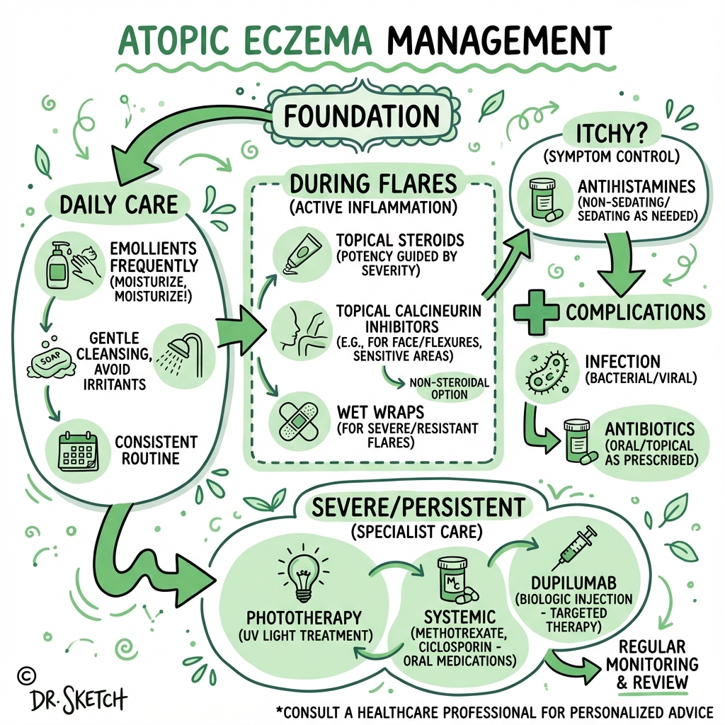
8. Management
Management Principles
- Stepwise Approach: Escalate treatment based on severity (mild → moderate → severe)
- Emollient Foundation: All patients, all severities, all the time
- Treat Flares Aggressively: Early potent topical therapy prevents progression
- Proactive Maintenance: Twice-weekly topical anti-inflammatory to previously affected areas (reduces flare frequency)
- Trigger Identification/Avoidance: Individualized (not all patients have identifiable triggers)
- Multidisciplinary: Dermatology, allergy, psychology, specialist nurses
- Patient Education: Essential for adherence and treatment success
Treatment Algorithm
┌─────────────────────────────────────────────────────┐
│          ALL SEVERITIES: FOUNDATION                 │
│  • Emollients 250-500g/week (leave-on + soap subs) │
│  • Trigger avoidance (irritants, allergens)         │
│  • Education (eczema schools, nurse support)        │
└─────────────────────────────────────────────────────┘
                          │
                          ▼
┌─────────────────────────────────────────────────────┐
│                MILD AD (BSA less than 10%, IGA 1-2)          │
│  • Mild topical corticosteroid (hydrocortisone 1%)  │
│  • Consider topical calcineurin inhibitor (face)    │
│  • Frequency: Once daily during flares              │
└─────────────────────────────────────────────────────┘
                          │
                          ▼ (If inadequate control)
┌─────────────────────────────────────────────────────┐
│              MODERATE AD (BSA 10-30%, IGA 3)        │
│  • Moderate-potent TCS (clobetasone, betamethasone) │
│  • Topical calcineurin inhibitors (tacrolimus 0.1%) │
│  • Topical PDE4 inhibitor (crisaborole)             │
│  • Topical JAK inhibitor (ruxolitinib 1.5%)         │
│  • Proactive maintenance (TCS 2×/week)              │
│  • Consider phototherapy (NB-UVB)                   │
└─────────────────────────────────────────────────────┘
                          │
                          ▼ (If inadequate control)
┌─────────────────────────────────────────────────────┐
│             SEVERE AD (BSA > 30%, IGA 4)             │
│  FIRST-LINE SYSTEMIC:                               │
│  • Dupilumab 600mg loading → 300mg SC Q2W           │
│  • JAK inhibitors:                                  │
│    - Baricitinib 2-4mg PO daily                     │
│    - Upadacitinib 15-30mg PO daily                  │
│    - Abrocitinib 100-200mg PO daily                 │
│                                                      │
│  SECOND-LINE SYSTEMIC:                              │
│  • Cyclosporine 2.5-5mg/kg/day (max 16 weeks)       │
│  • Methotrexate 10-25mg weekly                      │
│  • Azathioprine 1-3mg/kg/day                        │
│  • Mycophenolate mofetil 2-3g/day                   │
│                                                      │
│  AVOID:                                             │
│  • Oral corticosteroids (rebound flares)            │
└─────────────────────────────────────────────────────┘
Topical Therapies
1. Emollients (Foundation of All AD Management)
Evidence: Cochrane review confirms emollients reduce flares, steroid requirement, and TEWL. [5]
Prescription Quantities:
- Infants: 250g/week
- Children: 250-500g/week
- Adults: 500-1000g/week
- Severe AD: May require > 1000g/week
Types:
| Type | Examples | Indications | Notes |
|---|---|---|---|
| Light Lotions | Aveeno, E45 lotion | Mild xerosis, daytime use, hairy areas | Quickly absorbed; less occlusive |
| Creams | Epimax, Diprobase, Cetraben | Moderate xerosis, general use | 50:50 oil:water; good balance |
| Ointments | Emulsifying ointment, 50:50 liquid paraffin/white soft paraffin | Severe xerosis, lichenified areas, night use | Greasy; maximum occlusion; NO preservatives (lower allergy risk) |
| Soap Substitutes | Aqueous cream (controversial), emollient shower gels | Washing | Avoid traditional soaps (alkaline, barrier-disrupting) |
Application Technique:
- Frequency: At least 2-3 times daily (more in winter, dry climates)
- Quantity: Liberal—"slather, don't rub"
- Direction: Smooth in direction of hair growth (prevents folliculitis)
- Timing: Within 3 minutes after bathing ("soak and seal")
- Gap: Apply emollient 15-30 minutes BEFORE topical corticosteroids (allows absorption; TCS then "seals" emollient)
Common Errors:
- Under-prescribing (50-100g tubes last days, not weeks)
- Applying after TCS (should be before, or 30min after)
- Using soap (strips barrier)
Clinical Pearl: "Fingertip Unit" (FTU) Dosing: One FTU = amount of ointment from tip of adult index finger to first crease (~0.5g). Covers area of 2 adult palms.
| Body Area | Emollient FTUs (per application) |
|---|---|
| Face + neck | 2.5 FTUs |
| One arm | 3 FTUs |
| One leg | 6 FTUs |
| Trunk (front) | 7 FTUs |
| Trunk (back) | 7 FTUs |
Adult twice-daily full-body application = ~50 FTUs/day = 25g/day = 175g/week minimum.
2. Topical Corticosteroids (TCS)
Evidence: Level 1a—RCTs confirm efficacy in reducing inflammation, pruritus, and flare severity. [5,6]
Potency Ladder (UK Classification):
| Potency | Examples | Indications | Sites |
|---|---|---|---|
| Mild | Hydrocortisone 0.5%, 1%, 2.5% | Children, face, genitals, flexures | Safe for prolonged use on face |
| Moderate | Clobetasone butyrate 0.05% (Eumovate), fluocinolone 0.0625% | Body, limbs (not face/flexures) | Most commonly used for body eczema |
| Potent | Betamethasone valerate 0.1% (Betnovate), mometasone 0.1% | Thick lichenified plaques, body/limbs | NOT for face/flexures |
| Very Potent | Clobetasol propionate 0.05% (Dermovate) | Severe lichenified plaques, palms/soles | Short courses only (max 2-4 weeks); dermatologist supervision |
Dosing:
- Frequency: Once daily (as effective as twice daily for most TCS)
- Quantity: Fingertip units (see emollient table; TCS uses ~25-30% the amount of emollient)
- Duration:
- "Flare Treatment: Continue until lesions clear (typically 7-14 days)"
- "Proactive Maintenance: Twice weekly to previously affected sites (reduces flares by 60-70%)"
Common Prescribing Errors:
- Using mild TCS on lichenified plaques (under-treatment)
- Using potent TCS on face (skin atrophy risk)
- Prolonged very potent TCS without specialist input
- Stopping TCS too early (rebound flare)
Side Effects:
| Side Effect | Risk Factors | Prevention |
|---|---|---|
| Skin Atrophy | Potent/very potent TCS on face, flexures; prolonged use (> 6-12 weeks continuous) | Use appropriate potency; twice-weekly maintenance (NOT daily long-term) |
| Striae | Flexures, groin | Avoid potent TCS in flexures |
| Telangiectasia | Face | Avoid potent TCS on face |
| Perioral Dermatitis | Potent TCS on face | Use mild TCS on face; consider TCIs |
| Systemic Absorption | Very potent TCS on large BSA in infants | Limit use; monitor growth |
| Tachyphylaxis | Theoretical (rarely clinically significant) | Rotate TCS potencies; use proactive maintenance |
Steroid Phobia: Major barrier to adherence. Address proactively:
- Explain appropriate TCS use is safe
- Reassure: short bursts (1-2 weeks) and proactive maintenance (2×/week) do NOT cause significant atrophy
- Emphasize: undertreated eczema causes MORE harm (sleep loss, infection, QoL) than appropriate TCS use
3. Topical Calcineurin Inhibitors (TCIs)
Mechanism: Inhibit calcineurin → block T-cell activation → reduce IL-2, Th2 cytokines
Agents:
- Tacrolimus 0.03% (children), 0.1% (adults): More potent
- Pimecrolimus 1%: Less potent; suitable for mild-moderate AD
Indications:
- Face/Eyelids: Preferred over TCS (no atrophy risk)
- Flexures, Genitals: Steroid-sparing
- Maintenance Therapy: Proactive twice-weekly application (like TCS)
- Patients with steroid phobia
Efficacy: Tacrolimus 0.1% ≈ moderate-potent TCS; pimecrolimus ≈ mild TCS
Dosing: Twice daily (during flares); twice weekly (proactive maintenance)
Side Effects:
- Burning/Stinging: 40-60% (transient, first 3-7 days, resolves)
- Theoretical Cancer Risk: FDA black box warning (2006) based on animal studies and rare post-transplant lymphomas. NO EVIDENCE in AD patients—extensive post-marketing surveillance (20+ years, millions of patients) shows NO increased cancer risk. [17]
- Alcohol Flushing: Transient facial flushing with alcohol intake (disulfiram-like reaction)
Contraindications: Active infection, immunocompromised (transplant, HIV with low CD4)
Clinical Pearl: TCI Black Box Controversy: The FDA black box warning (2006) has created unwarranted fear. Multiple long-term studies (APPLES trial, European registries) with > 10 years follow-up show NO increased cancer risk (skin or lymphoma) in AD patients using TCIs. [17] They are safe and highly effective, especially for face/eyelids where TCS carry atrophy risk. Counsel patients that concern is theoretical, not evidence-based.
4. Topical PDE4 Inhibitor: Crisaborole 2%
Mechanism: Phosphodiesterase-4 inhibition → ↑ cAMP → reduced inflammation
Indication: Mild-to-moderate AD, age ≥2 years
Efficacy: IGA 0/1 in ~30-50% (less effective than moderate TCS)
Dosing: Twice daily
Advantages: Steroid-sparing; minimal side effects; safe for face
Disadvantages: Expensive; application site stinging (50%); moderate efficacy
Role: Second-line for mild AD in patients preferring non-steroid option
5. Topical JAK Inhibitor: Ruxolitinib 1.5% Cream
Mechanism: Janus kinase (JAK) 1/2 inhibition → blocks JAK-STAT signaling (downstream of IL-4, IL-13, IL-31)
Indication: Mild-to-moderate AD, age ≥12 years (FDA/EMA approved 2021-2022)
Efficacy: IGA 0/1 in ~50-75% (superior to vehicle; comparable to moderate-potent TCS)
Dosing: Twice daily
Advantages: Non-steroid; rapid itch relief (within days); safe for face
Side Effects: Application site reactions (10-20%), nasopharyngitis
Role: Emerging second-line for moderate AD; steroid-sparing
6. Wet Wrap Therapy (Occlusive Therapy)
Indications: Severe acute flares; children with extensive involvement
Method:
- Apply emollient ± diluted TCS to affected areas
- Cover with wet bandages/garments (dampen with warm water)
- Cover with dry layer (e.g., dry bandages, tubular bandages)
- Leave overnight or 2-4 hours
Efficacy: Rapid control of severe flares; increases TCS penetration (can use lower potency with occlusion)
Caution: Risk of systemic TCS absorption if potent TCS used under occlusion; monitor for infection (warm moist environment)
Setting: Often initiated during hospital admission or specialist nurse clinics
Systemic Therapies
1. Dupilumab (Dupixent) — IL-4Rα Monoclonal Antibody
Mechanism: Blocks IL-4 and IL-13 signaling (dual inhibitor of type 2 inflammation) → reduces Th2 inflammation, restores barrier function, reduces itch [10]
Indication:
- Moderate-to-severe AD inadequately controlled by topical therapy OR
- Topical therapy inadvisable (extensive BSA, quality of life impact)
- Age: ≥6 months (FDA/EMA approved across all age groups)
Dosing:
- Adults: 600mg SC loading dose (two 300mg injections) → 300mg SC every 2 weeks
- Adolescents/Children: Weight-based dosing
Efficacy (SOLO 1 and SOLO 2 Trials): [10]
- EASI-75 (75% reduction in EASI score): 70-75% at week 16
- IGA 0/1: 36-38% at week 16
- Itch Reduction: 50% reduction in pruritus NRS (numeric rating scale) by week 2-4
- Sustained Response: Efficacy maintained at 52 weeks and beyond
Onset: Itch improvement within 2-4 weeks; visible improvement by 8-12 weeks
Side Effects:
- Conjunctivitis: 10-20% (usually mild; managed with artificial tears or topical steroids)
- Injection Site Reactions: 10%
- Eosinophilia: Transient (not clinically significant)
- Arthralgia: Rare
Contraindications: Hypersensitivity; parasitic infections (treat helminth infections first)
Monitoring: Baseline eosinophils (if severely elevated, investigate); no routine monitoring required (safe profile)
Role: First-line systemic therapy for moderate-to-severe AD (revolutionized AD management; safer than traditional immunosuppressants)
2. JAK Inhibitors (Oral Small Molecules)
Mechanism: Janus kinase (JAK) inhibition → blocks JAK-STAT signaling pathways downstream of multiple cytokines (IL-4, IL-13, IL-31, IFN-γ, IL-22) → broad immunosuppression [11]
Approved Agents:
| Agent | JAK Selectivity | Dosing | Approval |
|---|---|---|---|
| Baricitinib (Olumiant) | JAK1/JAK2 | 2-4mg PO daily | EMA 2020, FDA 2022 (≥18y) |
| Upadacitinib (Rinvoq) | JAK1 preferential | 15-30mg PO daily | FDA/EMA 2021 (≥12y) |
| Abrocitinib (Cibinqo) | JAK1 selective | 100-200mg PO daily | FDA/EMA 2021 (≥12y) |
Efficacy (Phase 3 Trials): [11]
- EASI-75: 60-85% (higher with higher doses)
- IGA 0/1: 40-60%
- Rapid Onset: Itch reduction within 1-2 weeks; EASI improvement by 2-4 weeks (faster than dupilumab)
Comparison to Dupilumab:
- Faster Onset: JAK inhibitors work within 2-4 weeks (vs 8-12 weeks for dupilumab)
- Broader Immunosuppression: Target multiple pathways (vs IL-4/IL-13 only)
- Oral vs Injectable: Convenience varies by patient preference
- Side Effect Profile: Different safety considerations (see below)
Side Effects:
| Side Effect | Frequency | Notes |
|---|---|---|
| Infections | 10-20% (mostly URTI, nasopharyngitis) | Screen for TB, HBV/HCV; avoid in active infection |
| Acne | 10-15% (especially upadacitinib) | Dose-dependent; manage with topical retinoids |
| Headache | 5-10% | Usually mild |
| Nausea | 5-10% | Take with food |
| CPK Elevation | 5-10% (usually asymptomatic) | Monitor CPK; dose-dependent |
| Thromboembolism (VTE) | Rare (less than 1%); FDA Black Box Warning | Increased risk in RA trials (older population, higher dose); less clear in AD; avoid if VTE history |
| Malignancy | Rare; FDA Black Box Warning | Observed in RA trials (older, longer duration); unclear in AD (younger population) |
| Lipid Abnormalities | 10-20% (increased LDL, total cholesterol) | Monitor lipids; usually not clinically significant |
Monitoring:
- Baseline: FBC, LFTs, lipid profile, HBV/HCV serology, TB screening (IGRA)
- During Treatment: FBC, LFTs at 4, 8, 12 weeks, then every 3 months; lipids at 8-12 weeks
Contraindications: Active TB, active HBV/HCV, malignancy, recent VTE, pregnancy
Role: Alternative first-line systemic therapy for moderate-to-severe AD; may be preferred if faster onset desired or injection aversion; requires shared decision-making regarding VTE/malignancy warnings (based on RA, not AD data)
3. Traditional Immunosuppressants (Second-Line)
| Agent | Mechanism | Dosing | Efficacy | Monitoring | Limitations |
|---|---|---|---|---|---|
| Cyclosporine | Calcineurin inhibitor → blocks T-cell activation | 2.5-5mg/kg/day PO (divided BID) | EASI-50: 50-60% at 8-12 weeks | BP, creatinine (baseline, 2-weekly); Nephrotoxicity, Hypertension | Max 16 weeks continuous (then taper); rebound flares common |
| Methotrexate | Folate antagonist → antiproliferative | 10-25mg PO/SC weekly + folic acid 5mg | EASI-50: 40-50% at 12 weeks | FBC, LFTs (baseline, monthly); Hepatotoxicity, Teratogenic | Slower onset (8-12 weeks); well-tolerated |
| Azathioprine | Purine analogue → antiproliferative | 1-3mg/kg/day PO | EASI-50: 40-60% at 12 weeks | FBC, LFTs (baseline, 2-weekly × 8 weeks, then monthly); TPMT Testing (reduced activity → toxicity) | Variable response; avoid if TPMT deficient |
| Mycophenolate Mofetil | Inosine monophosphate dehydrogenase inhibitor | 2-3g/day PO (divided BID) | EASI-50: 40-50% at 12 weeks | FBC, LFTs (baseline, monthly); Teratogenic | Less robust evidence in AD; off-label |
Role: Used if dupilumab/JAK inhibitors unavailable, contraindicated, or inadequate response; cyclosporine useful for rapid control ("rescue therapy") but cannot be used long-term; methotrexate most commonly used long-term traditional immunosuppressant
Avoid Oral Corticosteroids:
- Rebound Flares: 60-80% experience severe rebound after stopping
- Long-Term Harm: Growth suppression (children), osteoporosis, adrenal suppression, weight gain, metabolic effects
- NICE/AAD Guidelines: Avoid systemic steroids in chronic AD; reserve ONLY for acute severe flares (e.g., erythroderma) as short bridge to definitive systemic therapy [5,6]
4. Phototherapy (UV Light Therapy)
Types:
- Narrowband UVB (NB-UVB): First-line phototherapy (300-313nm wavelength)
- UVA1: Alternative (340-400nm)
- PUVA (Psoralen + UVA): Rarely used (higher malignancy risk)
Indications: Moderate-to-severe AD; patients preferring non-drug option; bridge to systemic therapy
Regimen: 3× weekly for 8-12 weeks (total 24-36 treatments)
Efficacy: EASI-50: 50-70% (comparable to traditional immunosuppressants)
Advantages: No systemic side effects, no monitoring required
Disadvantages:
- Logistics: 3× weekly visits (compliance burden)
- Acute Side Effects: Erythema (sunburn), pruritus (paradoxical itch worsening initially)
- Long-Term Risk: Photoaging, skin cancer risk (with prolonged use > 200 treatments lifetime)
Contraindications: Photosensitivity disorders (lupus, porphyria), skin cancer history, immunosuppression
Role: Second-line option; useful in patients avoiding systemic medications or awaiting systemic therapy access
Adjunctive Therapies
1. Dilute Bleach Baths
Rationale: Reduce S. aureus colonization (present in 70-90% of AD lesions)
Method:
- Add 1/4 to 1/2 cup household bleach (5-6% sodium hypochlorite) to full bathtub (150L) → final concentration 0.005%
- Soak for 10 minutes, 2-3× weekly
- Rinse with clean water; apply emollient immediately after
Evidence: RCTs show modest benefit in reducing infection signs and disease severity (particularly in patients with recurrent S. aureus infections) [18]
Safety: Well-tolerated; avoid if extensive excoriations (stinging)
2. Antimicrobial Therapy
Topical Antibiotics:
- Fusidic Acid 2% or Mupirocin 2%: Localized infection (impetigo, crusting)
- Apply TDS × 7-10 days
- Avoid Prolonged Use: Resistance risk (MRSA)
Oral Antibiotics:
- Flucloxacillin 500mg QDS × 7 days: First-line for widespread S. aureus infection
- Clarithromycin 500mg BD × 7 days: If penicillin allergy
- Doxycycline or Co-trimoxazole: If MRSA (send swab for sensitivities)
Prophylactic Antibiotics: NOT recommended routinely (resistance, no long-term benefit); reserve for recurrent infections
3. Antihistamines
Role: Limited efficacy for itch (AD itch is IL-31-mediated, not histamine-mediated)
Sedating Antihistamines (e.g., promethazine, hydroxyzine, chlorphenamine):
- May improve sleep (sedative effect)
- Use at night during severe flares
- NOT recommended long-term (tolerance)
Non-Sedating Antihistamines (cetirizine, loratadine, fexofenadine):
- No evidence for itch reduction in AD
- May help if concurrent urticaria or allergic rhinitis
4. Psychological Support
- CBT (Cognitive Behavioral Therapy): Reduces itch-scratch behavior, improves coping
- Habit Reversal Training: Addresses scratching as learned behavior
- Eczema School Programs: Multidisciplinary education (improves QoL, adherence)
- Sleep Hygiene: Cool bedroom, cotton bedding, sedating antihistamine PRN
Trigger Avoidance
Irritants:
- Avoid soaps, detergents, fragrances
- Use soap substitutes (emollient washes)
- Wear cotton clothing (avoid wool, synthetics)
- Avoid prolonged water exposure (wet work—use vinyl gloves)
Allergens (if proven by testing and clinical correlation):
- House Dust Mite: Mattress/pillow covers, hot wash bedding (> 60°C), reduce soft furnishings
- Foods: Elimination ONLY if proven allergy (positive SPT + clinical history of reactions); avoid indiscriminate elimination (nutritional harm)
- Pet Dander: Consider rehoming if severe allergic sensitization (difficult decision; discuss with family)
Environmental:
- Humidify dry environments (winter heating)
- Avoid overheating (sweating triggers itch)
- Minimize stress (flares associated with psychological stress)
Exam Detail: Common Viva Question: "A mother asks whether she should remove all dairy from her child's diet to help eczema. How do you respond?"
Model Answer: "I would explain that indiscriminate dietary elimination is not recommended and can cause nutritional harm, particularly in growing children. Food allergy is relevant in a subset of AD patients, but requires clinical correlation: (1) History of immediate reactions (urticaria, angioedema, anaphylaxis) after ingestion, OR (2) Failure to thrive with GI symptoms. If clinical suspicion exists, I would arrange formal allergy testing (skin prick test or specific IgE) and guided elimination trial with dietician support. However, if the child has no convincing history of food-related reactions, dietary restriction is unlikely to benefit and risks calcium/protein deficiency. The cornerstone of eczema management is intensive emollient therapy and appropriate use of topical anti-inflammatory treatments."
9. Complications
| Complication | Incidence | Presentation | Management |
|---|---|---|---|
| Eczema Herpeticum | 3-5% of AD patients | Widespread monomorphic vesicles/erosions ("punched-out"), fever, malaise; often perioral/periocular | Urgent IV aciclovir 10mg/kg TDS (oral if mild); HSV PCR; hospitalize moderate-severe [9] |
| Secondary Bacterial Infection (Impetiginization) | 70-90% colonized; 20-30% clinical infection | Golden crusting, pustules, weeping, rapidly spreading eczema | Topical fusidic acid/mupirocin (localized); oral flucloxacillin (widespread); swab for MRSA if recurrent |
| Erythroderma | less than 1% of AD | > 90% BSA involvement; systemic symptoms (hypothermia, high-output cardiac failure, hypoalbuminemia) | Hospitalize; wet wraps, systemic therapy (dupilumab, cyclosporine); monitor fluids/electrolytes |
| Skin Atrophy | 5-10% (with inappropriate TCS use) | Thinning, telangiectasia, striae (especially face, flexures) | Prevention: appropriate TCS potency/site-matching; switch to TCIs on face; most atrophy reversible after TCS cessation |
| Sleep Disruption | 60-80% | Nocturnal pruritus → sleep loss → daytime fatigue, poor concentration | Sedating antihistamine PRN (night); aggressive itch control (dupilumab, JAK inhibitors); sleep hygiene |
| Psychosocial Impact | 40-60% | Anxiety, depression, social withdrawal, school/work absenteeism | Psychological support, CBT; treat underlying eczema aggressively (QoL improves with disease control) |
| Growth Impairment | Rare (less than 2%) | Failure to thrive (infants/children) | Causes: chronic inflammation, restrictive diets, inadequate calorie intake; dietician input; optimize eczema control |
| Keratoconus | Rare | Atopic eye disease; chronic eye rubbing → corneal thinning | Ophthalmology referral; avoid eye rubbing; treat periorbital eczema |
| Cataract | Rare | Associated with severe chronic AD (not TCS-related unless prolonged periocular potent TCS) | Ophthalmology monitoring in severe AD |
Eczema Herpeticum: Detailed Management
Red Flag Presentation: Any AD patient with sudden eruption of vesicles/erosions + fever = eczema herpeticum until proven otherwise
Pathophysiology: Disseminated HSV-1 (most common) or HSV-2 infection through compromised skin barrier; driven by impaired antimicrobial peptide production and barrier defects (especially FLG mutations)
Clinical Features:
- Clusters of monomorphic vesicles (2-3mm) → erosions ("punched-out" appearance)
- Painful (unlike typical eczema itch)
- Fever, malaise, lymphadenopathy
- Often perioral, periocular (high risk for ocular HSV → keratitis)
Diagnosis:
- Clinical diagnosis → start treatment immediately (do NOT wait for results)
- HSV PCR from swab: Confirmatory (result in 24-48h)
- Tzanck smear (multinucleated giant cells): Less sensitive, outdated
Management:
- Mild (localized, no systemic symptoms): Oral aciclovir 400mg 5× daily × 7-10 days
- Moderate-Severe (widespread, fever):
- Hospitalize
- IV aciclovir 10mg/kg TDS × 7-10 days
- Fluid resuscitation if dehydrated
- Ophthalmology review if periocular (slit-lamp for keratitis)
- Continue topical emollients (avoid TCS until infection controlled)
- Monitor for secondary bacterial infection (often coexists)
Prognosis: Excellent if treated promptly; mortality 1-9% if untreated (rare now); recurrence 10-20% (consider suppressive aciclovir if recurrent: 400mg BD)
10. Prognosis & Natural History
Natural History
| Outcome | Percentage | Details |
|---|---|---|
| Resolution by Adolescence | 60-70% | Most childhood AD resolves or significantly improves by age 12-16 |
| Persistence into Adulthood | 30-40% | Continue to have active disease or relapses; often hand eczema predominates |
| Adult-Onset AD | 10-15% of adult AD | No childhood history; often more severe, less likely to remit |
| Atopic March | 60-80% develop allergic rhinitis; 30-40% develop asthma | Sequential development: AD (infancy) → food allergy → allergic rhinitis (school age) → asthma (adolescence) [4] |
Prognostic Factors
Good Prognosis (More Likely to Remit):
- Mild disease at onset
- Onset after age 2
- No family history of atopy
- No FLG mutations
- Isolated AD (no asthma/allergic rhinitis)
Poor Prognosis (More Likely to Persist):
- Severe early-onset disease (less than 6 months)
- FLG loss-of-function mutations
- Coexistent asthma and allergic rhinitis
- Female sex (adulthood)
- Hand eczema predominance
Quality of Life Impact
DLQI Scores (Dermatology Life Quality Index, 0-30 scale):
- Mild AD: 3-6
- Moderate AD: 7-12
- Severe AD: 12-18 (comparable to ischaemic heart disease, diabetes)
Domains Affected:
- Sleep (60-80%): Average 1-2 hours lost per night in moderate-severe AD [7]
- Work/School: 10-20 days lost per year in severe AD
- Social/Relationships: Embarrassment, avoidance of social situations
- Mental Health: Anxiety (30-40%), depression (20-30%)
Impact of New Therapies: Dupilumab and JAK inhibitors dramatically improve QoL—DLQI reductions of 8-12 points (from 15 → 3-7) translate to major improvement in all life domains [10,11]
Atopic March and Comorbidities
Atopic March: Sequential development of allergic diseases:
- AD in Infancy (0-2y)
- Food Allergy (0-3y): Cow's milk, egg, peanut (often outgrown)
- Allergic Rhinitis (3-8y): House dust mite, pollens
- Asthma (5-12y): Triggered by allergens, infection
Risk Factors for Atopic March:
- Early-onset severe AD
- Multiple food allergies
- FLG mutations
- Elevated IgE (> 1000 IU/mL)
Prevention Hypothesis: Early aggressive AD management (barrier restoration) may prevent or attenuate development of asthma/allergic rhinitis—under investigation (conflicting trial results)
Long-Term Comorbidities in Adult AD:
- Cardiovascular disease (chronic inflammation → ↑ CV risk)
- Mental health disorders (anxiety, depression, suicidality)
- Autoimmune diseases (vitiligo, alopecia areata, inflammatory bowel disease)
11. Evidence & Guidelines
Key Guidelines
| Guideline | Year | Key Recommendations |
|---|---|---|
| NICE NG190: Atopic Eczema in Under 12s | 2023 | Stepwise approach; emollients 250-500g/week; once-daily TCS; proactive maintenance; dupilumab for severe AD ≥6m [5] |
| AAAAI/ACAAI Atopic Dermatitis Guideline | 2023 | GRADE-based 25 recommendations; strong recommendation FOR emollients, TCS, dupilumab, JAK inhibitors; AGAINST routine bleach baths, dietary elimination [6] |
| European Guidelines (EuroGuiDerm) | 2022 | European consensus; emphasizes patient education, emollients as foundation, systemic therapy escalation [19] |
| British Association of Dermatologists (BAD) | 2017 (update pending) | UK-focused; practical guidance on TCS potency, TCIs, traditional immunosuppressants |
Landmark Trials
Dupilumab: SOLO 1 and SOLO 2 (2016) [10]
- Design: Phase 3 RCTs, n=1,379 adults with moderate-to-severe AD
- Intervention: Dupilumab 300mg SC Q2W vs placebo (± TCS)
- Primary Endpoint: IGA 0/1 and EASI-75 at week 16
- Results:
- "EASI-75: 51% (SOLO 1), 44% (SOLO 2) vs 15% placebo (pless than 0.001)"
- "IGA 0/1: 37% (SOLO 1), 36% (SOLO 2) vs 10% placebo (pless than 0.001)"
- "Pruritus NRS ≥4-point reduction: 36-41% vs 10% placebo"
- Safety: Conjunctivitis 10%, injection site reactions 10%; well-tolerated
- Conclusion: Dupilumab is highly effective and safe—established as first biologic for AD
JAK Inhibitors: BREEZE-AD7 (Baricitinib, 2020) and Measure Up 1/2 (Upadacitinib, 2021) [11]
-
Baricitinib (BREEZE-AD7):
- "EASI-75 at week 16: 48% (4mg) vs 23% placebo"
- "IGA 0/1: 16% vs 5% placebo"
- Rapid itch reduction (week 2)
-
Upadacitinib (Measure Up 1/2):
- "EASI-75 at week 16: 70-80% (15mg), 60-65% (30mg) vs 15% placebo"
- "IGA 0/1: 48-62% (dose-dependent) vs 8% placebo"
- Fastest onset (1-2 weeks for itch)
-
Conclusion: JAK inhibitors offer faster onset and higher EASI response rates than dupilumab; require monitoring (VTE, malignancy warnings)
Bleach Baths: Northwestern Study (2009) [18]
- Design: RCT, n=31 children with moderate-severe AD
- Intervention: Dilute bleach baths (0.005%) 2×/week + intranasal mupirocin vs plain water baths
- Result: Greater reduction in affected BSA and severity in bleach bath group (pless than 0.001)
- Conclusion: Modest benefit; adjunctive therapy for recurrent S. aureus infections
Evidence Strength Summary
| Intervention | Evidence Level | Strength of Recommendation |
|---|---|---|
| Emollients | 1a (Cochrane systematic review) | Strong FOR |
| Topical Corticosteroids | 1a (multiple RCTs) | Strong FOR |
| Topical Calcineurin Inhibitors | 1a (multiple RCTs) | Strong FOR (especially face/flexures) |
| Dupilumab | 1a (SOLO 1/2, multiple RCTs) | Strong FOR (moderate-severe AD) |
| JAK Inhibitors | 1a (multiple RCTs) | Strong FOR (moderate-severe AD) |
| Proactive Maintenance TCS/TCI | 1a (RCTs) | Strong FOR (reduces flares 60-70%) |
| Phototherapy (NB-UVB) | 1b (RCTs) | Weak FOR (second-line) |
| Dilute Bleach Baths | 1b (small RCTs) | Weak FOR (adjunctive) |
| Oral Antihistamines (Itch) | 1a | Strong AGAINST (ineffective for AD itch) |
| Routine Dietary Elimination | 2b | Strong AGAINST (unless proven allergy) |
| Oral Corticosteroids (Chronic) | 5 (expert opinion) | Strong AGAINST (rebound flares) |
12. Examination Focus
Viva Voce Scenarios
Scenario 1: Pathophysiology Deep Dive
Examiner: "Explain the role of filaggrin in atopic dermatitis pathogenesis."
Model Answer: "Filaggrin is a key structural protein in the stratum corneum, encoded by the FLG gene located in the epidermal differentiation complex on chromosome 1q21. Its primary function is aggregating keratin filaments during terminal differentiation, and it is subsequently degraded into natural moisturizing factors—amino acids, urocanic acid, and pyrrolidone carboxylic acid.
Loss-of-function mutations (such as R501X and 2282del4 in Europeans) are found in 20-30% of AD patients and confer a 3-5 fold increased risk. The consequences are multifold: (1) Impaired stratum corneum formation increases transepidermal water loss, causing xerosis; (2) Reduced NMF further worsens dryness; (3) Loss of acidic degradation products elevates skin pH, which increases serine protease activity and further disrupts the barrier; (4) Elevated pH reduces activity of antimicrobial peptides, increasing susceptibility to S. aureus and HSV colonization; (5) Enhanced allergen penetration activates underlying immune cells, triggering type 2 inflammation.
Critically, FLG mutations are necessary but not sufficient—only 40% with mutations develop AD, indicating immune dysregulation (Th2) is equally important. Conversely, 70-80% of AD patients have normal FLG, demonstrating the 'outside-in' (barrier-driven) and 'inside-out' (immune-driven) models both contribute."
Scenario 2: Management Escalation
Examiner: "A 28-year-old with moderate-to-severe AD has failed topical treatments. BSA involvement is 40%, DLQI is 16, and sleep is severely disrupted. Discuss systemic treatment options."
Model Answer: "This patient has moderate-to-severe AD with significant quality of life impairment, warranting systemic therapy. I would discuss the following options:
First-Line Systemic: Dupilumab
- IL-4 receptor alpha antagonist, blocks IL-4 and IL-13 signaling (type 2 inflammation)
- Dosing: 600mg loading dose, then 300mg SC every 2 weeks
- Efficacy: 70-75% achieve EASI-75 at week 16 (SOLO trials); itch improves by 2-4 weeks, visible improvement by 8-12 weeks
- Safety: Excellent profile—conjunctivitis 10-20% (managed with artificial tears), injection site reactions. No monitoring required.
- Advantages: Targeted, safe long-term, no immunosuppression concerns
Alternative First-Line: JAK Inhibitors (Baricitinib, Upadacitinib, Abrocitinib)
- Oral small molecules blocking JAK-STAT signaling (broad spectrum)
- Efficacy: EASI-75 in 60-85%; faster onset (1-2 weeks for itch, 2-4 weeks for EASI improvement)
- Safety: URTI, acne, headache; FDA black box warnings for VTE and malignancy (based on RA data, not AD)
- Monitoring: FBC, LFTs, lipids (baseline and periodic)
- Advantages: Oral route (patient preference), rapid onset
- Disadvantages: Requires monitoring, safety warnings (though risk appears low in young AD populations)
I would present both options with shared decision-making, highlighting dupilumab's established safety profile versus JAK inhibitors' faster onset. If the patient prioritizes rapid itch relief (due to severe sleep disruption) and is comfortable with monitoring, a JAK inhibitor may be preferred. If long-term safety and minimal monitoring are priorities, dupilumab is ideal.
Second-Line Options (if biologics/JAK inhibitors unavailable or ineffective):
- Cyclosporine 2.5-5mg/kg/day (rapid onset but max 16 weeks; requires BP/creatinine monitoring)
- Methotrexate 15-25mg weekly (slower onset, well-tolerated, requires FBC/LFT monitoring)
- Phototherapy NB-UVB (3×/week for 8-12 weeks; no systemic SE but logistical burden)
I would AVOID oral corticosteroids due to rebound flares upon cessation."
Scenario 3: Eczema Herpeticum Emergency
Examiner: "A 6-year-old child with known eczema presents to A&E with sudden onset of painful vesicular rash on the face and neck, fever 38.5°C, and lethargy. Describe your immediate management."
Model Answer: "This presentation is highly suspicious for eczema herpeticum—a dermatological emergency. My immediate approach:
Immediate Assessment:
- ABCs: Ensure airway, breathing, circulation stable; assess hydration (may be dehydrated from fever/reduced intake)
- Full examination: Extent of vesicular eruption (look for monomorphic punched-out erosions, clustered vesicles); periocular involvement (risk of keratitis)
- Systemic signs: Fever, lymphadenopathy, lethargy (suggests systemic HSV infection)
Immediate Management:
- Start IV Aciclovir 10mg/kg TDS immediately—do NOT wait for viral swab results (clinical diagnosis)
- Take viral swab from vesicle base for HSV PCR (confirmatory)
- Admit to pediatric ward (moderate-severe disease warrants admission)
- IV fluids if dehydrated or reduced oral intake
- Analgesia (paracetamol, ibuprofen—lesions painful)
- Ophthalmology review if periocular involvement (slit-lamp examination for HSV keratitis)
- Continue emollients; hold topical corticosteroids until infection controlled
Monitoring:
- Clinical response (lesion crusting, new lesion formation cessation, fever resolution within 24-48h)
- Renal function (aciclovir nephrotoxicity—ensure adequate hydration)
- Swab result (confirm HSV; if negative and no improvement, consider alternative diagnosis—varicella, enterovirus)
Transition to Oral Therapy:
- Once improving, can switch to oral aciclovir 400mg 5× daily (complete 7-10 days total)
Education:
- Explain HSV dissemination through compromised barrier
- Advise avoidance of contact with individuals with active cold sores
- Consider suppressive aciclovir (400mg BD) if recurrent eczema herpeticum
Prognosis: Excellent with prompt treatment; untreated mortality 1-9%, but now rare due to early recognition and aciclovir."
OSCE/PACES Stations
Station 1: Counseling on Topical Steroid Use
Task: "This mother is concerned about using steroid creams on her child's face. Address her concerns."
Key Points to Cover:
- Acknowledge concerns (steroid phobia is common and understandable)
- Explain appropriate TCS use is safe:
- Mild steroids (hydrocortisone 1%) on face are very low risk
- Short bursts (7-14 days during flares) do NOT cause atrophy
- Proactive maintenance (twice weekly) prevents flares without long-term daily use
- Explain undertreated eczema causes MORE harm:
- Sleep disruption → developmental/behavioral issues
- Infection risk (HSV, S. aureus)
- Psychological impact, quality of life
- Discuss steroid-sparing alternatives for face:
- Tacrolimus 0.03% (no atrophy risk; can use long-term)
- Emollients are the foundation (250g+/week)
- Shared decision-making: "My goal is to control your child's eczema safely. I will prescribe the mildest effective steroid, use it short-term for flares, and switch to tacrolimus if we need prolonged facial treatment. Does this address your concerns?"
Station 2: Explaining the Atopic March
Task: "This patient asks why their child developed asthma after having eczema. Explain."
Key Points:
- Define atopic march: Sequential development of allergic diseases (AD → food allergy → allergic rhinitis → asthma)
- Epidemiology: 60-80% of children with AD develop allergic rhinitis; 30-40% develop asthma
- Mechanism hypotheses:
- Sensitization Through Skin: Allergen exposure through compromised skin barrier → systemic sensitization → respiratory allergy (outside-in)
- Shared Genetic/Immune Basis: Type 2 inflammation (Th2/IL-4/IL-13) affects multiple organs (skin, airways)
- Epithelial Barrier Dysfunction: Skin (filaggrin) and airway (epithelial tight junctions) share similar barrier defects
- Does treating eczema prevent asthma? "This is an active area of research. Some studies suggest early aggressive eczema treatment (barrier restoration) might reduce asthma risk, but results are mixed. What we know for certain is that controlling eczema improves your child's quality of life now, and we should monitor for asthma symptoms (wheeze, cough, breathlessness) as part of routine care."
13. Patient/Layperson Explanation
What is Atopic Eczema?
Atopic eczema (also called atopic dermatitis or eczema) is a common skin condition that causes dry, itchy, red, and inflamed patches of skin. It often runs in families and is linked to other allergic conditions like asthma and hay fever (this is called the "atopic triad").
Eczema affects about 1 in 5 children and 1 in 10 adults. It usually starts in early childhood (often before age 2), and while many children "grow out of it" by their teenage years, some people continue to have eczema into adulthood.
What Causes Eczema?
Eczema is caused by a combination of:
-
Skin Barrier Problem: Your skin's outer layer (like a brick wall) is damaged, so it can't hold moisture in or keep irritants and allergens out. This is partly due to genes (especially a gene called filaggrin).
-
Immune System Overreaction: Your immune system overreacts to triggers (like soaps, dust mites, or stress), causing inflammation and itching.
-
The Itch-Scratch Cycle: Itching leads to scratching, which damages the skin further, causing more inflammation and more itching. It's often called "the itch that rashes."
Why Does It Matter?
- Intense Itching: The itch can be unbearable, especially at night, disrupting sleep for you and your family.
- Infections: Scratching can break the skin, allowing bacteria (like Staphylococcus) or viruses (like herpes simplex) to cause infections.
- Quality of Life: Eczema can affect confidence, school or work, and social life.
How Is Eczema Treated?
Treatment focuses on repairing the skin barrier, reducing inflammation, and controlling itch:
1. Moisturizers (Emollients) — The Foundation
- Use large amounts every day (at least 250-500 grams per week)—think of them like "food for your skin"
- Apply at least 2-3 times daily, especially after bathing
- Types: Light lotions (daytime), thick creams, greasy ointments (night)
- Why It Works: Locks in moisture, repairs the skin barrier, reduces flares
2. Steroid Creams — For Flare-Ups
- Used during flare-ups (red, itchy patches)
- Mild steroids for face, stronger for body
- Apply once daily for 7-14 days until eczema clears
- Safety: Short-term use is safe; long-term daily use (months) on face can thin skin—but your doctor will guide you
- "Steroid Phobia": Many people worry about steroids, but appropriate use does NOT cause harm. Undertreated eczema causes MORE problems (infection, sleep loss).
3. Other Creams
- Tacrolimus or pimecrolimus (calcineurin inhibitors): Non-steroid creams safe for face and long-term use
- Crisaborole or ruxolitinib: Newer non-steroid options
4. Avoiding Triggers
- Use soap-free washes (emollient washes)
- Avoid harsh soaps, perfumes, rough fabrics (wool)
- Keep nails short to reduce scratch damage
- Identify and avoid allergens (if proven, e.g., dust mites, pets)
5. Treatments for Moderate-to-Severe Eczema
If creams aren't enough, your doctor may offer:
- Dupilumab (Dupixent): An injection every 2 weeks that blocks inflammation signals (highly effective, safe)
- JAK Inhibitor Pills: Oral tablets (baricitinib, upadacitinib) that work faster than dupilumab
- Light Therapy (Phototherapy): UV light treatment 3 times per week
- Other Medications: Tablets like cyclosporine or methotrexate (used less often now that dupilumab is available)
When to Seek Urgent Help
[!CAUTION] Go to A&E or call your doctor urgently if you have:
- Sudden widespread blisters or sores with fever (could be eczema herpeticum—a herpes infection)
- Eczema covering most of your body (> 90%)
- Yellow crusting, oozing, or pus (bacterial infection)
- Severe pain or eczema not improving with treatment
Will Eczema Get Better?
- Children: 60-70% of children with eczema see it improve or disappear by their teenage years
- Adults: 30-40% continue to have eczema into adulthood, but it can usually be well-controlled with treatment
- Atopic March: Children with eczema are more likely to develop hay fever (60-80%) and asthma (30-40%) as they grow—your doctor will monitor for this
Key Takeaways
- Moisturize, Moisturize, Moisturize: Use large amounts daily—this is the most important treatment
- Treat Flares Early: Use steroid creams as prescribed to get control quickly
- Don't Be Afraid of Steroids: Used correctly, they are safe and essential
- New Treatments Available: If eczema is severe, ask about dupilumab or JAK inhibitors—they can be life-changing
- Watch for Infections: Yellow crusting or sudden blisters need medical attention
14. References
Primary Guidelines
-
Nutten S. Atopic dermatitis: global epidemiology and risk factors. Ann Nutr Metab. 2015;66 Suppl 1:8-16. doi:10.1159/000370220. PMID: 25925336
-
Weidinger S, Beck LA, Bieber T, Kabashima K, Irvine AD. Atopic dermatitis. Nat Rev Dis Primers. 2018;4(1):1. doi:10.1038/s41572-018-0001-z. PMID: 29930242
-
Stefanovic N, Irvine AD. Filaggrin and beyond: New insights into the skin barrier in atopic dermatitis and allergic diseases, from genetics to therapeutic perspectives. Ann Allergy Asthma Immunol. 2024;132(2):187-195. doi:10.1016/j.anai.2023.09.009. PMID: 37758055
-
Dharmage SC, Lowe AJ, Matheson MC, Burgess JA, Allen KJ, Abramson MJ. Atopic dermatitis and the atopic march revisited. Allergy. 2014;69(1):17-27. doi:10.1111/all.12268. PMID: 24117677
-
National Institute for Health and Care Excellence. Atopic eczema in under 12 s: diagnosis and management (NG190). 2023. Available at: https://www.nice.org.uk/guidance/ng190
-
Chu DK, Schneider L, Asiniwasis RN, et al. Atopic dermatitis (eczema) guidelines: 2023 American Academy of Allergy, Asthma and Immunology/American College of Allergy, Asthma and Immunology Joint Task Force on Practice Parameters GRADE- and Institute of Medicine-based recommendations. Ann Allergy Asthma Immunol. 2024;132(3):274-312. doi:10.1016/j.anai.2023.11.009. PMID: 38108679
-
Silverberg JI, Garg NK, Paller AS, Fishbein AB, Zee PC. Sleep disturbances in adults with eczema are associated with impaired overall health: a US population-based study. J Invest Dermatol. 2015;135(1):56-66. doi:10.1038/jid.2014.325. PMID: 25078665
-
Dalgard FJ, Svensson Å, Gieler U, et al. Itch and mental health in dermatological patients across Europe: a cross-sectional study in 13 countries. J Invest Dermatol. 2020;140(3):568-573. doi:10.1016/j.jid.2019.05.034. PMID: 31228440
-
Mackley CL, Adams DR, Anderson B, Miller JJ. Eczema herpeticum: a dermatologic emergency. Dermatol Nurs. 2002;14(5):307-310. PMID: 12387862
-
Simpson EL, Bieber T, Guttman-Yassky E, et al. Two Phase 3 Trials of Dupilumab versus Placebo in Atopic Dermatitis. N Engl J Med. 2016;375(24):2335-2348. doi:10.1056/NEJMoa1610020. PMID: 27690741
-
Bieber T, Simpson EL, Silverberg JI, et al. Abrocitinib versus Placebo or Dupilumab for Atopic Dermatitis. N Engl J Med. 2021;384(12):1101-1112. doi:10.1056/NEJMoa2019380. PMID: 33761207
-
Eckert L, Gupta S, Amand C, Gadkari A, Mahajan P, Gelfand JM. Impact of atopic dermatitis on health-related quality of life and productivity in adults in the United States: An analysis using the National Health and Wellness Survey. J Am Acad Dermatol. 2017;77(2):274-279.e3. doi:10.1016/j.jaad.2017.04.019. PMID: 28606712
-
Drucker AM, Wang AR, Li WQ, Sevetson E, Block JK, Qureshi AA. The Burden of Atopic Dermatitis: Summary of a Report for the National Eczema Association. J Invest Dermatol. 2017;137(1):26-30. doi:10.1016/j.jid.2016.07.012. PMID: 27616422
-
Kantor R, Silverberg JI. Environmental risk factors and their role in the management of atopic dermatitis. Expert Rev Clin Immunol. 2017;13(1):15-26. doi:10.1080/1744666X.2016.1212660. PMID: 27417220
-
Kong HH, Oh J, Deming C, et al. Temporal shifts in the skin microbiome associated with disease flares and treatment in children with atopic dermatitis. Genome Res. 2012;22(5):850-859. doi:10.1101/gr.131029.111. PMID: 22310478
-
He H, Bissonnette R, Wu J, et al. Tape strips detect distinct immune and barrier profiles in atopic dermatitis and psoriasis. J Allergy Clin Immunol. 2021;147(1):199-212. doi:10.1016/j.jaci.2020.05.048. PMID: 32709423
-
Siegfried EC, Jaworski JC, Hebert AA. Topical calcineurin inhibitors and lymphoma risk: evidence update with implications for daily practice. Am J Clin Dermatol. 2013;14(3):163-178. doi:10.1007/s40257-013-0020-1. PMID: 23703375
-
Huang JT, Abrams M, Tlougan B, Rademaker A, Paller AS. Treatment of Staphylococcus aureus colonization in atopic dermatitis decreases disease severity. Pediatrics. 2009;123(5):e808-814. doi:10.1542/peds.2008-2217. PMID: 19403473
-
Wollenberg A, Kinberger M, Arents B, et al. European guideline (EuroGuiDerm) on atopic eczema - part I: systemic therapy. J Eur Acad Dermatol Venereol. 2022;36(9):1409-1431. doi:10.1111/jdv.18345. PMID: 35980214
Medical Disclaimer: MedVellum content is for educational purposes and clinical reference. Clinical decisions should account for individual patient circumstances.
Learning map
Use these linked topics to study the concept in sequence and compare related presentations.
Prerequisites
Start here if you need the foundation before this topic.
- Skin Barrier Function
- Type 2 Immunity
Differentials
Competing diagnoses and look-alikes to compare.
- Contact Dermatitis
- Seborrhoeic Dermatitis
- Psoriasis
Consequences
Complications and downstream problems to keep in mind.
- Eczema Herpeticum
- Asthma
- Allergic Rhinitis