EM · Biliary disease
Biliary disease — biliary colic, acute cholecystitis and ascending cholangitis
Also known as Biliary colic · Acute cholecystitis · Ascending cholangitis · Acute cholangitis · Gallstone disease · Charcot triad
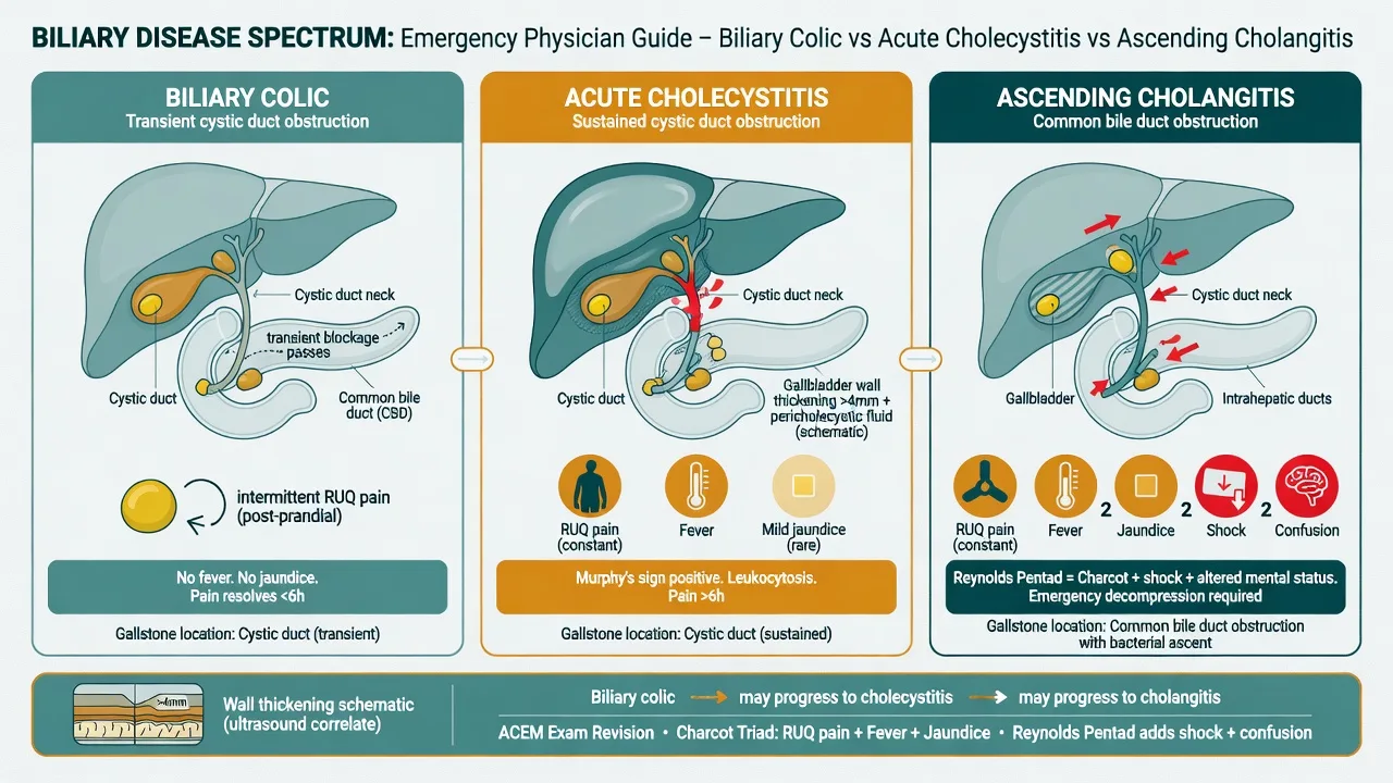
The biliary disease spectrum — biliary colic, the transient cystic duct obstruction with self-limiting post-prandial RUQ pain managed by elective cholecystectomy; acute cholecystitis, the sustained obstruction with RUQ tenderness, fever, a positive Murphy sign, ultrasound wall thickening over 3 mm and pericholecystic fluid, managed with analgesia, ceftriaxone 2 g IV plus metronidazole 500 mg IV, and early laparoscopic cholecystectomy; and ascending cholangitis, the obstructed infected biliary tree with Charcot triad and Reynolds pentad, managed with antibiotics, fluid resuscitation and urgent ERCP decompression. ACEM-primary, globally tagged.
On this page & tools
Your progress
Saved locally on this device.
5 MCQs with explanations
Target exams
Red flags
Related topics
- Acute pancreatitis
- Acute abdominal pain — the emergency department approach
- Sepsis and septic shock — the emergency department approach
- Upper gastrointestinal bleed
- Acute coronary syndromes (STEMI, NSTEMI and unstable angina)
- Ectopic pregnancy
- Point-of-care ultrasound: biliary and renal (and the incidental AAA)
Biliary disease is a spectrum defined by where in the biliary tree a gallstone obstructs and how the body responds to that obstruction. The Fellowship candidate must hold three distinct conditions in mind at once — biliary colic (transient cystic duct obstruction, self-limiting), acute cholecystitis (sustained cystic duct obstruction with gallbladder inflammation) and acute (ascending) cholangitis (obstruction of the common bile duct with infection and sepsis) — because the disposition, the urgency and the drug doses diverge sharply across them. The emergency task is to place each patient correctly on the spectrum at the bedside, exclude the lethal mimics, and escalate the obstructed septic patient to urgent biliary drainage before organ failure develops.[1][2]
Definition and classification
Biliary colic is symptomatic uncomplicated gallstone disease: a gallstone transiently impacts the cystic duct or Hartmann pouch during a post-prandial gallbladder contraction, generates visceral pain that typically lasts one to six hours, and then dislodges or passes, so the pain resolves spontaneously and there is no gallbladder inflammation or infection. Acute cholecystitis is sustained obstruction of the cystic duct producing gallbladder wall inflammation — chemical and ischaemic injury first, bacterial superinfection second — with fever, right upper quadrant tenderness and a positive Murphy sign. Roughly 90 per cent is calculous (a stone); about 10 per cent is acalculous, arising in the critically ill, the septic, the burned or the fasting patient on total parenteral nutrition, and carrying a much higher rate of gangrene and perforation. Acute cholangitis is bacterial infection of an obstructed bile duct, classically ascending from the duodenum, producing the systemic inflammatory and septic response that defines this as a medical and procedural emergency.[1][2]
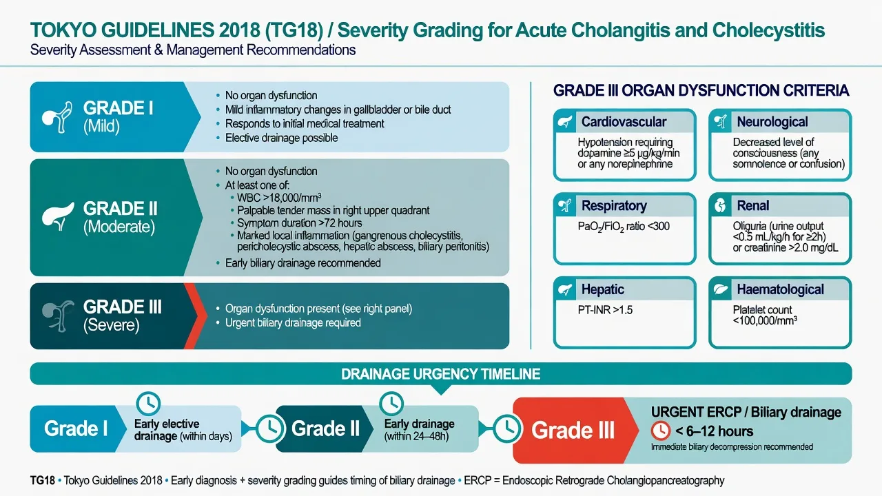
The Tokyo Guidelines (TG18) grade severity consistently across the spectrum. Acute cholecystitis is mild (Grade I) when there is no organ dysfunction and no marked local inflammation, moderate (Grade II) when there is marked local inflammation (gangrene, abscess, pericholecystic gas, perforation) or systemic inflammation that does not respond to initial treatment, and severe (Grade III) when there is organ dysfunction requiring supportive care.[2] Acute cholangitis is graded the same way: severe (Grade III) cholangitis is defined by organ dysfunction — cardiovascular (hypotension requiring vasopressors), neurological (disturbed consciousness), respiratory (oxygen required), renal (oliguria or creatinine over 2 mg/dL), hepatic (INR over 1.5) or haematological (platelet count under 100) — and mandates urgent biliary drainage.[1]
Epidemiology and risk
Gallstones are present in 10 to 15 per cent of adults in Western populations; most remain asymptomatic, but each year roughly 1 to 3 per cent of carriers become symptomatic. About 80 per cent of stones are cholesterol stones, formed when bile becomes supersaturated with cholesterol and nucleation occurs in a sluggish gallbladder; the remainder are pigment stones (black, from haemolysis and cirrhosis; brown, from biliary stasis and bacterial infection of bile). The traditional risk profile is captured, loosely, by the five F's. [1]
The 5 F's — risk factors for cholesterol gallstones
5 F's
Oestrogen increases cholesterol saturation of bile
Rising prevalence with age
Oestrogen and progesterone; multiparity
Increased biliary cholesterol secretion
Historical marker of higher-risk populations
The clinically useful risk list runs wider than the mnemonic. Rapid weight loss (including post-bariatric surgery), oestrogen therapy and oral contraceptives, terminal ileal disease (Crohn's, resection) impairing bile-salt reabsorption, cirrhosis, total parenteral nutrition, diabetes mellitus, and the haemoglobinopathies (sickle cell, hereditary spherocytosis, thalassaemia) for pigment stones all raise risk. Certain ethnicities — North American Pima Indians, South Americans, Northern Europeans — have markedly higher gallstone prevalence. A family history roughly doubles individual risk. [1]
Pathophysiology
Cholesterol stones form when bile is supersaturated with cholesterol relative to its bile-salt and phospholipid content, and when gallbladder hypomotility allows nucleation into crystals that aggregate into stones. Pigment stones form when unconjugated bilirubin precipitates — classically from chronic haemolysis (black pigment) or from bacterial deconjugation of bilirubin in an obstructed, infected duct (brown pigment). [1]
[1]In cholecystitis the obstructed gallbladder becomes inflamed through three overlapping mechanisms: chemical injury from concentrated bile and lysolecithin, ischaemic injury from venous and lymphatic obstruction as the wall distends, and bacterial infection in roughly half (Escherichia coli, Klebsiella, Enterococcus, anaerobes). Untreated, the natural history is progression through gangrene (most often at the fundus, the least vascular part), perforation (localised into a pericholecystic abscess, or free into the peritoneum producing biliary peritonitis), and empyema. In cholangitis the raised intraductal pressure forces bacteria-laden bile into the systemic circulation via the hepatic sinusoids and lymphatics, so bacteraemia is common and septic shock develops quickly: blood cultures are positive in roughly a third of patients. [1]
Clinical presentation
Biliary colic presents as severe, gripping right upper quadrant or epigastric pain that begins an hour or two after a meal (classically a fatty meal, through cholecystokinin release), builds to a plateau over minutes to an hour, lasts one to six hours, and then gradually resolves as the stone dislodges. Nausea and vomiting are common. The patient is afebrile, may be writhing during the attack, and is entirely well and non-tender between attacks — the resolution of pain and the absence of fever distinguish it from cholecystitis. Presentation atypical or referral pain to the right scapular tip or interscapular area is common. [1]
Acute cholecystitis begins like colic but the pain does not resolve — it persists beyond six hours, and fever, anorexia, nausea and right upper quadrant tenderness develop. The classic bedside sign is Murphy sign: the examiner's hand is placed at the costal margin in the right upper quadrant, the patient is asked to breathe in deeply, and the sign is positive when inspiration is arrested by pain as the inflamed gallbladder contacts the examining hand; the same manoeuvre on the left is negative. The sensitivity is high but it falls in the elderly, the diabetic and the chronically ill. Mild jaundice may occur from concomitant common-duct stones or from Mirizzi syndrome, but frank jaundice should always raise the question of cholangitis. [1]
Acute cholangitis presents with the Charcot triad — right upper quadrant pain, fever (often with rigors) and jaundice — in roughly 50 to 70 per cent of cases. The full Reynolds pentad adds hypotension and a depressed conscious level and marks severe, suppurative disease. Presentation in the elderly or diabetic is frequently atypical: confusion may be the dominant feature, pain minimal, and fever absent, so any septic patient with obstructive liver enzymes and biliary dilation has cholangitis until proven otherwise. Hypotension, oliguria, a raised lactate and a falling conscious level herald septic shock and demand immediate resuscitation and urgent biliary drainage. [1]
The clinical markers — Charcot and Reynolds
Differential diagnosis
Right upper quadrant pain overlaps with several lethal and several benign mimics, and the first task is to exclude a perforation, an inferior myocardial infarction, a septic source in the biliary tree and acute pancreatitis before attributing the picture to simple colic. [1]
Acute cholangitis
- RUQ pain, fever with rigors, jaundice (Charcot triad)
- Reynolds pentad adds hypotension and confusion — severe disease
- Obstructive LFTs: raised bilirubin, ALP, GGT; dilated CBD on ultrasound
- Blood cultures positive in ~30 %; needs urgent ERCP
Acute cholecystitis
- Persistent RUQ pain over six hours with fever
- Positive Murphy sign; RUQ tenderness and guarding
- Ultrasound: stones, wall over 3 mm, pericholecystic fluid
- Bilirubin usually near-normal; jaundice suggests a CBD stone
Biliary colic
- Post-prandial RUQ pain lasting one to six hours, self-limiting
- Afebrile, non-tender between attacks
- Stones on ultrasound but normal wall and no fluid
- Managed as an outpatient with elective cholecystectomy
Acute pancreatitis
- Severe epigastric pain radiating straight through to the back
- Lipase three times the upper limit of normal
- Often gallstone-related; can coexist with cholangitis
- CT shows peripancreatic fat stranding
Acute viral or alcoholic hepatitis
- RUQ pain and tenderness with marked transaminitis
- Predominantly AST and ALT in the hundreds to thousands
- Viral serology, alcohol history, drug history
- Ultrasound normal; bilirubin may be raised but no duct dilation
Perforated peptic ulcer
- Sudden severe epigastric pain becoming generalised
- Rigid, board-like abdomen; loss of liver dullness
- Erect chest X-ray or CT shows free gas
- Lipase and bilirubin usually normal
Inferior myocardial infarction
- Epigastric pain, nausea, diaphoresis in the older patient
- ECG shows inferior ST elevation (II, III, aVF)
- Always obtain an ECG in epigastric or RUQ pain
- Bilirubin, ALP and lipase normal
Right lower lobe pneumonia
- Pleuritic right-sided chest pain with fever and cough
- Pleural rub or crackles at the right base
- Chest X-ray shows right lower lobe consolidation
- Abdominal examination comparatively benign
Other considerations include hepatic abscess (continuing fever, right pleuritic pain, a collection on imaging, often in the immunocompromised or post-biliary-sepsis patient), pyelonephritis or nephrolithiasis (pain radiating to the groin, urinalysis, no jaundice), Fitz-Hugh-Curtis syndrome (perihepatitis from pelvic inflammatory disease — the young woman with RUQ pleuritic pain), acute appendicitis (especially in pregnancy where the appendix is displaced upward), and Mirizzi syndrome (a stone impacted in the cystic duct or Hartmann pouch compressing the common hepatic duct, producing obstructive jaundice with an intact common duct). [1]
Bedside assessment
Begin with airway, breathing and circulation — the patient with ascending cholangitis may be in established septic shock within the first hour of presentation. Give oxygen, attach monitoring, establish two large-bore intravenous cannulae, and obtain intravenous access for fluid and vasoactive drugs. A focused history establishes the pain pattern (onset after food, duration, persistence or resolution), the presence of fever, rigors, jaundice, dark urine and pale stools, and the risk factors (family history, recent weight loss, oestrogen use, known gallstones, haemoglobinopathy). Examination looks for jaundice, the scratch marks of chronic pruritus, right upper quadrant tenderness, a positive Murphy sign, guarding or peritonism (perforation), and signs of chronic liver disease. In the older or diabetic patient, explicitly assess the conscious level and the haemodynamics for the early features of Reynolds pentad. An explicit screen for organ dysfunction — oxygen saturation, respiratory rate, blood pressure, urine output, conscious level and lactate — determines whether this is a ward, high-dependency or intensive-care admission, and whether biliary drainage is an emergency rather than an urgent task. [1]
Investigations
Send blood for a full blood count (leukocytosis in cholecystitis and cholangitis), urea and electrolytes, liver function tests, coagulation, C-reactive protein, amylase or lipase (to exclude pancreatitis), glucose and a venous lactate. The liver function tests are the diagnostic pivot: a predominantly obstructive picture (raised bilirubin, alkaline phosphatase and gamma-glutamyl transferase) with right upper quadrant pain and fever points to a biliary source, while very high aspartate aminotransferase and alanine aminotransferase (in the hundreds or thousands) with obstructive change is "gallstone hepatitis" — a stone lodged at the ampulla — and may precede clinical pancreatitis. Take blood cultures before antibiotics in any febrile or septic patient; they are positive in roughly a third of cholangitis. Check a lipase to exclude pancreatitis, and obtain an ECG and troponin in any older patient with epigastric or right upper quadrant pain to exclude an inferior myocardial infarction. [1]
Abdominal ultrasound is the first-line imaging test for all three conditions. It identifies gallstones (echogenic foci with acoustic shadowing), measures gallbladder wall thickening over 3 mm, detects pericholecystic fluid, elicits a sonographic Murphy sign (maximal tenderness over the sonographically identified gallbladder), and measures the common bile duct diameter (normally up to about 6 mm in the adult, increasing by roughly 1 mm per decade after 60). A duct over 7 mm with jaundice suggests choledocholithiasis. Ultrasound is operator-dependent and limited by bowel gas, but it is fast, bedside-available and radiation-free. Magnetic resonance cholangiopancreatography (MRCP) is the non-invasive test of choice for the common-duct stone when ultrasound is equivocal or the duct is dilated. Computed tomography is reserved for the complicated picture — suspected perforation, empyema, gas in the gallbladder wall (emphysematous cholecystitis), or when the diagnosis is unclear — and for the patient in whom pancreatitis is also suspected. A HIDA scan (hepatobiliary iminodiacetic acid scintigraphy) is the gold standard for suspected acalculous cholecystitis: non-filling of the gallbladder confirms cystic duct obstruction even when no stone is seen. [1]
[1]Severity grading — the Tokyo Guidelines
The Tokyo Guidelines 2018 (TG18) provide the diagnostic criteria and severity grading used worldwide and in the Fellowship exam.[1][2] The diagnostic criteria for acute cholecystitis require one local sign (Murphy sign, right upper quadrant mass, pain or tenderness), one systemic sign (fever, raised inflammatory marker, raised white cell count) and confirmatory imaging (ultrasound, CT or HIDA). The criteria for acute cholangitis require evidence of systemic inflammation, cholestasis (raised bilirubin, ALP, GGT) and biliary obstruction (dilated duct, stone, stricture) or imaging confirming the cause. Charcot triad alone is no longer required to diagnose, but its presence strongly supports the diagnosis.
TG18 severity grading of acute cholangitis
For cholecystitis the same three grades apply: mild disease has no organ dysfunction and no marked local complication and settles with supportive care; moderate disease has marked local inflammation (gangrene, abscess, emphysematous change, perforation) and needs early cholecystectomy; severe disease has organ dysfunction demanding intensive care and cholecystostomy or cholecystectomy once stabilised. The operational point for the emergency clinician is identical across both conditions: organ dysfunction defines the severe patient, and the severe patient needs urgent biliary drainage. [1]
Immediate management and resuscitation
The management diverges sharply by diagnosis. Biliary colic is managed with analgesia (paracetamol 1 g orally, or a non-steroidal such as diclofenac 75 mg intramuscularly or ibuprofen 400 mg orally, with an opioid such as morphine 5 to 10 mg intravenously for severe pain), an antiemetic (ondansetron 4 mg intravenously or metoclopramide 10 mg intravenously), and discharge with surgical outpatient follow-up for an elective laparoscopic cholecystectomy. The patient must be warned of the return precautions — new fever, persistent pain, jaundice or dark urine, all of which signal progression to cholecystitis or cholangitis. [1]
Acute cholecystitis is admitted. Give nothing by mouth, establish intravenous access, give intravenous fluids (a balanced crystalloid such as Hartmann solution or Plasma-Lyte, 1 to 1.5 mL per kilogram per hour, titrated to urine output), and provide analgesia (morphine 2.5 to 5 mg intravenously titrated, or fentanyl 50 to 100 micrograms intravenously) and an antiemetic (ondansetron 4 mg intravenously). Start antibiotics as soon as cultures are drawn — the Tokyo Guidelines first-line regimen for mild to moderate disease is ceftriaxone 2 g intravenously once daily plus metronidazole 500 mg intravenously every eight hours.[3] For severe disease, penicillin allergy, or where local resistance is high, escalate to piperacillin-tazobactam 4.5 g intravenously every eight hours, or a carbapenem (meropenem 1 g intravenously every eight hours) in the septic or previously-instrumented patient. The antibiotics cover the typical biliary flora — the Gram-negative aerobes Escherichia coli, Klebsiella and Enterobacter, the Enterococci, and the anaerobes (Bacteroides, Clostridium) when disease is severe or there has been previous biliary surgery.
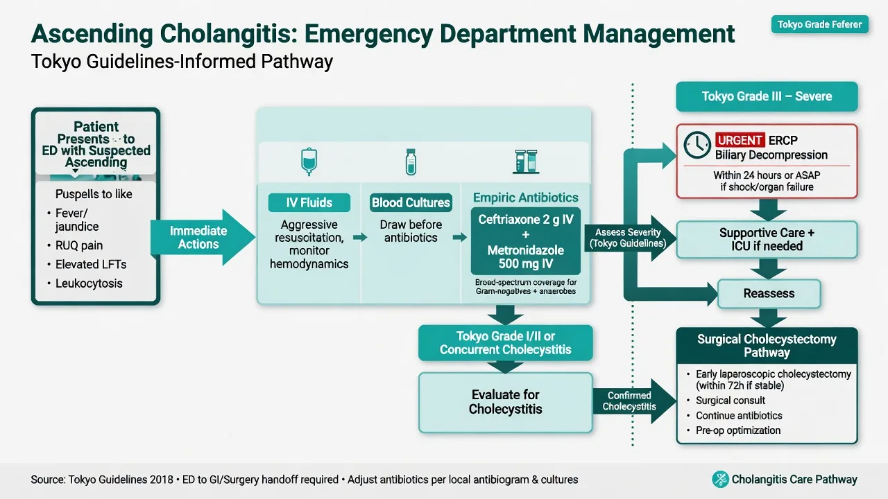
Acute cholangitis is a resuscitation and drainage emergency. Apply the sepsis six within the first hour: oxygen, two large-bore cannulae, give a 20 to 30 mL per kilogram crystalloid bolus (balanced crystalloid) titrated to mean arterial pressure and urine output, take blood cultures and a lactate before antibiotics, give broad-spectrum antibiotics within the hour (ceftriaxone 2 g intravenously plus metronidazole 500 mg intravenously, or piperacillin-tazobactam 4.5 g intravenously for severe disease), start a noradrenaline infusion for refractory septic shock (titrated to mean arterial pressure over 65 mmHg), and catheterise to monitor urine output. Broad-spectrum antibiotics alone do not relieve the obstruction — the definitive treatment of severe cholangitis is urgent biliary decompression by ERCP, ideally within 12 hours of grade III disease and within 24 to 48 hours of grade II. ERCP achieves sphincterotomy, stone extraction and stent placement across the obstruction, and reduces mortality dramatically. When ERCP fails or is unavailable, percutaneous transhepatic biliary drainage (PTBD) or surgical drainage are the fallback. [1]
[1]Definitive management and disposition
Biliary colic is referred for an elective laparoscopic cholecystectomy; surgery can be delayed safely, but recurrence is common and roughly 1 to 3 per cent of colic patients develop cholecystitis, cholangitis or pancreatitis each year. A patient with one attack is counselled to return immediately if fever, persistent pain or jaundice develop. [1]
Acute cholecystitis is managed with admission, antibiotics and early laparoscopic cholecystectomy within 72 hours of symptom onset. The Cochrane review of early versus delayed cholecystectomy for acute cholecystitis found that early surgery reduced complications, shortened hospital stay and did not increase conversion to open operation compared with a delayed cholecystectomy weeks later.[4] The 72-hour window matters because beyond it the inflammatory adhesions become dense and the conversion rate rises. For the high-risk surgical candidate — severe comorbidity, advanced age, or the acalculous patient in intensive care — a percutaneous cholecystostomy drains the obstructed gallbladder and deflates the sepsis, with cholecystectomy deferred or avoided entirely. Antibiotics are continued until the systemic inflammation resolves (typically five to seven days) and the patient is fit for surgery.
Acute cholangitis is managed by resuscitation, antibiotics and urgent ERCP. Grade III (severe) cholangitis goes to intensive care with the biliary drain as the central intervention; Grade II is drained early (within 24 to 48 hours); Grade I may settle on antibiotics alone but most centres drain to remove the source. After drainage and recovery, the gallbladder is removed electively if it still contains stones, to prevent recurrence. Acalculous cholecystitis in the critically ill is managed by percutaneous cholecystostomy as the first intervention. Emphysematous cholecystitis (gas in the gallbladder wall, classically in the diabetic older man from Clostridium or gas-forming Gram-negatives) is treated with broad-spectrum antibiotics covering anaerobes and an emergency cholecystectomy because of the high rate of gangrene and perforation. [1]
Complications and pitfalls
Gallbladder complications include gangrene (ischaemic necrosis, most often at the fundus), perforation (localised into a pericholecystic abscess or free into the peritoneum causing biliary peritonitis), empyema (a gallbladder filled with pus — effectively an abscess, treated with drainage), and emphysematous cholecystitis. Biliary-duct complications include cholangitis and gallstone pancreatitis (a stone obstructing the ampulla, often after passing through the cystic duct). Two rarer phenomena are exam favourites: Mirizzi syndrome, in which a stone impacted in the cystic duct or Hartmann pouch externally compresses the common hepatic duct to cause obstructive jaundice, and gallstone ileus, in which a large stone erodes through a cholecystoduodenal fistula and lodges at the terminal ileum causing small-bowel obstruction — the Rigler triad of pneumobilia, small-bowel obstruction and an ectopic gallstone on imaging makes the diagnosis. [1]
The common pitfalls invert good care. Treating persistent fever and tenderness as biliary colic misses cholecystitis. Drawing blood cultures after antibiotics lowers the yield and removes the microbiological guide. Delaying ERCP in severe cholangitis while waiting for the gastroenterology review kills patients — organ dysfunction mandates drainage within hours. Missing an inferior myocardial infarction in the older patient with epigastric pain and gallstones is avoided by an ECG on every such patient. Missing acalculous cholecystitis in the intensive-care patient with sepsis of unknown source is avoided by ultrasound (and HIDA when the picture is equivocal) in any ventilated patient with new fever and right upper quadrant collection. Overlooking emphysematous cholecystitis in the diabetic delays emergency surgery. [1]
Prognosis and disposition
Biliary colic is benign — discharge with elective surgical referral and return precautions. Acute cholecystitis settles on antibiotics and early cholecystectomy in over 95 per cent of cases; mortality is under 1 per cent in the otherwise well, rising in the elderly and the acalculous intensive-care patient. Acute cholangitis carries a mortality that rises sharply with severity — modern series report overall mortality around 2 to 5 per cent but up to 10 to 30 per cent in severe disease with organ dysfunction, and the single intervention that transforms outcome is timely biliary drainage.[1] Disposition follows severity: biliary colic to home; mild to moderate cholecystitis to a surgical ward with a plan for early cholecystectomy; severe cholecystitis or any cholangitis with organ dysfunction to high-dependency or intensive care with the drainage pathway activated.
Special populations
Pregnancy: biliary colic and cholecystitis are common in pregnancy because of the oestrogen-driven rise in biliary cholesterol. The first episode of colic is managed conservatively; recurrent symptomatic disease or cholecystitis in the second trimester is treated with laparoscopic cholecystectomy, which is safe in pregnancy and preferable to the risks of progression to pancreatitis or preterm labour from ongoing sepsis. The elderly present atypically — confusion rather than localised pain, minimal fever, an unremarkable white cell count — and tolerate untreated sepsis poorly; a high index, early imaging and early drainage are essential. Acalculous cholecystitis is more common in this group. The critically ill patient (sepsis, burns, major surgery, trauma, prolonged fasting, total parenteral nutrition) develops acalculous cholecystitis from gallbladder ischaemia and stasis; ultrasound or HIDA confirms, and percutaneous cholecystostomy is the first intervention. The diabetic is at particular risk of emphysematous cholecystitis and of severe, rapidly progressive infection. Children with gallstones usually have an underlying cause — haemolytic disease (sickle cell, spherocytosis), a biliary anomaly, or total parenteral nutrition — and are managed along the same algorithm. [1]
Evidence and regional guidelines
The contemporary framework is the Tokyo Guidelines 2018 (TG18), which unified the diagnostic criteria and severity grading for acute cholecystitis and cholangitis, codified the antibiotic regimens by severity, and formalised the timing of biliary drainage (urgent for severe, early for moderate).[1][2][3] The practice-defining surgical evidence is the Cochrane review of early versus delayed laparoscopic cholecystectomy for acute cholecystitis, which established that surgery within the first 72 hours reduces complications and length of stay without raising the conversion rate.[4] The WSES 2022 guidelines for acute calculous cholecystitis reinforce the same principles, and the NICE (United Kingdom) guideline on gallstone disease endorses early laparoscopic cholecystectomy and risk-stratifies the surgical candidate. Across all guidelines, the three fixed points are: biliary colic is managed electively; acute cholecystitis is admitted, given antibiotics and operated early; and acute cholangitis is resuscitated, given antibiotics and drained urgently.
ANZ practice note. Australasian emergency practice follows the Tokyo Guidelines. Ceftriaxone 2 g intravenously daily plus metronidazole 500 mg intravenously every eight hours is the standard empirical regimen for community-acquired biliary sepsis, escalating to piperacillin-tazobactam for severe disease or the healthcare-associated case. ERCP is the drainage modality of choice and is available around the clock in the larger centres; smaller and rural hospitals rely on transfer or, where transfer is delayed, percutaneous cholecystostomy by radiology to bridge the septic patient. Laparoscopic cholecystectomy within 72 hours of acute cholecystitis is the surgical standard. Sepsis resuscitation follows the Surviving Sepsis Campaign bundle, with early antibiotics and a low threshold for early goal-directed therapy in the cholangitis patient. [1]
Resuscitation and drainage pathways
The two time-critical pathways — cholangitis and cholecystitis — converge on the same principle: relieve the obstruction. The flowcharts below encode the Tokyo Guidelines 2018 initial management sequence and the Surviving Sepsis Campaign hour-1 bundle.[5][6]
Acute cholangitis — the sepsis-to-drainage pathway (ED to ERCP)
0 to 15 min — recognise sepsis and resuscitate
ABCDE, high-flow oxygen, two large-bore cannulae, full monitoring. Identify Charcot triad (RUQ pain, fever with rigors, jaundice) and screen hard for Reynolds pentad (add hypotension and confusion). Take venous lactate — a raised lactate with hypotension is grade III until proven otherwise and activates the drainage timer.
15 to 45 min — cultures, lactate and antibiotics within the hour
Draw two sets of blood cultures and a bile culture (if instrumented) BEFORE antibiotics. Give empiric ceftriaxone 2 g IV plus metronidazole 500 mg IV, or piperacillin-tazobactam 4.5 g IV for severe or healthcare-associated disease. Send FBC, U&E, LFTs, coagulation, CRP, glucose, amylase/lipase. Catheterise for hourly urine output.
45 to 60 min — ultrasound and source confirmation
Bedside or formal RUQ ultrasound for CBD diameter (over 6 to 7 mm, plus 1 mm per decade after sixty) and obstruction. Cross-check against obstructive LFTs (raised bilirubin, ALP, GGT). MRCP if ultrasound equivocal and the patient is stable; do NOT delay drainage for MRCP in grade III disease.
0 to 6 h — fluid resuscitation and vasopressors
Give 30 mL/kg balanced crystalloid bolus titrated to MAP over 65 mmHg and urine output over 0.5 mL/kg/h. Add noradrenaline for refractory septic shock. Correct coagulopathy (vitamin K, fresh frozen plasma) only if it would delay ERCP.
Urgent (grade III) or 24 to 48 h (grade II) — biliary decompression
ERCP with sphincterotomy, stone extraction and nasobiliary drain or stent is first-line. Severe (grade III) cholangitis with organ dysfunction is drained urgently, ideally within 12 hours. Percutaneous transhepatic biliary drainage (PTBD) is the fallback when ERCP fails or is unavailable.
Post-drainage — ICU, de-escalate antibiotics, plan cholecystectomy
Continue antibiotics for four to seven days post-drainage, de-escalate to culture-directed therapy. Arrange interval laparoscopic cholecystectomy if the gallbladder still contains stones to prevent recurrence. Address the cause — stones, stricture, malignancy, stent occlusion.
Acute cholecystitis — the ED-to-theatre pathway (within 72 hours)
0 to 30 min — recognise and risk-stratify
Persistent RUQ pain beyond six hours with fever distinguishes cholecystitis from colic. Elicit Murphy sign and confirm severity: mild (no organ dysfunction, no local complication), moderate (gangrene, abscess, emphysematous change, perforation), severe (organ dysfunction). Run a NEWS/MEWS and an ECG on every older patient.
30 to 60 min — analgesia, fluids and antibiotics
NBM, IV access, balanced crystalloid. Morphine 2.5 to 5 mg IV titrated for pain; ondansetron 4 mg IV. Start TG18 first-line antibiotics once cultures are drawn: ceftriaxone 2 g IV daily plus metronidazole 500 mg IV every eight hours for mild to moderate disease.
1 to 2 h — ultrasound confirmation
Confirm at least two of: gallstones, wall thickening over 3 mm, pericholecystic fluid, sonographic Murphy sign, gallbladder distension. Measure CBD; a bilirubin over 30 to 40 micromol/L or a dilated duct prompts MRCP to exclude choledocholithiasis before theatre.
Admit and fix the surgical window
Admit under the surgical team. Book laparoscopic cholecystectomy WITHIN 72 HOURS of symptom onset — early surgery reduces complications and length of stay without increasing conversion. Beyond the window, dense inflammatory adhesions raise the open-conversion rate.
High-risk or unstable — cholecystostomy instead
For the elderly, frail, acalculous or critically ill patient unsuitable for surgery, percutaneous cholecystostomy drains the obstructed gallbladder and deflates the sepsis. Interval cholecystectomy follows once the patient recovers, or is avoided entirely.
Antibiotic duration and follow-up
Continue antibiotics until systemic inflammation settles (typically five to seven days). Emphysematous or gangrenous cholecystitis needs broader cover (anaerobes, gas-formers) and an emergency operation, not a 72-hour window.
Pharmacology in depth — antibiotic selection
The biliary tree is colonised by enteric Gram-negatives and enterococci, with anaerobes joining when disease is severe, gangrenous or post-instrumentation. The Tokyo Guidelines 2018 stratify empiric therapy by severity, local resistance and allergy.[3] The aim is to cover Escherichia coli, Klebsiella, Enterobacter and Enterococcus, adding anaerobic cover (Bacteroides, Clostridium) for severe, complicated or healthcare-associated infection.
Mild cholecystitis / colic
- Ceftriaxone 2 g IV daily PLUS metronidazole 500 mg IV every 8 h
- Oral equivalent (co-amoxiclav 625 mg TDS) for the discharged or low-severity patient
- Duration 5 to 7 days or until systemic inflammation settles
- Blood cultures before the first dose
Moderate cholecystitis / cholangitis
- Ceftriaxone 2 g IV daily PLUS metronidazole 500 mg IV every 8 h (first line)
- Piperacillin-tazobactam 4.5 g IV every 8 h for healthcare-associated or severe local disease
- Add anaerobic cover for gangrene, empyema, perforation
- Send blood AND bile cultures if instrumented
Severe (Grade III) / septic shock
- Piperacillin-tazobactam 4.5 g IV every 6 to 8 h, OR
- Meropenem 1 g IV every 8 h for prior instrumentation, ESBL risk, or ICU-acquired infection
- Surviving Sepsis hour-1 bundle — antibiotics within 60 min
- De-escalate to culture-directed therapy post-drainage
Penicillin allergy
- Ciprofloxacin 400 mg IV every 12 h PLUS metronidazole 500 mg IV every 8 h
- Or a carbapenem (meropenem) for severe disease with anaphylaxis history
- Clarithromycin-based regimens are inadequate for severe sepsis
- Document the reaction type before choosing
Emphysematous cholecystitis
- Piperacillin-tazobactam 4.5 g IV every 8 h OR meropenem 1 g IV every 8 h
- Mandatory anaerobic cover (Clostridium, gas-forming Gram-negatives)
- Emergency cholecystectomy, not a 72-hour window
- High rate of gangrene and perforation
Drainage and source control
Definitive treatment of any obstructed biliary tree is to relieve the obstruction. The modality depends on where the stone sits, the patient's stability, and what is locally available.[1][5]
ERCP (first-line for choledocholithiasis / cholangitis)
- Endoscopic sphincterotomy, stone extraction, stent or nasobiliary drain
- Diagnostic and therapeutic in one procedure; low mortality
- Ideal within 12 h for grade III cholangitis, 24 to 48 h for grade II
- Risks: pancreatitis (3 to 5 %), bleeding, perforation, sedation in the septic patient
Percutaneous transhepatic biliary drainage (PTBD)
- Fallback when ERCP fails, is unavailable, or anatomy is altered (Billroth II, Roux-en-Y)
- Drains an obstructed duct above a high stricture or hilar tumour
- Risks: bleeding, bile leak, cholangitis flare, pneumothorax
- Bridge to definitive surgery or stenting
Percutaneous cholecystostomy
- First-line for acalculous cholecystitis in the critically ill and the frail elderly
- Drains the obstructed gallbladder and deflates the sepsis when surgery is too risky
- Bedside or radiology-guided; rapid source control
- Definitive cholecystectomy deferred or avoided entirely
Early laparoscopic cholecystectomy
- Definitive for acute calculous cholecystitis within 72 h of onset
- Cochrane evidence: fewer complications, shorter stay, no rise in conversion
- Also clears the gallbladder to prevent recurrent colic, cholangitis, pancreatitis
- Conversion to open if dense adhesions, gangrene, Mirizzi, unclear anatomy
Surgical bile duct exploration
- Reserved for the large or impacted CBD stone when ERCP fails
- Combined with cholecystectomy and T-tube placement
- Higher morbidity than ERCP; rare in modern practice
- Consider in centres without ERCP and a deteriorating septic patient
Landmark trial evidence
The Fellowship candidate must be able to quote the trials that define modern biliary practice — the diagnostic and severity framework (Tokyo Guidelines), the timing of surgery (Cochrane), the resuscitation standard (Surviving Sepsis Campaign) and the contemporary synthesis of grey areas.[1][2][3][4][6][7]
Tokyo Guidelines 2018 — diagnostic criteria and severity grading of acute cholangitis
Journal of Hepato-Biliary-Pancreatic Sciences
PMID 29032610
Key finding
The consensus update that unified the diagnostic criteria (systemic inflammation plus cholestasis plus biliary obstruction) and the three-grade severity scale for acute cholangitis, defining grade III by organ dysfunction (cardiovascular, neurological, respiratory, renal, hepatic, haematological) and mandating urgent biliary drainage.
Practice change
The framework that drives the timing of ERCP — severe disease is drained urgently (within hours), moderate disease early (24 to 48 h), and the exam answer to severity grading in biliary sepsis.
Tokyo Guidelines 2018 — diagnostic criteria and severity grading of acute cholecystitis
Journal of Hepato-Biliary-Pancreatic Sciences
PMID 29032636
Key finding
Defined the diagnostic criteria for acute cholecystitis (one local sign, one systemic sign, confirmatory imaging) and the three-grade severity scale, with grade III defined by organ dysfunction and grade II by marked local inflammation (gangrene, abscess, emphysematous change, perforation).
Practice change
Provides the ultrasound criteria (stones, wall over 3 mm, pericholecystic fluid, sonographic Murphy) and the severity-driven management: early cholecystectomy for moderate, cholecystostomy or surgery after stabilisation for severe.
Early versus delayed laparoscopic cholecystectomy for acute cholecystitis (Cochrane)
Cochrane Database of Systematic Reviews
PMID 23813477
Key finding
A systematic review of randomised trials showing that laparoscopic cholecystectomy within the first week of acute cholecystitis reduced total complications, wound infections and bile leaks, shortened hospital stay, and did not increase the conversion-to-open rate compared with delayed surgery weeks later.
Practice change
The evidence base for the 72-hour early-cholecystectomy rule taught in every emergency and surgical textbook — early is safe and superior.
Surviving Sepsis Campaign 2021 — international guidelines
Critical Care Medicine
PMID 34643578
Key finding
The international consensus bundle for sepsis and septic shock, recommending empiric broad-spectrum antibiotics within one hour, a 30 mL/kg crystalloid bolus, vasopressors to a MAP of 65 mmHg, lactate-guided resuscitation and rapid source control.
Practice change
The resuscitation backbone for grade III cholangitis — antibiotics and fluids buy time, but the bundle explicitly demands early source control, which for the biliary tree means ERCP.
Clinical update on acute cholecystitis and biliary pancreatitis (EClinicalMedicine)
EClinicalMedicine
PMID 39469538
Key finding
A contemporary review reconciling the Tokyo Guidelines with current evidence on timing of cholecystectomy, the role of cholecystostomy in the frail elderly, antibiotic de-escalation, and the grey areas around same-admission versus interval surgery for complicated disease.
Practice change
Reinforces early laparoscopic cholecystectomy within 72 h and clarifies when percutaneous drainage is preferable to surgery in the high-risk patient.
TG18 Grade I (mild)
- Cholangitis: responds to initial fluids and antibiotics; no organ dysfunction
- Cholecystitis: no organ dysfunction, no marked local complication
- Management: antibiotics, supportive care; drainage may be elective
- Mortality low (under 2 to 5 %)
TG18 Grade II (moderate)
- Cholangitis: does not respond to initial therapy
- Cholecystitis: marked local inflammation (gangrene, abscess, gas, perforation)
- Management: early biliary drainage within 24 to 48 h; early cholecystectomy
- Mortality intermediate
TG18 Grade III (severe)
- Defined by organ dysfunction: hypotension, confusion, hypoxia, oliguria, INR over 1.5, platelets under 100
- Management: ICU, Surviving Sepsis bundle, URGENT drainage (ideally within 12 h)
- ERCP as soon as the patient is resuscitated; PTBD if ERCP fails
- Mortality 10 to 30 % despite modern care
High-yield bedside and exam pearls
[1] [1] [1] [1] [1] [1] [1] [1] [1] [1]Exam pearls
- The spectrum in one line: colic is transient cystic duct obstruction and self-limiting; cholecystitis is sustained obstruction with fever and a positive Murphy sign; cholangitis is an obstructed infected duct with Charcot triad or Reynolds pentad.
- Charcot triad is right upper quadrant pain, fever (often with rigors) and jaundice; Reynolds pentad adds hypotension and confusion. The full pentad marks severe suppurative cholangitis.
- Murphy sign is inspiratory arrest on right upper quadrant palpation — high sensitivity for cholecystitis, lower in the elderly and diabetic.
- Ultrasound criteria: stones, wall over 3 mm, pericholecystic fluid, sonographic Murphy, gallbladder distension. A common duct over 6 to 7 mm with jaundice suggests a retained stone.
- Antibiotics for cholecystitis and mild cholangitis: ceftriaxone 2 g IV daily plus metronidazole 500 mg IV every eight hours (TG18 first line). Escalate to piperacillin-tazobactam 4.5 g IV every eight hours for severe disease.
- Acute cholecystitis: admit, NBM, fluids, analgesia, antibiotics, and early laparoscopic cholecystectomy within 72 hours — the Cochrane evidence shows fewer complications and shorter stay, no rise in conversion.
- Severe cholangitis is defined by organ dysfunction and is treated with urgent ERCP within hours, not a routine gastroenterology review.
- Antibiotics do not cure an obstructed infected duct — drainage is the definitive treatment.
- Gallstone hepatitis — AST and ALT in the hundreds or thousands with obstructive change — is a stone at the ampulla, and may precede pancreatitis.
- Emphysematous cholecystitis (gas in the wall, usually diabetic) needs broad-spectrum antibiotics covering anaerobes and emergency cholecystectomy.
- Always do an ECG in epigastric or right upper quadrant pain in the older patient — inferior myocardial infarction is the lethal mimic.
- Mirizzi syndrome and gallstone ileus (Rigler triad of pneumobilia, small-bowel obstruction, ectopic gallstone) are the classic exam rarer presentations. [1]
Exam practice
SAQ — Acute cholangitis with Reynolds pentad: the sepsis-to-drainage pathway
10 minutes · 10 marks
A 74-year-old woman with known gallstones is brought to the emergency department by her family with a 24-hour history of right upper quadrant pain, rigors and progressive drowsiness. On arrival she is confused (GCS 13), jaundiced and cool peripherally: temperature 39.1 degrees C, heart rate 128, blood pressure 82/48 (MAP 59), respiratory rate 28, SpO2 94 per cent on room air. The abdomen is tender in the right upper quadrant with a positive Murphy sign. Venous gas shows pH 7.28, lactate 4.1 mmol/L, bilirubin 96 micromol/L, ALP 380, ALT 110, INR 1.6, white cell count 24.5 x 10^9/L, creatinine 175 micromol/L. Bedside ultrasound shows a 9 mm common bile duct with an echogenic shadowing focus at the distal end and intrahepatic duct dilatation.
SAQ — Biliary colic versus acute cholecystitis: differentiation and disposition
10 minutes · 10 marks
A 42-year-old woman presents to the emergency department four hours after the onset of severe right upper quadrant pain that began an hour after a fatty takeaway meal. She has had two similar but shorter episodes over the past six months that settled spontaneously. Today the pain has not eased. She is afebrile (temperature 37.2 degrees C), heart rate 96, blood pressure 124/78, respiratory rate 18, SpO2 99 per cent on room air. The abdomen is locally tender in the right upper quadrant with a positive Murphy sign. Bilirubin 14 micromol/L, ALP 110, ALT 45, lipase 40 (normal), white cell count 11.8 x 10^9/L, CRP 22. Bedside ultrasound shows multiple gallstones, gallbladder wall thickness 4.5 mm, a thin rim of pericholecystic fluid and a sonographic Murphy sign; the common bile duct measures 5 mm.
Red flags
[1]References
- [1]Kiriyama S, Kozaka K, Takada T, et al. Tokyo Guidelines 2018: diagnostic criteria and severity grading of acute cholangitis (with videos) J Hepatobiliary Pancreat Sci, 2018.PMID 29032610
- [2]Yokoe M, Hata J, Takada T, et al. Tokyo Guidelines 2018: diagnostic criteria and severity grading of acute cholecystitis (with videos) J Hepatobiliary Pancreat Sci, 2018.PMID 29032636
- [3]Gomi H, Solomkin JS, Takada T, et al. Tokyo Guidelines 2018: antimicrobial therapy for acute cholangitis and cholecystitis J Hepatobiliary Pancreat Sci, 2018.PMID 29090866
- [4]Gurusamy KS, Davidson C, Gluud C, Davidson BR. Early versus delayed laparoscopic cholecystectomy for people with acute cholecystitis Cochrane Database Syst Rev, 2013.PMID 23813477
- [5]Miura F, Okamoto K, Takada T, et al. Tokyo Guidelines 2018: initial management of acute biliary infection and flowchart for acute cholangitis J Hepatobiliary Pancreat Sci, 2018.PMID 28941329
- [6]Evans L, Rhodes A, Alhazzani W, et al. Executive Summary: Surviving Sepsis Campaign: International Guidelines for the Management of Sepsis and Septic Shock 2021 Crit Care Med, 2021.PMID 34643578
- [7]Fugazzola P, Podda M, Tian BW, et al. Clinical update on acute cholecystitis and biliary pancreatitis: between certainties and grey areas EClinicalMedicine, 2024.PMID 39469538