EM · Procedural & diagnostic ED skills
Point-of-care ultrasound: biliary and renal (and the incidental AAA)
Also known as POCUS biliary · POCUS renal · Point-of-care ultrasound right upper quadrant · Bedside gallbladder ultrasound · Hydronephrosis ultrasound · Emergency renal ultrasound · RUQ ultrasound
Point-of-care ultrasound of the biliary tree, the kidneys and the incidental abdominal aortic aneurysm — the right-upper-quadrant scan for gallstones (hyperechoic with acoustic shadowing), gallbladder wall thickening over 3 millimetres, the sonographic Murphy sign and the common bile duct over 7 millimetres; the renal scan for hydronephrosis (graded mild to severe), stones and cysts; and the aorta measured in every older flank-pain patient to catch the aneurysm that masquerades as colic. Technique with the curvilinear probe and an appropriate depth; the differentials (the non-visualised post-prandial gallbladder, bowel gas, ascites); and the diagnostic accuracy (POCUS rules cholecystitis in, it does not rule it out). ACEM-primary, globally tagged.
On this page & tools
Your progress
Saved locally on this device.
Target exams
Red flags
Related topics
- Biliary disease — biliary colic, acute cholecystitis and ascending cholangitis
- Renal colic and nephrolithiasis
- Abdominal aortic aneurysm (ruptured and intact)
- Focused Assessment with Sonography in Trauma (FAST and E-FAST)
- Acute abdominal pain — the emergency department approach
- Acute pancreatitis
- Acute kidney injury
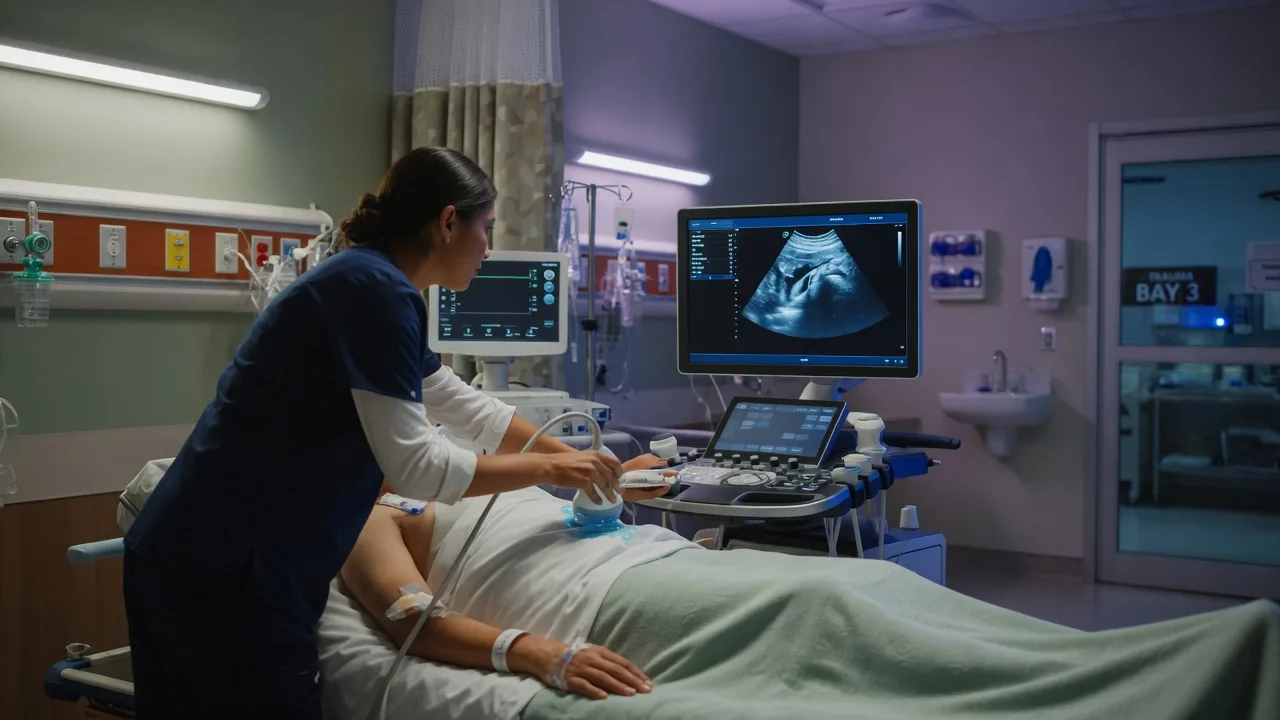
Point-of-care ultrasound (POCUS) of the biliary tree and the kidneys is a focused, clinician-performed bedside examination that answers three questions at once in the patient with abdominal or flank pain: is there evidence of gallbladder disease, is there hydronephrosis suggesting obstruction, and — the safety net that catches the lethal mimic — is there an abdominal aortic aneurysm. It is rapid, repeatable, free of ionising radiation and performed by the treating clinician, and it sits beside the history and the examination rather than waiting for the radiology department. Its power is as a rule-in tool: a gallbladder with stones, wall thickening and a sonographic Murphy sign strongly supports acute cholecystitis, and moderate-to-severe hydronephrosis predicts a larger, intervention-worthy stone. Its limitation — which the Fellowship candidate must hold at the front of the mind — is that a negative bedside scan never excludes disease, because a contracted post-prandial gallbladder, bowel gas and a stone lodged without upstream dilation all read falsely negative.[1][5]
Definition and the principle
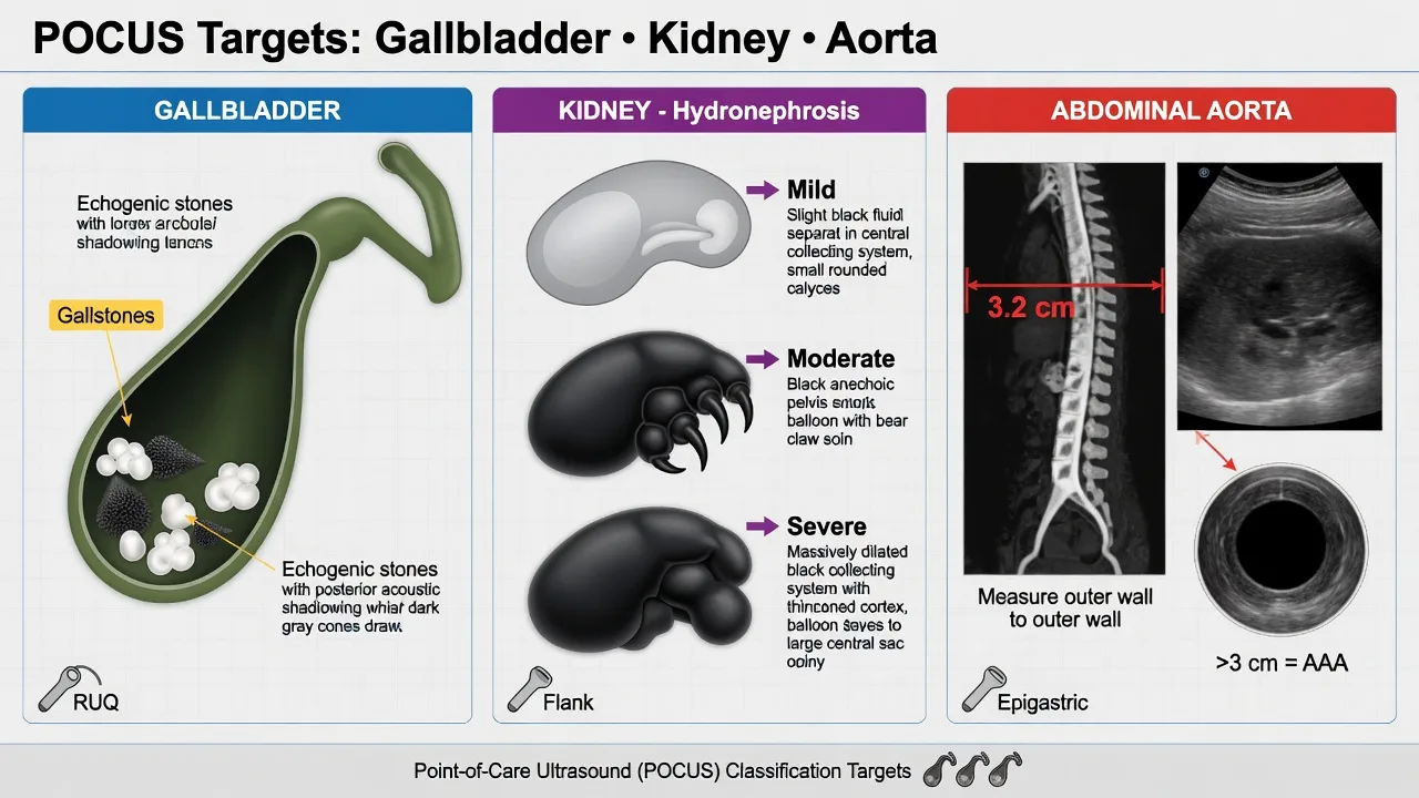
The biliary–renal POCUS is a goal-directed, rule-in examination built around three anatomical targets. The gallbladder is interrogated for stones (echogenic foci with clean acoustic shadowing), wall thickening (over 3 millimetres is abnormal), pericholecystic fluid and the sonographic Murphy sign (maximal tenderness elicited by the probe pressing directly on the visualised gallbladder).[2] The common bile duct is measured at the porta hepatis; an upper limit of around 4 millimetres widens by roughly 1 millimetre per decade after the age of 60, and a duct over 7 millimetres with jaundice and fever points to obstruction and ascending cholangitis. The kidneys are imaged in the long axis for hydronephrosis — graded mild (dilation of the renal pelvis and calyces with preserved parenchyma), moderate (ballooned calyces with thinning) and severe (cortical loss) — and for stones (echogenic with shadowing) and simple cysts (anechoic, well-defined, with posterior enhancement). The aorta is measured in the transverse plane at the renal hilum in every older flank-pain patient, because a diameter over 3 centimetres defines an aneurysm and a leak or rupture masquerades as renal colic.[4][5]
Indications
The biliary scan is indicated in the patient with right-upper-quadrant pain, fever, deranged liver enzymes or a clinical suspicion of acute cholecystitis or biliary colic — particularly the patient who is pregnant, where radiation is to be avoided. The renal scan is indicated in suspected renal colic (acute flank or loin pain radiating to the groin, with haematuria), where bedside hydronephrosis guides analgesia, predicts the larger stone and triages urology referral; it is the first-line imaging choice in pregnancy, recurrent stone-formers and the child, replacing CT KUB.[5] The aortic measurement is indicated in any patient over 50 with abdominal or flank pain in whom an aneurysm is a live differential, and it is built into the renal and biliary scan as a matter of routine so that the lethal mimic is not missed.
Contraindications — when not to delay
There is no absolute contraindication to a non-invasive bedside scan. The contraindication is to delaying a definitive intervention for the scan. The shocked patient with a tender pulsatile abdomen goes to theatre or CT, not to a prolonged POCUS; the septic cholangitis patient goes to decompression and antibiotics. An indeterminate scan in the morbidly obese patient, the patient with extensive bowel gas or the uncooperative agitated patient is documented as indeterminate and escalated to formal imaging, never falsely reported as negative. [1]
Differential diagnosis — the mimics and the false results
The Fellowship candidate must hold two questions at once: what could mimic the pathology, and what could generate a false result. Both explain most of the errors the scan produces. [1]
Biliary — stone + wall + Murphy
- Acute cholecystitis until proven otherwise; admit, analgesia, antibiotics, surgical review
- POCUS sensitivity ~70 to 75 per cent, specificity ~90 per cent — a strong rule-in, never a rule-out
- A negative scan with high suspicion goes to formal ultrasound or CT
- Combine with TG18 criteria: local + systemic + imaging = definite diagnosis
GB wall thickening — non-biliary causes
- Hypoalbuminaemia, hepatitis, congestive cardiac failure and chronic cholecystitis all thicken the wall without infection
- Post-prandial contracted gallbladder reads thick-walled and tender — always ask about recent food
- Correlate wall thickness with the systemic picture, never with one measurement alone
- Sludge (low-level echoes without shadowing) is a common incidental finding, not itself cholecystitis
Hydronephrosis — present but no stone
- Pregnancy hydronephrosis (right more than left), full bladder, prior obstruction, congenital PUJ obstruction
- Renal sinus fat can mimic mild dilation — scan with an empty bladder and in two planes
- An obstructed infected system (pyonephrosis) shows echogenic debris within the dilation
- Severity of hydronephrosis predicts stone size and urology need, not the diagnosis
Flank pain — the aneurysm
- Measure the aorta on every older renal or biliary scan — ruptured AAA presents identically to renal colic
- Aorta over 3 cm is aneurysmal; over 5.5 cm or tender and rapidly expanding is a surgical emergency
- Never attribute haematuria in the older patient to a stone until the aorta is cleared
- If visualised, call vascular, resuscitate, and image with CT only if stable
The false-negative causes deserve emphasis, because a falsely reassuring scan is the dangerous error. The contracted post-prandial gallbladder is thick-walled, tender and hard to see, so the biliary scan is best performed fasting. Bowel gas over the gallbladder fossa and the common bile duct obscures both, and repositioning the patient (left lateral decubitus, deep inspiration to bring the liver down) is the rescue manoeuvre. A stone in the cystic duct or neck may produce pathology the wall signs take hours to catch. On the renal side, a stone lodged without upstream dilation reads falsely negative for hydronephrosis, and the sensitivity of bedside POCUS for the stone itself is only around 70 per cent.[5][6]
Relevant anatomy and landmarks
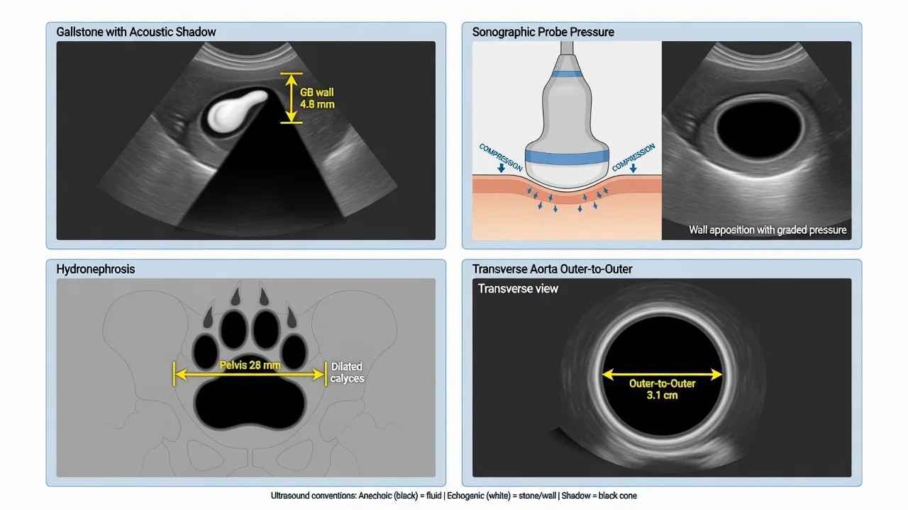
The gallbladder lies in the right upper quadrant, in the interlobar fissure (the main lobar fissure) between the right and the left liver lobes, anterior to the kidney. It is best found by placing the probe in the midclavicular line at the costal margin in a sagittal plane, asking the patient to inspire deeply to drive the liver (and the gallbladder) down beneath the ribs. The gallbladder appears as a pear-shaped anechoic (black) sac; the wall is the thin echogenic lining, and the wall thickness is measured in the anterior (non-dependent) wall, where reverberation artefact are least. The common bile duct runs anterior to the portal vein at the porta hepatis (the "Mickey Mouse" sign — the head is the portal vein, the ears are the bile duct and the hepatic artery), and is measured in its widest dimension.[4]
The kidneys lie in the retroperitoneum, the right lower than the left because of the liver. Each is approached in the posterior axillary line (coronal plane), the probe marker towards the head, with the liver (right) or the spleen (left) used as an acoustic window. The kidney appears as the echogenic renal sinus (the fat and the collecting system) surrounded by the hypoechoic cortex; hydronephrosis distends the central renal pelvis and the calyces with anechoic urine, confluent with the cortex. The aorta lies just left of midline in the epigastrium, anterior to the spine; in transverse it is the round pulsatile structure to the left of the larger, compressible inferior vena cava, and it is measured outer wall to outer wall in the anteroposterior dimension. [1]
Equipment and machine setup
A curvilinear probe (low-frequency 2 to 5 megahertz) is the workhorse for all three targets — the depth and the penetration it provides image the adult gallbladder, kidney and aorta that a high-frequency linear probe cannot reach. The depth is set initially at 15 to 18 centimetres for the adult abdomen so the target organ fills the screen, then adjusted to bring the region of interest to the mid-screen; the renal scan often needs a deeper setting than the gallbladder. The gain is set so that the urine in the bladder and the bile in the gallbladder are genuinely black (anechoic) and the parenchyma is mid-grey — too much gain fills anechoic fluid with artefact and hides small stones and mild hydronephrosis. The abdominal preset is selected; the scan is in two-dimensional B-mode. The probe marker convention is fixed (marker to the head for sagittal and coronal, to the patient's right for transverse) so images are reproducible. A phased-array probe is an acceptable alternative that fits the intercostal spaces better for the gallbladder when ribs obstruct the curvilinear.[2]
The normal measurements and the cutoffs
Patient preparation and positioning
The patient is examined supine, fully exposed from the xiphisternum to the pubis. For the gallbladder the patient is rolled into the left lateral decubitus position and asked to take a deep inspiration — this rotates the gallbladder away from the bowel gas and drives it beneath the costal margin beneath the acoustic window of the liver. For the kidneys the patient remains supine but the probe is moved to the flank and the patient may be rolled slightly to the contralateral side; deep inspiration brings the kidneys down. For the aorta the patient is supine with the knees flexed to relax the abdominal wall. A full bladder helps the pelvic view but distends the renal pelvis and can mimic mild hydronephrosis, so the renal scan is interpreted with the bladder state noted. Consent is implicit in the emergency; the scan is non-invasive and adds no radiation. [1]
Stepwise technique — the structured scan
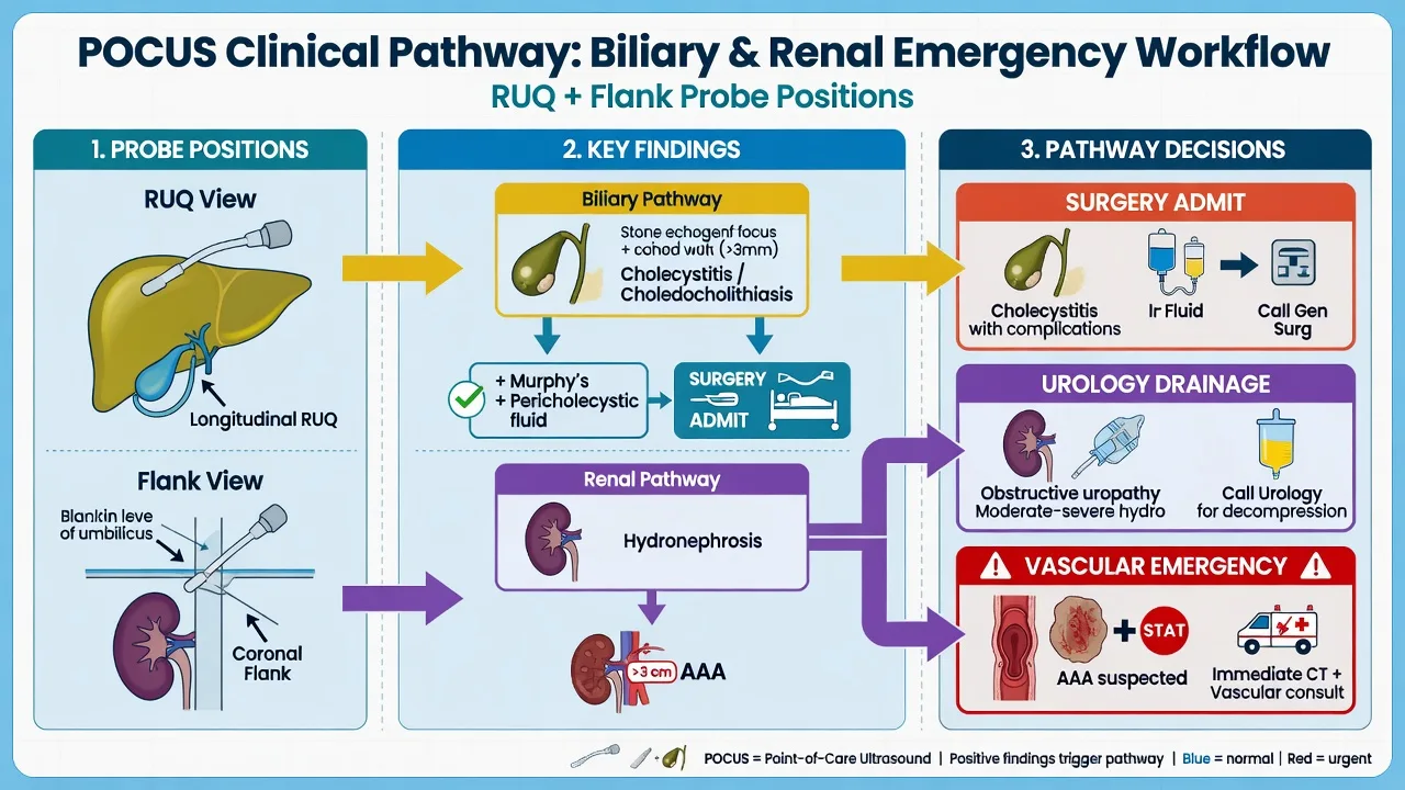
The scan is run to a fixed sequence so that no target is missed under pressure. The discipline is to image every structure in two planes (sagittal/sagittal-oblique and transverse for the gallbladder and the aorta; coronal long-axis and transverse for the kidney), to measure at the widest point, and to correlate the still image with the live finding (the Murphy sign, the respiratory collapse of the IVC). [1]
The biliary, renal and aortic scan, in order
- Gallbladder, sagittal. Place the curvilinear probe at the right costal margin in the midclavicular line, marker to the head. Ask the patient to inspire deeply. Sweep medially and laterally to find the pear-shaped anechoic sac. Identify stones (echogenic foci with clean shadowing), measure the anterior wall thickness, look for pericholecystic fluid, and elicit the sonographic Murphy sign (maximal tenderness on probe pressure over the visualised gallbladder).
- Gallbladder, transverse. Rotate 90 degrees to confirm the findings through the fundus, body and neck, and to catch a stone in the cystic duct or the neck that a single plane might miss.
- Common bile duct. Identify the portal vein and the "Mickey Mouse" sign at the porta hepatis; measure the duct (anterior wall to anterior wall) in its widest dimension and compare with the age-adjusted upper limit.
- Right kidney, coronal. Move the probe to the right posterior axillary line, marker to the head. Image the kidney in long axis through the liver window; grade any hydronephrosis and look for stones and cysts.
- Left kidney, coronal. Mirror the approach on the left (spleen window); the left kidney is often harder to see because of bowel gas over the splenic flexure.
- Aorta, transverse. Move to the epigastrium, marker to the patient's right. Identify the aorta (left, pulsatile, round) and the IVC (right, compressible). Measure the aorta outer-to-outer at the level of the renal hilum and the proximal segment; sweep from the diaphragm to the bifurcation to exclude an aneurysm. [1]
The two structural errors are to declare a single hard window "negative" when it was simply not seen, and to read the aorta as cleared without measuring it in the older flank-pain patient. Each target is reported as positive, negative or indeterminate, and the indeterminate is honestly labelled so it does not falsely reassure. [1]
Sonographic findings reproduced — the named criteria
The Tokyo Guidelines 2018 (TG18) diagnostic criteria for acute cholecystitis are reproduced here because the bedside scan supplies the imaging component (C) that the criteria require.[4]
TG18 diagnostic criteria for acute cholecystitis — definite diagnosis requires one item from A, one from B, and C: [1]
- A — local signs: Murphy's sign; right upper quadrant mass, pain or tenderness.
- B — systemic signs: fever; elevated inflammatory markers (C-reactive protein, white cell count).
- C — imaging findings: findings characteristic of acute cholecystitis on ultrasound (or CT, HIDA). [1]
The sonographic features of acute cholecystitis that supply C are, in descending order of yield: gallstones (especially an impacted stone in the neck or cystic duct), a positive sonographic Murphy sign, gallbladder wall thickening over 3 millimetres (best in the fasted patient), gallbladder distension (over 4 by 10 centimetres) and pericholecystic fluid. The combination of stones and a sonographic Murphy sign carries a positive predictive value of around 92 per cent.[2][3]
Hydronephrosis grading — the bedside renal scan grades dilation to predict the stone and the need for urology: [1]
- Mild — separation of the central renal sinus echo complex; calyces not blunted.
- Moderate — ballooning of the calyces with confluent anechoic spaces; cortical thinning begins.
- Severe — gross dilation with marked cortical loss; the kidney may appear as a multicystic structure. [1]
Moderate-to-severe hydronephrosis has a specificity of around 94 per cent for a stone over 5 millimetres, whereas mild or absent hydronephrosis does not exclude a stone.[5][6]
Diagnostic accuracy — the evidence
The biliary POCUS meta-analysis of Wu and colleagues pooled the emergency-physician-performed gallbladder scan and reported a sensitivity of around 70 to 75 per cent and a specificity of around 90 per cent for acute cholecystitis, comparable to radiology-performed ultrasound.[1] The foundational studies of Ralls (the sonographic Murphy sign) and Bree confirmed that the sign is highly useful when positive and combined with stones, but is operator-dependent and blunted by analgesia.[2][3] The renal POCUS meta-analysis of Wong and colleagues pooled five studies (over 1,700 patients) and reported a sensitivity of around 70 per cent and a specificity of around 75 per cent for hydronephrosis as a marker of a stone — too low to rule out nephrolithiasis, but useful to triage analgesia, predict the larger stone and avoid CT in the right patient.[5] The single-centre studies of Sibley and Al-Balushi confirmed the moderate accuracy and the operator-dependence, and warned against using the scan as a stand-alone rule-out.[6][7]
Resuscitation and the drugs that surround the scan
The POCUS itself requires no drug, but it is performed within the resuscitation that does. The Fellowship candidate must state the doses for the analgesia and the antibiotics that surround the biliary and the renal scan. [1]
The drugs that surround the biliary and renal scan
Diclofenac (75 milligrams intramuscularly, or 100 milligrams per rectum) is the first-line analgesic for ureteric colic — the systematic review confirms the NSAID is at least as effective as the opioid with fewer adverse effects.[5] Morphine (5 milligrams intravenously, titrated in 1 to 2 milligram increments) is added for refractory colic or biliary pain, blunting the catecholamine surge at the cost of sedation and hypotension that are titrated against. For the patient with acute cholecystitis or cholangitis, analgesia is paired with broad-spectrum antibiotics — piperacillin-tazobactam 4.5 grams intravenously (or the local equivalent) covering the gram-negative and anaerobic biliary flora, with the dose escalated to sepsis-dose coverage if the patient is shocked. The patient with pyonephrosis (the infected, obstructed, hydronephrotic kidney) gets antibiotics and urgent urological decompression, not medical expulsive therapy — tamsulosin has no role in the infected system.
Complications — the procedural and the interpretive
The scan is non-invasive and carries no procedural complications beyond probe pressure on a tender abdomen. The real complications are interpretive — the wrong disposition driven by a misread scan. The false positive over-calls wall thickening (the contracted post-prandial gallbladder, hepatitis, cardiac failure) or mild hydronephrosis (the full bladder, the pregnant patient, renal sinus fat), sending a patient to an unnecessary cholecystectomy or CT. The false negative withholds treatment from the patient with a contracted or gas-obscured gallbladder, a stone lodged without upstream dilation, or a missed aneurysm. The indeterminate scan reported as negative is the classic trap, and the rescue is honest reporting plus escalation to formal imaging when the clinical suspicion is high.[1][5]
Pitfalls and practical tips
The pitfalls invert the discipline. Scanning a recently fed patient — the contracted, thick-walled, tender gallbladder that mimics cholecystitis; always ask about the last meal. Measuring the dependent posterior wall of the gallbladder — the near-field reverberation over-reads; measure the anterior wall. Over-calling hydronephrosis with a full bladder — decompress and re-scan. Mistaking the IVC for the aorta (or vice versa) — the aorta is left, round, pulsatile and non-compressible; the IVC is right, oval, phasic and compressible. Forgetting the aorta in the older flank-pain patient — the aneurysm that masquerades as renal colic and kills in the CT queue. Accepting a single hard window as negative — reposition, roll the patient, return to it. Reading wall thickening alone as cholecystitis — combine the stones, the Murphy sign and the systemic picture (the TG18 criteria). The practical tips are the mirror: scan fasting, measure the anterior wall, decompress the bladder for the renal view, identify the aorta every time, scan in two planes, and treat the patient — not the scan. [1]
Post-procedure care and disposition
The disposition hinges on the scan combined with the clinical picture. Acute cholecystitis (stones plus wall thickening plus Murphy sign, with fever and inflammatory markers) is admitted for analgesia, antibiotics and early laparoscopic cholecystectomy, with surgical review and a low threshold for a hepatobiliary referral. Ascending cholangitis (the dilated common bile duct with jaundice, fever and the Charcot or Reynolds pentad) is a sepsis emergency — antibiotics, resuscitation and urgent biliary drainage (ERCP) within hours, not days. Renal colic with mild hydronephrosis and a stable, afebrile patient is managed with analgesia, hydration as tolerated, a stone filter and outpatient urology; the larger stone with moderate-to-severe hydronephrosis is referred for urology. Pyonephrosis (the infected, obstructed, hydronephrotic kidney) is the urological emergency needing urgent decompression by ureteric stent or percutaneous nephrostomy. The aneurysm discovered incidentally is referred to vascular surgery — the asymptomatic aneurysm over 5.5 centimetres for elective repair, the tender or rapidly expanding aneurysm as a surgical emergency. [1]
Special populations
The pregnant patient is the prime indication for POCUS over CT — biliary disease is the most common non-obstetric surgical emergency in pregnancy, and the renal scan replaces CT KUB for suspected colic. The gravid uterus displaces viscera and the relative hydronephrosis of pregnancy (right greater than left) can falsely inflate the renal scan, so the scan is interpreted with the gestation noted. The elderly patient carries the aneurysm risk that mandates the aortic measurement on every flank-pain scan, and the atypical presentation (confusion, falls) that lowers the threshold to scan for cholecystitis. The paediatric patient is scanned with a smaller probe and a shallower depth, and the renal POCUS is the first-line imaging for the child with suspected colic to avoid radiation. The obese patient is the technically limited scan, honestly labelled indeterminate where the windows fail and escalated to CT when the suspicion warrants it. [1]
Evidence and regional guidelines
The diagnostic accuracy of the biliary POCUS is established in the meta-analysis of Wu and colleagues, which confirmed that the emergency-physician-performed scan approaches the accuracy of radiology ultrasound for gallbladder disease, with the caveat that a negative scan never rules out cholecystitis.[1] The foundational work on the sonographic Murphy sign was performed by Ralls and extended by Bree, establishing the sign as a useful rule-in when combined with stones, but operator-dependent and blunted by analgesia.[2][3] The TG18 diagnostic criteria codify the imaging component that the bedside scan supplies.[4] The renal accuracy is established in the meta-analysis of Wong and colleagues and the single-centre studies of Sibley and Al-Balushi, all confirming moderate sensitivity and the rule-in, never rule-out, role of the bedside hydronephrosis scan.[5][6][7] The contemporary framework is embedded in the American College of Emergency Physicians and the American Institute of Ultrasound in Medicine consensus guidelines, the regional training of the Australasian College for Emergency Medicine and the Royal College of Radiologists, and the AAA screening programmes of the United Kingdom and Australia — all of which place the bedside scan beside the history and the examination as a repeatable, clinician-performed triage tool, and warn against accepting a single negative scan as clearance.
ANZ practice note. The biliary and renal POCUS are core emergency medicine skills in the ACEM curriculum, performed by the treating clinician as part of the abdominal assessment, and reinforced by the ACEM and the Australasian Society for Ultrasound in Medicine training. The biliary scan triages the patient with right-upper-quadrant pain to admission, antibiotics and cholecystectomy when positive, and to formal imaging when the suspicion persists despite a negative scan. The renal scan replaces CT KUB in pregnancy, in the child and in the recurrent stone-former, and grades hydronephrosis to predict the larger stone and the urology referral. The aortic measurement is built into every older flank-pain scan as a safety net. The STONE score and the diclofenac-first analgesic strategy are the standard ANZ approach to suspected renal colic, with the bedside scan guiding rather than replacing the clinical decision. [1]
Exam pearls
- POCUS rules cholecystitis in, never out — sensitivity around 70 to 75 per cent and specificity around 90 per cent; combine stones, the sonographic Murphy sign and the wall over 3 millimetres for the highest yield.[1]
- The sonographic Murphy sign plus stones carries a positive predictive value of around 92 per cent — but the sign is operator-dependent and blunted by analgesia, so perform it before the opioid.[2]
- Measure the common bile duct every time — the upper limit is around 4 millimetres plus 1 millimetre per decade after 60, and a duct over 7 millimetres with fever and jaundice is cholangitis until drained.[4]
- A non-visualised gallbladder is not a negative scan — a contracted post-prandial gallbladder, bowel gas, deep habitus and a previous cholecystectomy all hide the organ; reposition, re-scan, and escalate.[1]
- Hydronephrosis neither confirms nor excludes a stone — sensitivity around 70 per cent; a stone lodged without upstream dilation reads falsely negative, and the absence of hydronephrosis never excludes obstruction.[5]
- Measure the aorta on every older renal and biliary scan — the ruptured aneurysm masquerades as renal colic, and the bedside aortic measurement is the safety net that catches it.[5]
Exam practice
SAQ — Biliary POCUS for suspected acute cholecystitis
10 minutes · 10 marks
A 52-year-old woman (body mass index 32) presents at 03:00 with eight hours of constant right-upper-quadrant pain radiating to the right scapula, with nausea and two episodes of vomiting. She is febrile at 38.6 degrees Celsius, heart rate 108, blood pressure 128/76, respiratory rate 20, SpO2 97 per cent on room air. On examination there is marked right-upper-quadrant tenderness with a positive clinical Murphy sign and voluntary guarding. She ate a meal four hours ago. Bloods: WCC 15.2, CRP 88, ALT 110, ALP 220, GGT 145, bilirubin 28 micromol/L, lipase normal. You perform a focused biliary point-of-care ultrasound with the curvilinear probe.
SAQ — Renal POCUS with the incidental abdominal aortic aneurysm
10 minutes · 10 marks
A 68-year-old man who is a current 50-pack-year smoker presents with acute left loin pain radiating to the groin, macroscopic haematuria and one episode of vomiting. He is afebrile, heart rate 96, blood pressure 168/96, respiratory rate 18, SpO2 96 per cent on room air. The abdomen is soft with left loin tenderness and no peritonism. You perform a focused renal and aortic point-of-care ultrasound with the curvilinear probe.
Red flags
[1]References
- [1]Wu X, Liu H, Liu D, et al. The Accuracy of Point-of-Care Ultrasound in the Detection of Gallbladder Disease: A Meta-analysis Acad Radiol, 2024.PMID 37838525
- [2]Ralls PW, Halls J, Lapin SA, et al. Prospective evaluation of the sonographic Murphy sign in suspected acute cholecystitis J Clin Ultrasound, 1982.PMID 6804512
- [3]Bree RL, Ralls PW, Babb JW, et al. Further observations on the usefulness of the sonographic Murphy sign in the evaluation of suspected acute cholecystitis J Clin Ultrasound, 1995.PMID 7730462
- [4]Yokoe M, Hata J, Takada T, et al. Tokyo Guidelines 2018: diagnostic criteria and severity grading of acute cholecystitis (with videos) J Hepatobiliary Pancreat Sci, 2018.PMID 29032636
- [5]Wong C, Teitge B, Younger A, Dukleska K, Miffin-Khan P, Li D, Henneberry R, Hall P, Andolfatto G, May S, Andruchow J, Zehtabchi S, Bhatt M, Worster A. The Accuracy and Prognostic Value of Point-of-care Ultrasound for Nephrolithiasis in the Emergency Department: A Systematic Review and Meta-analysis Acad Emerg Med, 2018.PMID 29427476
- [6]Sibley S, Karounis A, Pauls M, Dionne J, Lynch T. Point-of-care ultrasound for the detection of hydronephrosis in emergency department patients with suspected renal colic Ultrasound J, 2020.PMID 32507905
- [7]Al-Balushi A, Al-Hinai F, Al-Kalbani L, Al-Mufarji F, Sankhla P, Kharusi K, Pashankar R. The Accuracy of Point-of-Care Ultrasound Performed by Emergency Physicians in Detecting Hydronephrosis in Patients with Renal Colic Sultan Qaboos Univ Med J, 2022.PMID 36072079