EM · The deadly ECG patterns
The deadly ECG patterns — the emergency department recognition and immediate management
Also known as Lethal ECG patterns · STEMI equivalents · Occlusion myocardial infarction · Brugada pattern · Sodium-channel-blocker cardiotoxicity
The emergency department ECG patterns that immediately change disposition or trigger an antidote — the STEMI territories (anterior, inferior, lateral, posterior, right ventricular), the STEMI-equivalents that represent an acutely occluded artery without meeting ST-elevation thresholds (de Winter T waves, Wellens syndrome, hyperacute T waves, Sgarbossa criteria in left bundle branch block and paced rhythm), the Brugada type-1 coved pattern, the hyperkalaemia progression from peaked T to sine wave, the long QT over 500 ms and torsades de pointes, the pulmonary embolism right-heart-strain pattern, the broad-complex ventricular tachycardia, and the sodium-channel-blocker wide-QRS with an R-prime in aVR. Includes the ECG-driven drug doses — adenosine 6 mg for SVT, amiodarone 300 mg for VT, magnesium 2 g for torsades, calcium chloride 10 mL of 10% for hyperkalaemia, sodium bicarbonate for TCA toxicity. ACEM-primary, globally tagged.
On this page & tools
Your progress
Saved locally on this device.
8 MCQs with explanations
Target exams
Red flags
Related topics
- The 12-lead ECG — the systematic emergency department interpretation approach
- Acute coronary syndromes (STEMI, NSTEMI and unstable angina)
- Tachyarrhythmias in the emergency department
- Bradyarrhythmias and atrioventricular block in the emergency department
- Electrolyte emergencies — potassium and sodium
- Tricyclic antidepressant poisoning (emergency department diagnosis and management)
- Pulmonary embolism (acute, in the emergency department)
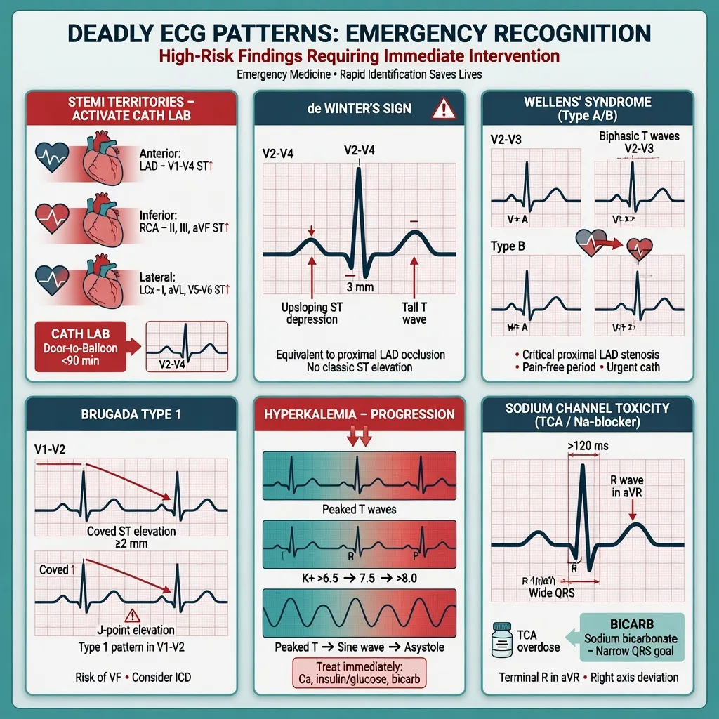
A small, finite set of electrocardiographic patterns carry an immediate treatment implication — the catheter-lab activation, the antidote, the defibrillation, or the avoidance of a contraindicated drug. These are the deadly patterns. The Fellowship examiner will present one in a written stem, an OSCE resuscitation station, a data-viva, or a structured clinical discussion, and the candidate is expected to recognise it on the trace, name the underlying mechanism, state the disposition, and give the drug with the dose, route, and rationale. This topic catalogues the deadly patterns by mechanism — the acutely occluded coronary artery, the channelopathy, the electrolyte derangement, the right-heart-strain state, the ventricular tachyarrhythmia, and the sodium-channel-blocker cardiotoxicity — and gives the ECG-to-drug sequence for each.[1][5]
Why a pattern is "deadly"
A deadly ECG pattern is one of three things. It is the only sign of an occluded coronary artery in a patient who still has salvageable myocardium — the de Winter and Wellens patterns, the posterior and right-ventricular infarct, and the Sgarbossa-positive left bundle branch block or paced rhythm all represent an acute or threatened occlusion that does not reach the standard ST-elevation threshold and is therefore missed if the trace is read literally. It is the precursor to a ventricular arrhythmia — the Brugada type-1 pattern, the long QT, and the hyperkalaemic sine wave all predict imminent polymorphic VT, torsades, or asystole. Or it is the arrhythmia or the toxic state itself — ventricular tachycardia, torsades de pointes, and the wide-QRS sodium-channel-blocker state, where the wrong drug (verapamil, an AV-node blocker, a class Ia or Ic antiarrhythmic) kills the patient. The recognition step is binary — present or not — and the management step is a small committed set of doses. [1]
The STEMI territories — the occlusion that meets threshold
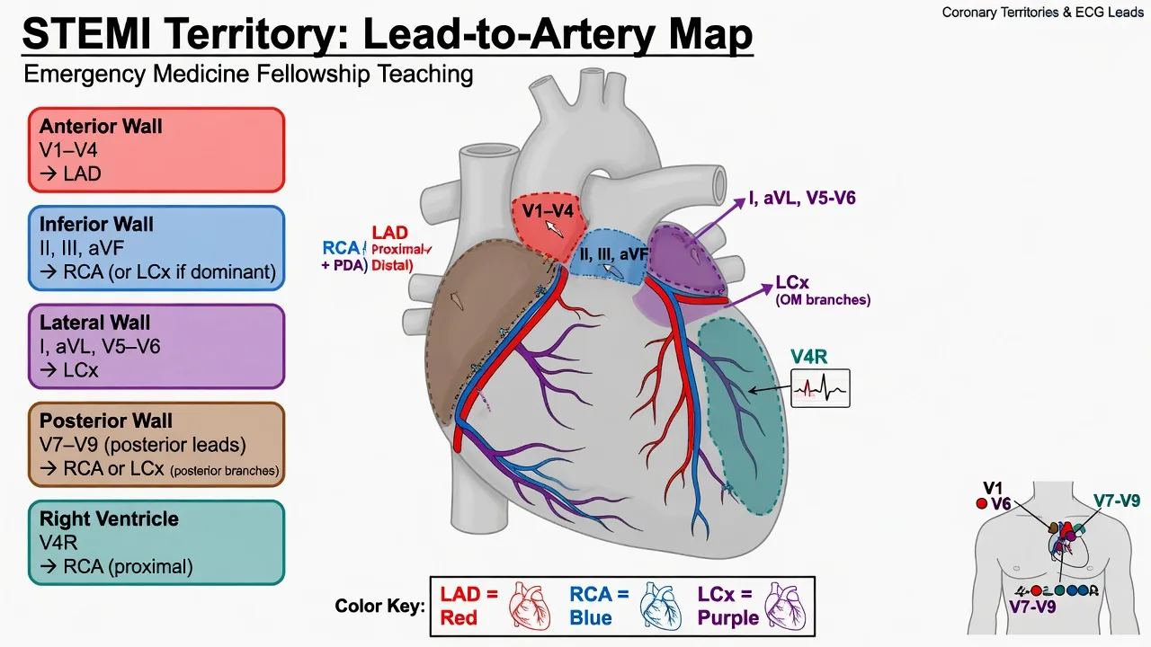
ST-elevation myocardial infarction is the prototype deadly pattern, and the territory localises the artery. Anterior infarction (ST elevation in V1 to V4) is the left anterior descending territory; a proximal occlusion produces extensive anterior loss, cardiogenic shock, ventricular fibrillation, and a risk of left-ventricular aneurysm and rupture. Inferior infarction (ST elevation in II, III, aVF, with the elevation in III greater than in II favouring the right coronary artery) is the right coronary or the circumflex; it carries a high risk of atrioventricular block and right-ventricular involvement, and a bradycardia or a hypotensive response to nitrates is a clue. Lateral infarction (I, aVL, V5, V6) is the circumflex and is frequently under-recognised because the absolute ST elevation is modest. Posterior infarction is the mirror image — horizontal ST depression in V1 to V3 with a tall R wave and an upright T wave, confirmed by direct ST elevation in posterior leads V7 to V9; it is the territory most often missed because it is read as "anterior ischaemia" rather than what it is, a posterior occlusion.[2] Right-ventricular infarction accompanies about a third of inferior STEMIs and is identified by ST elevation in right-precordial lead V4R; it is preload-dependent, nitrates precipitate profound hypotension, and the treatment is a fluid bolus with the patient monitored for pulmonary congestion.
[1]The STEMI-equivalents — occlusion that does not meet threshold
The standard STEMI threshold misses a substantial minority of acutely occluded arteries, which is why the modern concept of occlusion myocardial infarction (OMI) has replaced strict millimetre counting. Five patterns must be hunted for in any trace from a patient with chest pain or an anginal equivalent, because each represents an acutely or imminently occluded coronary artery. [1]
Hyperacute T waves are the earliest electrical sign of transmural ischaemia, appearing within minutes of occlusion and before ST elevation develops. They are broad-based, bulky, and asymmetric, and they dwarf the preceding R wave in the affected leads; in anterior ischaemia they appear in V2 to V4. They are missed because they are interpreted as "normal variant tall T" or hyperkalaemia — the differentiator is the bulky asymmetric morphology and the chest pain. [1]
De Winter T waves are a proximal LAD occlusion equivalent. The pattern is upsloping ST depression at the J point of 1 to 3 millimetres in the anterior precordials (V1 to V6, most striking in V2 to V3), with tall, prominent, symmetrical T waves and no ST elevation, and it is persistent rather than evolutionary.[1] It is treated as a STEMI and the catheter lab is activated — waiting for ST elevation to develop costs myocardium.
Wellens syndrome is the signature of critical proximal LAD stenosis in a patient whose chest pain has resolved by the time of the ECG — the resting trace is recorded in a pain-free window, which is precisely why it is missed. The two patterns are type A, deeply biphasic T waves in V2 and V3 (and often V1 to V4), and type B, deeply inverted symmetrical T waves in the same leads; both show preserved R waves and no pathological Q, and no or minimal ST elevation. The cardinal error is to send the patient for a stress test, which precipitates an acute occlusion and ventricular fibrillation. The correct management is urgent coronary angiography. [1]
Sgarbossa criteria apply to left bundle branch block and to a paced rhythm, where the standard ST-elevation thresholds are useless because the broad QRS itself displaces the ST segment. The original three Sgarbossa criteria, adapted and now refined by the Smith-modified third criterion, identify occlusion in this population with high specificity.[2] Concordant ST elevation — the ST vector points the same way as the dominant QRS — is the most specific (≈98 per cent). Concordant ST depression in V1 to V3 is the second. The third is excessively discordant ST elevation, defined by the original criterion as at least 5 millimetres, and by the Smith-modified criterion as a ratio of ST elevation to the depth of the S wave of at least 25 per cent (one quarter), which corrects for the depth of the underlying QRS.
The Sgarbossa and Smith-modified Sgarbossa criteria
Hyperacute equivalents in paced rhythm use the same Sgarbossa logic, and the paced Sgarbossa criteria carry comparable specificity to the LBBB version.[2]
The Brugada pattern — the coved type-1 trace
Brugada syndrome is an autosomal-dominent sodium-channel channelopathy (most commonly a loss-of-function SCN5A mutation) that produces a characteristic right-precordial pattern and a risk of polymorphic ventricular tachycardia and sudden cardiac death, typically in young and middle-aged men. Three patterns are described in V1 to V3, but only the type-1 pattern is diagnostic of the syndrome. [1]
The type-1 pattern is a coved ST elevation of at least 2 millimetres in at least one right-precordial lead (V1 to V3, recorded in standard or higher intercostal spaces), descending into a negative T wave. The type-2 pattern is a saddle-back ST elevation of at least 2 millimetres with a high takeoff, a trough of at least 1 millimetre, and a positive or biphasic T wave. The type-3 pattern is either morphology with ST elevation under 2 millimetres. Type-2 and type-3 are not diagnostic; they are converted to a type-1 by sodium-channel-blocker provocation (ajmaline, flecainide, procainamide) when the diagnosis is suspected. A spontaneous type-1 with symptoms (syncope, nocturnal agonal breathing, documented VT/VF) or a family history of sudden death under 45 confers a high arrhythmic risk and is an indication for an implantable cardioverter-defibrillator. [1]
[1]Hyperkalaemia — the progression from peaked T to sine wave
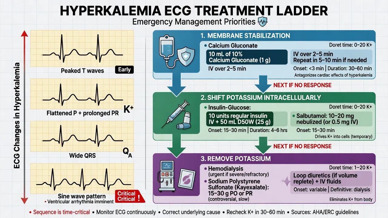
Hyperkalaemia is the electrolyte emergency with the most predictable ECG evolution, and the trace is the first — and often the only — investigation available before the laboratory returns. The progression reflects rising extracellular potassium and the inactivation of cardiac sodium channels.[3]
The hyperkalaemia ECG progression
PEAKED
The earliest sign — narrow, symmetric, pointed T waves (different from the bulky asymmetrical hyperacute T of ischaemia)
PR interval lengthens and the P wave amplitude drops as atrial conduction slows
The P wave disappears, with a junctional or sinoventricular escape rhythm
The QRS broadens diffusely as ventricular conduction slows
Ventricular escape rhythms and conduction blocks emerge
A smooth sine-wave tracing merging QRS and T, progressing to asystole or ventricular fibrillation
The treatment ladder matches the trace. Membrane stabilisation with calcium comes first in any patient with a widened QRS, a sine wave, or haemodynamic instability — calcium chloride 10 mL of 10 per cent (6.8 mmol) intravenously over 5 to 10 minutes, repeated as needed; calcium gluconate 30 mL of 10 per cent is an alternative if a central line is not available. Potassium shift into cells follows: insulin 10 units with 50 mL of 50 per cent dextrose intravenously (the dextrose prevents hypoglycaemia, monitor glucose for several hours), salbutamol 10 to 20 mg nebulised (a beta-2 agonist that drives potassium into cells), and sodium bicarbonate if there is coexistent acidosis. Potassium removal is then achieved with a gastrointestinal cation-exchange resin or, definitively, haemodialysis in the refractory or the renal-failure patient. ECG changes that resolve with calcium confirm the diagnosis at the bedside. [1]
Long QT and torsades de pointes
A long QT is the substrate for torsades de pointes, the characteristic polymorphic broad-complex tachycardia on a prolonged QT with a twisting of the QRS axis around the baseline, often in short self-terminating runs ("salvos") that can degenerate to ventricular fibrillation. The corrected QT is calculated by Bazett's formula (QTc equals QT divided by the square root of the R-to-R interval in seconds). The thresholds are over 440 milliseconds in men and over 460 milliseconds in women for abnormal, and over 500 milliseconds for high torsades risk. The risk is multiplicative — long QT plus a trigger (a premature ventricular beat, an electrolyte disturbance, a bradycardia) produces the arrhythmia. [1]
The acquired causes of a long QT
Torsades is treated with magnesium sulphate 2 g intravenously over 10 to 15 minutes regardless of the serum magnesium, withdrawal of the offending drug, and potassium correction to a high-normal level (4.5 to 5.0 mmol/L). A sustained or recurrent torsades is managed by overdrive pacing (transvenous or transcutaneous at a rate of 100 to 120 to shorten the QT) and isoprenaline for the bradycardia-dependent form. A defibrillator is applied from the outset because degeneration to ventricular fibrillation is treated with an unsynchronised shock.[4]
Pulmonary embolism — the right-heart-strain trace
The ECG is neither sensitive nor specific for pulmonary embolism, but in the right patient it carries the diagnosis. The commonest finding is sinus tachycardia. The classic but uncommon finding is the S1Q3T3 pattern (an S wave in lead I, a Q wave in lead III, and an inverted T wave in lead III), described by McGinn and White in 1935; it reflects acute right-heart strain with right-axis deviation and is most often seen with a massive or submassive embolus. The most diagnostically useful findings are new T-wave inversion in V1 to V4 (right-heart strain), right-axis deviation, a right-bundle-branch-block pattern (new or rate-related), an S wave in V1 with an R-prime, and new atrial fibrillation. The changes resolve as the embolus lyses or is lysed. The ECG in suspected pulmonary embolism is most valuable for excluding the alternative diagnosis (an anterior or inferior STEMI) and for risk-stratification — a trace showing right-heart strain plus haemodynamic compromise points to a massive embolus that may need thrombolysis or embolectomy. [1]
Ventricular tachycardia — the broad-complex tachycardia to assume
A regular broad-complex tachycardia (QRS at least 120 milliseconds, rate over 100) is ventricular tachycardia until proven otherwise, and the burden of proof is on the dissenting clinician. The features that confirm VT are the AV dissociation sign — independent P waves marching through the broad QRS complexes, visible on a long rhythm strip — the capture beat (a narrow normally-conducted QRS appearing among the broad ones, capturing the moment the AV node conducts through), and the fusion beat (a hybrid QRS that is a summation of a conducted and a ventricular beat). Concordance of the precordial QRS — all positive or all negative from V1 to V6 — and an extreme axis (northwest) also favour VT. The morphological criteria of Brugada and Griffith apply to V1 and V6 to distinguish VT from SVT with aberrancy. [1]
The stable regular broad-complex tachycardia receives amiodarone 300 mg intravenously over 20 to 60 minutes followed by an infusion (900 mg over 24 hours); the alternative for a clearly monomorphic stable VT is procainamide. The unstable regular broad-complex tachycardia — hypotension, shock, ischaemic chest pain, acute heart failure, or altered consciousness — is treated with synchronised cardioversion. The cardiac arrest dose of amiodarone for shockable VT or VF is 300 mg as an intravenous bolus, with a further 150 mg if required. Verapamil is absolutely contraindicated in any broad-complex tachycardia: it is a negative inotrope that has caused cardiovascular collapse and death when given for what was in fact VT. [1]
Sodium-channel-blocker toxicity — the wide QRS with R-prime in aVR
A wide QRS in the poisoned patient is sodium-channel blockade until proven otherwise, and the trace carries both the diagnosis and the antidote. The causative agents are the tricyclic antidepressants, the class Ia antiarrhythmics (quinidine, procainamide, disopyramide), the class Ic antiarrhythmics (flecainide, propafenone), the antihistamines (diphenhydramine), the antimalarials (chloroquine, hydroxychloroquine), carbamazepine, lamotrigine, lacosamide, and cocaine — all sodium-channel blockers, and all treatable with the same antidote.[5]
[1]The antidote is sodium bicarbonate: a 1 to 2 milliequivalent per kilogram intravenous bolus of 8.4 per cent sodium bicarbonate (typically 50 to 100 mL), repeated every few minutes until the QRS narrows (under 100 milliseconds), the R-prime in aVR resolves, the blood pressure improves, and the serum pH is between 7.45 and 7.55. It works by providing a sodium load (overcoming the channel blockade), alkalinising the blood (which traps the drug off the sodium channel), and correcting the acidosis that itself worsens channel blockade. Hyperventilation to a target pH of 7.45 to 7.55 augments the alkalinisation in the intubated patient. The unstable or arresting sodium-channel-blocker patient is treated with high-dose sodium bicarbonate, and intralipid is considered for the refractory case (the local cardiac-arrest protocol for the lipophilic sodium-channel blockers — bupivacaine, verapamil, and the local-anaesthetic-class drugs). [1]
Differential diagnosis — the patterns that masquerade as each other
The deadly patterns overlap on the trace, and the differential is the difference between the right antidote and a fatal misclassification. [1]
Hyperacute T (ischaemia)
- Broad-based, bulky, asymmetric; dwarfs the preceding R wave
- Accompanied by chest pain; in the territory leads (anterior V2–V4)
- Often the earliest sign of an LAD occlusion before ST elevation
- Treat as a STEMI — catheter-lab activation, dual antiplatelet, heparin
Peaked T (hyperkalaemia)
- Narrow, symmetric, pointed — a "tented" morphology
- Accompanied by PR prolongation, P-wave flattening, QRS widening
- Diffuse rather than territorial; the patient may be in renal failure or on a potassium-sparing drug
- Treat with calcium chloride 10 mL of 10%, then insulin-dextrose and salbutamol
Early repolarisation (benign)
- Concave ST elevation, most marked in V4; J-point notch; tall T waves
- Stable over time; common in young fit men; no reciprocal change
- Does not progress and is not territorial
- Recognise and do not treat — but exclude occlusion if any doubt
De Winter T waves (LAD occlusion)
- Upsloping ST depression at the J point with tall symmetrical T in V1–V3
- Persistent rather than evolutionary; a proximal LAD occlusion equivalent
- Often the only sign — do not wait for ST elevation
- Catheter-lab activation as a STEMI
Wellens syndrome (critical LAD)
- Deep biphasic (type A) or deeply inverted (type B) T waves in V2–V3
- Recorded when chest pain has RESOLVED; preserved R waves, no Q
- Do NOT stress-test — risk of acute occlusion and VF
- Urgent angiography for critical proximal LAD stenosis
Brugada type-1 vs phenocopy
- Coved ST elevation ≥2 mm in V1–V3 with negative T wave
- Type-1 with syncope, family history, or documented VT = the syndrome
- Phenocopy resolves when the precipitant (fever, hyperkalaemia, ischaemia) is treated
- Refer suspected syndrome to electrophysiology; treat the precipitant of the phenocopy
The two trace-level distinctions that cost the most lives when missed are hyperacute T (ischaemia) versus peaked T (hyperkalaemia) — giving an LAD occlusion to a hyperkalaemic patient as "STEMI" delays calcium, and giving thrombolysis to a hyperkalaemic peaked T precipitates arrest — and sodium-channel-blocker VT versus structural VT — treating a sodium-channel-blocker wide-QRS with amiodarone alone (without bicarbonate) delays the antidote, while treating a true VT with verapamil collapses the ventricle. [1]
Immediate management and resuscitation — the ECG-to-drug sequence
The deadly patterns are treated at the bedside from the trace, and the doses are committed to memory because the unstable patient cannot wait for a lookup. [1]
The ECG-driven emergency drug doses
A stable regular narrow-complex SVT is treated with vagal manoeuvres, then adenosine 6 mg as a rapid intravenous bolus followed by a flush, escalating to 12 mg then a further 12 mg if the rhythm persists. Adenosine transiently blocks the AV node and terminates the re-entrant circuit; warn the patient of the brief flush, dyspnoea and chest tightness, and avoid it in asthma and high-grade block. A stable monomorphic VT receives amiodarone 300 mg intravenously over 20 to 60 minutes, with the unstable rhythm cardioverted synchronised. Torsades on a long QT receives magnesium sulphate 2 g intravenously, withdrawal of the offending drug, and potassium correction. Hyperkalaemia with a wide QRS or a sine wave receives calcium chloride 10 mL of 10 per cent intravenously for membrane stabilisation, then insulin with dextrose and a beta-agonist to shift potassium. Sodium-channel-blocker cardiotoxicity receives sodium bicarbonate to narrow the QRS and alkalinise the serum. [1]
Subtypes and scenarios — the patient who already has a confounder
The patient with a left bundle branch block, a paced rhythm, or a known channelopathy is the highest-risk group for a missed deadly pattern, because the baseline trace is already abnormal. In left bundle branch block and paced rhythm, occlusion is judged only by the Sgarbossa or Smith-modified Sgarbossa criteria — concordant ST change or excessively discordant ST elevation.[2] In known Brugada syndrome with fever, treat the fever aggressively (it unblocks the type-1 pattern and the arrhythmic risk) and monitor. In the dialysis patient with chest pain, hyperkalaemia and ischaemia coexist — the peaked T of hyperkalaemia can precede and obscure the ST elevation of an infarct, so calcium is given for the wide QRS and serial ECGs are taken. In the pregnant patient, adenosine, a synchronised cardioversion, magnesium, calcium, and sodium bicarbonate are all safe; thrombolysis is relatively contraindicated but used for life-threatening PE or STEMI when percutaneous intervention is not available.
Complications and pitfalls
The recurring errors with the deadly patterns are: reading a trace literally and missing the occlusion-equivalent (de Winter, Wellens, posterior, RV, Sgarbossa-positive) because the ST-elevation threshold is not met; giving a nitrate to an inferior STEMI before recording V4R and precipitating hypotension in an RV infarct; sending a Wellens patient for a stress test; treating a regular broad-complex tachycardia with verapamil; giving an AV-node blocker (adenosine, verapamil, beta-blocker, digoxin) to pre-excited atrial fibrillation and accelerating conduction down the accessory pathway to ventricular fibrillation; giving amiodarone alone — without bicarbonate — to sodium-channel-blocker cardiotoxicity; reaching for thrombolysis before excluding hyperkalaemia in a patient with peaked T waves and renal failure; and stopping the read at the first abnormality, missing a second. The discipline is to finish the systematic read on every trace and to compare with the previous one. [1]
Special populations
In the elderly, atypical presentations (breathlessness, syncope, confusion) and baseline ECG abnormalities (bundle branch block, paced rhythm, old infarcts) obscure the deadly pattern; a high index and serial traces are mandatory. In women, the STEMI threshold in V2 to V3 is lower (1.5 millimetres vs 2.0 in men over 40), and ischaemic T-wave inversion may be misread as normal variant. In children and young athletes, early repolarisation and the juvenile T-wave pattern overlap with hyperacute and ischaemic T waves — the clinical context and a comparison with a baseline trace settle the matter. In renal failure and on dialysis, hyperkalaemia is the first assumption for any arrhythmia or conduction abnormality; calcium first, then ask why. [1]
Evidence and regional guidelines
The modern concept of occlusion myocardial infarction, of which the de Winter and Wellens patterns are the prototypes, has displaced strict millimetre ST-elevation counting as the operational standard for catheter-lab activation.[1] The Sgarbossa criteria and the Smith-modified third criterion are the accepted method for diagnosing occlusion in left bundle branch block and paced rhythm.[2] The hyperkalaemia ECG progression is reproducible but imperfect — a normal ECG does not exclude a high potassium, and a borderline trace should not delay calcium in the unwell hyperkalaemic patient.[3] The QT-prolonging effect of antipsychotics is dose- and route-dependent and has been quantified in meta-analysis.[4] The wide-QRS sodium-channel-blocker state is treated with sodium bicarbonate by the ACMT (American College of Medical Toxicology) and the ANZ tox guidelines; intralipid is the rescue therapy for the refractory case.[5]
ANZ practice note. The deadly-pattern recognition and the peri-arrest drug doses follow the ANZCOR advanced-life-support algorithm and the local cardiology and toxicology pathways. Adenosine 6 then 12 then 12 mg is the SVT escalation, amiodarone 300 mg intravenously is the stable-VT choice, magnesium 2 g is the torsades dose, calcium chloride 10 mL of 10 per cent is the hyperkalaemia membrane stabiliser, and sodium bicarbonate 1 to 2 mEq/kg is the sodium-channel-blocker antidote. Pre-excited atrial fibrillation is cardioverted and never given an AV-node blocker. The catheter lab is activated for any STEMI-equivalent (de Winter, Wellens, posterior, RV, Sgarbossa-positive) on the principle of occlusion myocardial infarction, not strict ST-elevation counting. The local toxicology service is consulted for any sodium-channel-blocker or polypharmacy cardiotoxic ingestion. [1]
Digoxin toxicity — the scooped ST and the "reverse tick"
Digoxin is the cardiac glycoside that produces a characteristic repolarisation abnormality even at therapeutic levels, and a constellation of arrhythmias and conduction blocks at toxic levels. The pathognomonic trace finding is the downsloping, "scooped" ST-segment depression (the "sagging" or "reverse tick" sign, likened to Salvador Dalí's moustache) — most evident in the lateral leads with a small or negative T wave; it is a digoxin effect, not by itself toxicity, but it is the pattern the examiner expects the candidate to recognise and to contextualise against the clinical picture. [1]
The lethal toxicity is far subtler than the scoop. Digoxin inhibits the sodium–potassium ATPase, raises intracellular calcium (the inotropic effect), and directly suppresses atrioventricular nodal conduction; toxicity therefore expresses as a heightened automaticity coupled with AV block — the classic rhythm is atrial tachycardia with block (an atrial rate of 150 to 250 with a 2:1 or variable AV response), but also bidirectional ventricular tachycardia (alternating QRS axis, beat to beat — virtually pathognomonic), bradycardia with premature ventricular complexes, and high-grade AV block. The single most useful bedside clue to life-threatening toxicity is hyperkalaemia — digoxin poisoning drives serum potassium up by inhibiting the Na⁺/K⁺ pump, and a potassium above 5.0 mmol/L in the setting of digoxin predicts a high mortality and is itself a trigger for the antidote. [1]
[1]The antidote is the digoxin-specific antibody (Fab) fragment — the original ovine Fab (Digibind / DigiFab), which binds circulating digoxin and is renally excreted as the inactive complex.[10] The indications for Fab are life-threatening arrhythmia (ventricular tachyarrhythmia, symptomatic bradycardia or high-grade AV block unresponsive to atropine), hyperkalaemia above 5.0 mmol/L in acute overdose (the Fab corrects the potassium by restoring pump function), haemodynamic instability, and a serum digoxin above 10 ng/mL in the acute ingestion or above 6 ng/mL in the chronic setting. The dose is calculated from the ingested dose or from the steady-state serum concentration using the published nomograms; where these are unknown, an empirical loading dose (typically 5 to 10 vials in the acute, life-threatening presentation) is given. Atrial tachyarrhythmias may transiently worsen as the AV block is reversed, and the Fab is given in a monitored setting. Magnesium sulphate, and the avoidance of calcium (calcium may potentiate the intracellular calcium overload — though this is debated, it is a sensible caution in the examiner's mind), round out the supportive management. Cardioversion of a digoxin-toxic rhythm risks asystole and is a last resort — correct with Fab first.
[1]Suspected digoxin toxicity — the ED sequence
Recognise the toxidrome
A patient on digoxin (or who has ingested it) with nausea and vomiting, visual disturbance (blurred or yellow-green vision, haloes), confusion, and an arrhythmia — especially atrial tachycardia with block, bidirectional VT, bradycardia with premature ventricular complexes, or high-grade AV block. A serum potassium above 5.0 mmol/L in the acute ingestion is the alarm bell.
Confirm and grade
Send a serum digoxin level (note that the level is most useful at steady state — 6 to 8 hours after an oral dose; a level drawn immediately after an acute overdose underestimates the burden), a potassium, a magnesium, a renal function, and a 12-lead with a long rhythm strip. Apply the indication checklist for the Fab.
Give the Fab if any criterion is met
Life-threatening arrhythmia (VT, symptomatic bradycardia, high-grade AV block unresponsive to atropine), potassium above 5.0 mmol/L in acute overdose, haemodynamic instability, or a level above 10 ng/mL acute (6 chronic). Dose by the ingested-dose or the serum-level nomogram; empirically 5 to 10 vials if the dose or level is unknown in a life-threatening presentation.
Correct the potassium and magnesium
The Fab reverses the hyperkalaemia as it restores the Na⁺/K⁺ pump — do not give intravenous calcium (the theoretical risk of "stone heart" from calcium in the setting of intracellular calcium overload), and reserve the insulin–dextrose for the patient in whom the Fab is delayed. Correct the hypomagnesaemia with magnesium sulphate, which also suppresses the digoxin-induced afterdepolarisations.
Support and avoid the traps
AVOID the precipitants — hypokalaemia, hypomagnesaemia, hypothyroidism, and renal failure all raise the effective digoxin level; the interacting drugs (amiodarone, verapamil, diltiazem, macrolides, amphotericin) do the same. De-escalate the cardioversion of a digoxin-toxic rhythm — it risks asystole; give the Fab first. Monitor for the rebound as the Fab is renally cleared in the patient with the renal failure.
Wolff–Parkinson–White and pre-excitation — the delta wave and the short PR
The Wolff–Parkinson–White (WPW) pattern is the electrocardiographic signature of an accessory atrioventricular pathway (the bundle of Kent) that bypasses the AV node and pre-excites the ventricles. The three features are the short PR interval (under 120 milliseconds), the delta wave (a slurred upstroke of the QRS from the premature depolarisation of a small portion of ventricle through the pathway ahead of the AV node), and the broadened QRS (the fusion of the accessory-pathway and the normal nodal depolarisation). The pattern is common (about 1 to 3 per 1000) and most carriers are asymptomatic; the concern is the pre-excited tachyarrhythmia — atrioventricular re-entrant tachycardia (AVRT) and, the lethal one, atrial fibrillation conducting rapidly down the accessory pathway. [1]
[1]The lethal rhythm is pre-excited atrial fibrillation. Atrial fibrillation conducts down the accessory pathway, which (unlike the AV node) has no rate-limiting property — the ventricular rate can exceed 250 per minute, the broad irregularly irregular QRS with the delta wave degenerates to ventricular fibrillation, and sudden death is the well-described outcome in the young WPW patient. The cardinal error is to give an AV-nodal-blocking drug (adenosine, verapamil, diltiazem, a beta-blocker, or digoxin) — these block the AV node, preferentially funnel the atrial fibrillation down the accessory pathway, and accelerate the ventricular rate to ventricular fibrillation. Pre-excited atrial fibrillation is treated by synchronised cardioversion if unstable, or by an intravenous antiarrhythmic that blocks the accessory pathway (procainamide, flecainide, or amiodarone) if stable; the AV-node blockers are absolutely contraindicated. [1]
WPW pre-excited AF (irregular broad)
- An irregularly irregular broad-complex tachycardia, rate often over 200–250, with a varying QRS morphology and width (the delta wave)
- The shortest pre-excited RR interval under 250 ms predicts the VF risk
- NEVER give adenosine, verapamil, diltiazem, beta-blocker, or digoxin — they funnel conduction down the accessory pathway
- Treat with synchronised cardioversion if unstable; procainamide, flecainide, or amiodarone if stable
VT (regular broad)
- A regular broad-complex tachycardia, rate over 100, QRS over 120 ms
- AV dissociation, capture and fusion beats confirm; concordance and extreme axis favour VT
- Give amiodarone 300 mg if stable, synchronised cardiovert if unstable
- Never give verapamil
SVT with aberrancy (regular broad)
- A regular broad-complex tachycardia with a bundle-branch-block morphology that matches a known aberrancy
- Onset in a patient with a known conduction defect; responds to vagal manoeuvres or adenosine
- Adenosine 6–12 mg terminates the AV-nodal-dependent tachycardia
- Assume VT until proven otherwise — the burden of proof is on the dissenting clinician
Pre-excited AF vs SVT
- The irregularity is the key — pre-excited AF is irregularly irregular; SVT is regular
- The QRS in pre-excited AF varies in width and morphology; in SVT it is fixed
- Adenosine is safe and diagnostic in the regular SVT; it is dangerous in the irregular pre-excited AF
- When in doubt and the patient is unstable, cardiovert
Ventricular tachycardia versus SVT with aberrancy — the Brugada algorithm
A regular broad-complex tachycardia is ventricular tachycardia until proven otherwise, and the burden of proof is on the clinician who claims an SVT with aberrancy. The morphological and the structural criteria that distinguish the two are codified in the Brugada algorithm (not to be confused with the Brugada syndrome), a four-step flow with a high sensitivity for the diagnosis of VT.[6]
The Brugada four-step algorithm — the broad-complex tachycardia decision
Step 1 — Absence of an RS complex in all precordial leads
If there is no RS complex (an R wave followed by an S wave) in any of the leads V1 to V6, the diagnosis is ventricular tachycardia. This is the first and the most specific step; if positive, the algorithm stops.
Step 2 — R-to-S interval over 100 milliseconds
If an RS complex is present, measure the interval from the onset of the R to the nadir of the S in any precordial lead. An interval over 100 milliseconds identifies ventricular tachycardia; stop.
Step 3 — AV dissociation
Look for the independent P waves, the capture beats, and the fusion beats. The presence of any of these confirms ventricular tachycardia; stop. (These are the most specific features but the least often seen — examine a long rhythm strip.)
Step 4 — The morphological criteria in V1 and V6
If steps 1 to 3 are negative, apply the morphology criteria. In a right-bundle-branch-block–like pattern (dominant R in V1), a notched or biphasic R in V1 (a triphasic RSR) and a notched downstroke of the S favour VT. In a left-bundle-branch-block–like pattern (dominant S in V1), a broad R wave or a notched downstroke of the S in V1 and a Q wave in V6 favour VT. If the morphology criteria are met, the diagnosis is VT; if all four steps are negative, the diagnosis is SVT with aberrancy.
The hyperkalaemia treatment ladder — the three-tiered response
The hyperkalaemia ECG progression is matched by a three-tiered treatment ladder that the candidate must recite by mechanism and by the dose: membrane stabilisation (to buy time), potassium shift (to lower the level), and potassium removal (for the definitive and the renal-failure patient).[3]
The hyperkalaemia three-tiered ladder — the ED sequence
Tier 1 — Membrane stabilisation (immediate)
Calcium chloride 10 mL of 10 per cent (6.8 mmol) intravenously over 5 to 10 minutes for any widened QRS, sine wave, or haemodynamic instability; it raises the threshold potential and stabilises the myocardium within minutes, and it does NOT lower the potassium — it buys time. Calcium gluconate 30 mL of 10 per cent (9 mmol) is the alternative if a central line is not available. Repeat as needed.
Tier 2 — Potassium shift into cells (next 15 to 30 minutes)
Insulin 10 units with 50 mL of 50 per cent dextrose intravenously (the dextrose prevents the hypoglycaemia; monitor the glucose for several hours for the late hypoglycaemia), salbutamol 10 to 20 mg nebulised (a beta-2 agonist that drives the potassium into the cells), and the sodium bicarbonate 1 to 2 mEq/kg if there is a coexistent acidosis. Each lowers the potassium by 0.5 to 1.0 mmol/L over 30 to 60 minutes; the effect is transient.
Tier 3 — Potassium removal (definitive)
The gastrointestinal cation-exchange resin (patiromer, sodium zirconium) for the milder and the outpatient; the haemodialysis for the refractory, the renal-failure, and the severe (potassium over 7 with the ECG changes, or over 6.5 refractory to the tier-2 measures). The dialysis is the definitive removal; the calcium and the insulin buy the time to get there.
The differential of the ST elevation — the territory that says the artery
The ST-elevation territory localises the occluded artery, and the candidate is expected to read the territory and to name the vessel. The five territories, with the vessel and the high-yield correlate, are: [1]
Anterior (V1–V4)
- The left anterior descending (LAD); a proximal occlusion is the extensive anterior and the highest-risk infarct
- Risk of VF, cardiogenic shock, left-ventricular aneurysm and rupture, and a new bundle-branch block
- ST elevation in V1 with the right-bundle-branch block and the PR depression suggests a proximal LAD / septal occlusion
- Reperfusion by the primary PCI; watch for the heart block and the pump failure
Inferior (II, III, aVF)
- The right coronary artery (elevation in III greater than in II favours the RCA) or the circumflex
- High risk of the AV block (the nodal artery comes off the RCA in 90 per cent) and the right-ventricular involvement
- Record the V4R before the nitrates; an RV infarct is preload-dependent
- Treat the RV infarct with the fluid; avoid the nitrates; reperfusion
Lateral (I, aVL, V5–V6)
- The circumflex (and the diagonal or the obtuse marginal); frequently under-recognised
- The absolute ST elevation is modest; the reciprocal change in the inferior leads is the clue
- A high lateral infarct (I and aVL) may show the reciprocal ST depression in II, III, aVF
- Reperfusion; the isolated high-lateral is the one most often missed
Posterior (V7–V9; mirror in V1–V3)
- The circumflex or the right coronary; confirmed by the direct ST elevation of 0.5 mm in V7–V9
- The mirror image in V1–V3: the horizontal ST depression, the tall R wave, the upright T wave
- Mistaken for "anterior ischaemia" — it is a posterior occlusion
- Reperfusion; record the posterior leads in any inferior or the isolated anterior ST depression
Right ventricular (V4R)
- The proximal right coronary artery; accompanies a third of the inferior STEMIs
- ST elevation of 1 mm or more in V4R; preload-dependent, nitrate-precipitated hypotension
- Treat with the fluid bolus, monitored for the pulmonary congestion; avoid the nitrates
- Reperfusion; the RV function usually recovers over days to weeks
The landmark evidence — the trials and the consensus papers
Smith et al — reversal of digoxin intoxication with Fab fragments
New England Journal of Medicine, 1976
A landmark series of 26 patients with advanced, life-threatening digoxin intoxication (ventricular tachyarrhythmias, high-grade AV block, hyperkalaemia) treated with the Fab fragments of digoxin-specific antibodies, with the reversal of the toxicity within 30 to 45 minutes of the infusion.
Key finding
The Fab fragments rapidly and completely reversed the life-threatening digoxin toxicity, including the ventricular arrhythmias and the conduction blocks; the effect was dramatic and durable.
Practice change
Established the digoxin-specific Fab as the antidote for the life-threatening digoxin toxicity, and it remains the standard of care — the indications, the dose nomograms, and the empiric dosing for the unknown ingestion all derive from this and the subsequent work.
Brugada et al — the differential diagnosis of a regular tachycardia with a wide QRS complex
Circulation, 1991
A retrospective analysis of 554 regular tachycardias with a wide QRS complex (over 120 milliseconds), with the derivation of a four-step algorithm (absence of an RS complex in the precordials, the R-to-S interval over 100 ms, the AV dissociation, and the morphological criteria in V1 and V6) to distinguish the ventricular tachycardia from the SVT with aberrancy.
Key finding
The four-step algorithm had a sensitivity of 98.7 per cent and a specificity of 96.5 per cent for the diagnosis of the ventricular tachycardia in the derivation cohort; the algorithm has been validated and refined in the subsequent studies.
Practice change
Codified the systematic approach to the broad-complex tachycardia and remains the bedside algorithm taught in the resuscitation courses; it does not replace the default rule of "treat as VT until proven otherwise" but it structures the proof.
de Zwaan, Bär and Wellens — the characteristic ECG pattern of the critical proximal LAD
American Heart Journal, 1982
A prospective angiographic study of patients with the unstable angina who showed a characteristic pattern of the deeply inverted or the biphasic T waves in the anterior precordials, with the preserved R waves and no pathological Q, recorded in the pain-free window.
Key finding
All patients had a critical (over 50 per cent) stenosis high in the left anterior descending coronary artery, with the preserved anterior wall motion; the progression to the extensive anterior infarction was the rule in the untreated.
Practice change
Defined the Wellens syndrome as the signature of the critical proximal LAD lesion, recorded pain-free, and the cardinal warning against the stress test — the angiography is the management.
Brugada and Brugada — right bundle branch block, persistent ST elevation and sudden cardiac death
Journal of the American College of Cardiology, 1992
A description of eight patients (from three families) with the structurally normal hearts, the right bundle branch block, the persistent ST elevation in V1 to V3, and the episodes of the ventricular fibrillation and the sudden cardiac death, predominantly in the young and the middle-aged men.
Key finding
The constellation defined a distinct clinical and the electrocardiographic syndrome (the eponymous Brugada syndrome), inherited as the autosomal dominant, with the high risk of the polymorphic VT and the sudden death.
Practice change
Established the Brugada syndrome as a recognised channelopathy and the precursor of the sudden cardiac death; the type-1 coved pattern is diagnostic, and the symptomatic patient or the family history warrants the implantable cardioverter-defibrillator.
Tzivoni et al — magnesium therapy for torsades de pointes
American Journal of Cardiology, 1984
A report of three patients with the torsades de pointes on a prolonged QT (one congenital, two drug-induced), treated with the intravenous magnesium sulphate.
Key finding
The magnesium sulphate 1 to 2 g intravenously terminated the torsades in all three patients within minutes, despite the normal serum magnesium levels; the recurrence was prevented by the continuation of the infusion.
Practice change
Established the intravenous magnesium as the first-line treatment for the torsades de pointes, regardless of the serum magnesium — the standard of care to this day.
Anselm, Baranchuk et al — the International Registry on Brugada Phenocopy
Pacing and Clinical Electrophysiology, 2019
A multinational registry analysis of the reported Brugada phenocopies (the type-1-pattern traces induced by the non-syndromic precipitants — the myocardial ischaemia, the fever, the electrolyte disturbance, the pulmonary embolism, the metabolic), with the categorisation and the outcomes.
Key finding
The Brugada phenocopy is a distinct entity from the Brugada syndrome; it resolves with the treatment of the precipitant and it does not, in the registry data, carry the same arrhythmic risk, though the long-term follow-up and the provocative testing are recommended.
Practice change
Codified the distinction between the syndrome and the phenocopy, sparing the patient with the reversible precipitant the unnecessary implantable cardioverter-defibrillator while ensuring the proper evaluation of the underlying cause.
SAQ — Hyperkalaemia ECG progression in the dialysis patient
10 minutes · 10 marks
A 68-year-old man with end-stage renal failure on haemodialysis presents to the ED with progressive weakness and palpitations after missing his last two dialysis sessions. He is bradycardic at 38 with a blood pressure of 88/52. His 12-lead ECG shows tall, narrow, symmetric T waves, flattened P waves, and a widened QRS at 140 ms approaching a sine-wave morphology. A venous gas returns a potassium of 7.8 mmol/L.
SAQ — Wellens syndrome and the STEMI-equivalents
10 minutes · 10 marks
A 58-year-old woman presents to the ED with intermittent central chest pain over the past 48 hours that resolved spontaneously an hour before arrival. She is pain-free and haemodynamically stable. Her 12-lead ECG shows deeply inverted symmetrical T waves in V2 and V3 (extending into V1 and V4), with preserved R waves and no pathological Q waves, and no ST elevation. Her high-sensitivity troponin is mildly elevated at 45 ng/L (upper limit of normal 20).
Additional red flags
[1]Exam pearls
- A regular broad-complex tachycardia is VT until proven otherwise — give amiodarone 300 mg if stable, cardiovert if unstable, never give verapamil.
- De Winter and Wellens are anterior STEMI-equivalents — both represent a critical proximal LAD lesion and are treated as a STEMI activation; Wellens is recorded pain-free.
- Sgarbossa in LBBB and paced rhythm — concordant ST elevation, concordant ST depression in V1–V3, or excessively discordant ST elevation (at least 5 mm original, or at least 25 per cent of S-wave depth Smith-modified).
- Hyperkalaemia peaked T is narrow and symmetric; hyperacute T of ischaemia is broad and bulky. Calcium chloride 10 mL of 10 per cent for the wide QRS.
- Long QT over 500 ms is high torsades risk — give magnesium 2 g, stop the offending drug, correct potassium.
- Brugada type-1 is coved ST elevation of at least 2 mm in V1–V3 with a negative T wave — only type-1 is diagnostic; consider a phenocopy from fever or hyperkalaemia.
- Sodium-channel-blocker wide QRS with R-prime in aVR — give sodium bicarbonate 1 to 2 mEq/kg to narrow the QRS.
- Inferior STEMI — record V4R before nitrates; treat the RV infarct with fluid.
- Always compare with the old ECG — a borderline change that is new is the one that matters.
- Pre-excited atrial fibrillation (WPW) — irregularly irregular broad-complex tachycardia; never give AV-node blockers (adenosine, verapamil, digoxin), they funnel conduction down the accessory pathway to VF — cardiovert if unstable, procainamide / flecainide / amiodarone if stable.
- Digoxin toxicity — the scoop / reverse-tick ST is an effect, not toxicity; toxicity is the arrhythmia (atrial tachycardia with block, bidirectional VT, AV block) with a potassium above 5.0 — give the Fab fragments, never IV calcium.
- WPW pseudo-infarction — the delta wave can mimic an inferior or lateral STEMI; recognise the short PR and the slurred upstroke before activating the lab.
- Posterior STEMI — horizontal ST depression, tall R wave, upright T wave in V1–V3 (the mirror image); confirm with V7–V9; treat as a STEMI.
- The hyperkalaemia treatment ladder — membrane stabilisation (calcium chloride 10 mL of 10 per cent), then shift (insulin–dextrose, salbutamol, bicarbonate), then removal (dialysis); the calcium does not lower the potassium.
- The Brugada algorithm for the broad-complex tachycardia — no RS complex in the precordials, R-to-S over 100 ms, AV dissociation, morphology in V1/V6; sensitive but treat as VT when in doubt. [1]
Red flags
[1]References
- [1]de Winter RW, Verouden NJW, Wellens HJJ, et al. Precordial junctional ST-segment depression with tall symmetric T-waves signifying proximal LAD occlusion, case reports of STEMI equivalence J Electrocardiol, 2016.PMID 26560436
- [2]Maloy KR, Bhardwaj A, Zimetbaum PJ, et al. Sgarbossa Criteria are Highly Specific for Acute Myocardial Infarction with Pacemakers West J Emerg Med, 2010.PMID 21079708
- [3]Quintero JA, Galvis LM, Saavedra AC, et al. Electrocardiographic Abnormalities in Patients with Hyperkalemia: A Retrospective Study in an Emergency Department in Colombia Open Access Emerg Med, 2024.PMID 38952854
- [4]Berling I, Isoardi KZ, Lopresti D, et al. QT interval prolongation in acute antipsychotic poisoning: systematic review and recommendations Clin Toxicol (Phila), 2025.PMID 41255343
- [5]Chua-Tuan JL, Hayes BD, Jani-Majewski P, et al. Cardiac sodium channel blockade after an intentional ingestion of lacosamide, cyclobenzaprine, and levetiracetam: Case report Clin Toxicol (Phila), 2015.PMID 25951877
- [6]Brugada P, Brugada J, Mont L, et al. A new approach to the differential diagnosis of a regular tachycardia with a wide QRS complex Circulation, 1991.PMID 2022022
- [7]Brugada P, Brugada J. Right bundle branch block, persistent ST segment elevation and sudden cardiac death: a distinct clinical and electrocardiographic syndrome. A multicenter report J Am Coll Cardiol, 1992.PMID 1309182
- [8]de Zwaan C, Bär FWHM, Wellens HJJ. Characteristic electrocardiographic pattern indicating a critical stenosis high in left anterior descending coronary artery in patients admitted because of impending myocardial infarction Am Heart J, 1982.PMID 6121481
- [9]Tzivoni D, Banai S, Schuger C, et al. Magnesium therapy for torsades de pointes Am J Cardiol, 1984.PMID 6695782
- [10]Smith TW, Haber E, Yeatman L, Butler VP. Reversal of advanced digoxin intoxication with Fab fragments of digoxin-specific antibodies N Engl J Med, 1976.PMID 943040
- [11]Anselm DD, Baranchuk A, et al. Link between Brugada phenocopy and myocardial ischemia: Results from the International Registry on Brugada Phenocopy Pacing Clin Electrophysiol, 2019.PMID 30924150