EM · Procedural & diagnostic ED skills
Eye, ENT and dental procedures in the emergency department
Also known as Ocular irrigation · Slit-lamp examination · Tonometry · Corneal foreign body removal · Nasal packing for epistaxis · Nasal foreign body removal · Dental avulsion replantation · Dry socket management
The ED eye, ENT and dental procedures cluster — slit-lamp and fluorescein staining for the corneal abrasion, tonometry for the acute angle-closure glaucoma screen, corneal foreign-body removal and the rust-ring drill, copious ocular irrigation for the chemical burn (the Morgan lens with 2 L of saline driven to a neutral pH), epistaxis control by silver-nitrate cautery and anterior/posterior packing (the Merocel, the Rapid Rhino, the Foley), nasal foreign-body extraction by the hook, the Katz catheter and positive pressure, the avulsed permanent tooth reimplanted within 30 minutes and stored in milk or saline, and the dry socket irrigated and dressed. The Fellowship candidate must hold each technique, its landmark, its contraindication and its complication at procedural recall.
On this page & tools
Your progress
Saved locally on this device.
Target exams
Red flags
The eye, ENT and dental procedures form the cluster of bedside techniques that the emergency physician performs without specialist delay — the slit-lamp examination, the fluorescein stain, the applanation tonometry, the corneal foreign-body removal and the chemical-eye irrigation on the ocular side; the epistaxis cautery and packing, the nasal foreign-body extraction on the ENT side; and the avulsed-tooth replantation and the dry-socket dressing on the dental side. The Fellowship candidate must hold each procedure as a structured encounter: a clear indication, a known contraindication, a stepwise technique with the right equipment, and the complication that defines a safe operator. Two principles unify the cluster. First, the chemical eye burn is the one diagnosis in which treatment precedes assessment — irrigation starts the moment the history is heard, and the slit-lamp comes later. Second, the open globe, the button battery and the avulsed tooth are all time-critical: a missed open globe is blinded by a tonometer, a missed battery perforates a septum, and a delayed replantation loses a tooth.[2][6][9]
Scope and indications
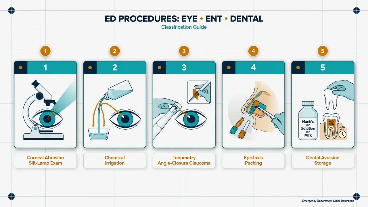
Each procedure answers a distinct ED question. The slit-lamp examination with fluorescein answers "is there a corneal epithelial defect, ulcer, foreign body or an anterior-chamber leak?" and is the cornerstone of ocular assessment. The tonometry answers "is the intraocular pressure raised?" and screens for the acute angle-closure attack. The corneal foreign-body removal relieves pain and prevents rust-ring infection. The ocular irrigation decontaminates a chemical burn and is triggered by any chemical exposure to the eye. The epistaxis procedure stops a nosebleed that has failed first-aid pressure. The nasal foreign-body extraction removes an object from a child's (or an adult's) nose. The dental avulsion replantation salvages a knocked-out permanent tooth, and the dry-socket dressing relieves the severe post-extraction pain of alveolar osteitis. The unifying test is that the question is mechanical or chemical and answerable at the bedside, with referral for the definitive ophthalmic, ENT or dental care once the immediate problem is controlled.[6][9]
Contraindications
The contraindications are procedure-specific but converge on the central question — is the globe intact? Tonometry and any procedure that presses on the eye are contraindicated when an open globe is suspected (a penetrating injury, a laceration, a teardrop pupil, a shallow anterior chamber, or a known intraocular foreign body). Corneal foreign-body removal at the slit-lamp is contraindicated in the uncooperative patient and the deep corneal foreign body threatening the anterior chamber (refer to ophthalmology). Silver-nitrate cautery for epistaxis is contraindicated on both sides of the nasal septum at the same visit (risk of septal perforation) and in the active posterior bleed that has no visible vessel. Nasal packing is relatively contraindicated in the severe bleeding disorder without correction support. Nasal foreign-body instrumentation is contraindicated when the object is a button battery and the operator cannot reach it within minutes — escalate to ENT. Dental replantation is contraindicated for a primary (deciduous) tooth (it ankyloses and damages the permanent successor) and for an avulsed permanent tooth with an extraoral dry time over 60 minutes (the periodontal ligament cells are dead and replantation will fail with resorption).[6][8]
Slit-lamp examination and fluorescein staining — the corneal abrasion
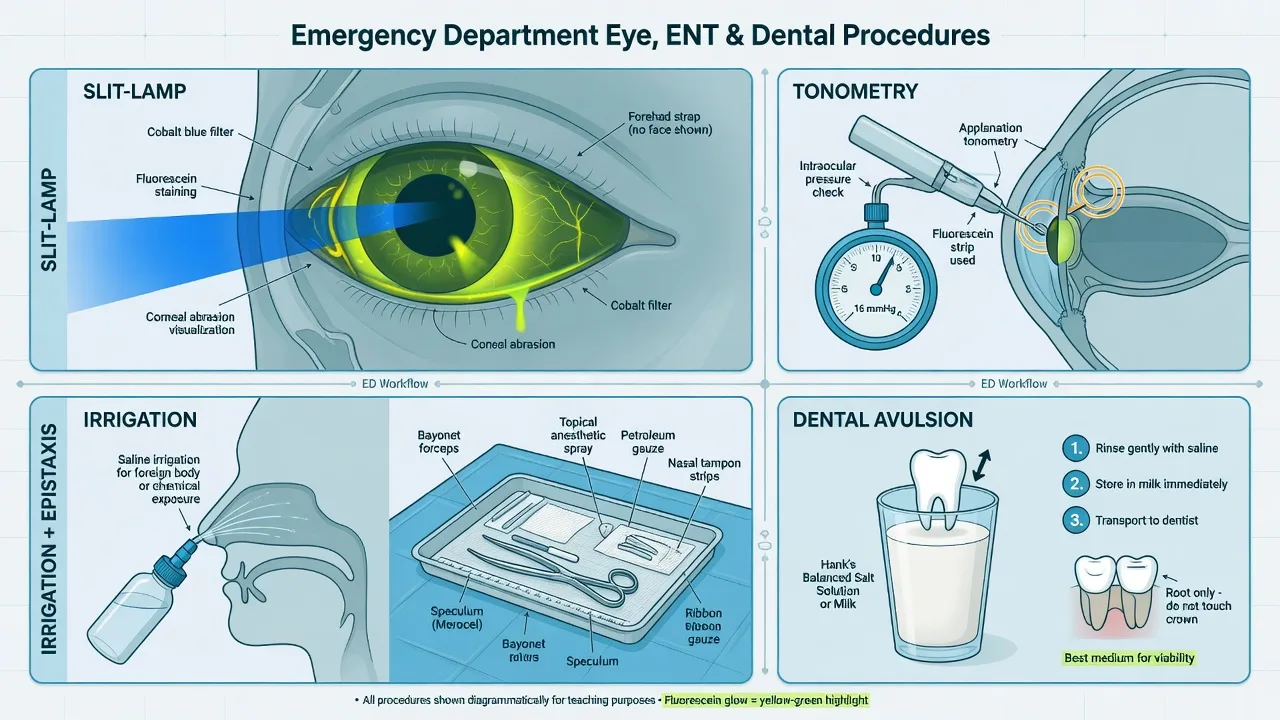
The slit-lamp is the binocular microscope that gives the magnified, illuminated view of the anterior segment. The candidate must run it through its three illuminations: diffuse (the wide beam for the lids, conjunctiva and the overall survey), slit beam (the optical section for the anterior-chamber depth and the lens) and cobalt blue (the excitation light for the fluorescein stain). Fluorescein is the orange dye that stains only where the corneal epithelium is lost — the abrasion, the ulcer and the denuded area — and it appears bright green under cobalt blue.[4]
The corneal abrasion presents with the foreign-body sensation, the tearing, the photophobia and the pain that is worse on blink. The fluorescein reveals a linear or a patchy green defect. Vertical linear abrasions on the upper cornea are the clue to a foreign body trapped under the upper lid, and the lid must be everted and swept — the forgotten subtarsal foreign body is the cause of the non-healing abrasion. The abrasion is managed with topical antibiotic (chloramphenicol or a fluoroquinolone for the contact-lens wearer), oral analgesia, and the instruction NOT to wear the contact lens. Patching does not improve healing or pain and is not recommended.[4] A contact-lens-related abrasion is never patched and is covered against Pseudomonas with a fluoroquinolone.
Tonometry — the acute angle-closure screen
Tonometry measures the intraocular pressure (IOP) by applanating (flattening) a known area of the cornea. The Goldmann applanation tonometer attached to the slit-lamp is the gold standard; the iCare rebound and the Tono-Pen are the portable handheld devices for the ED. The normal IOP is 10 to 21 mmHg, and the acute angle-closure attack produces a pressure typically above 40 mmHg.[5]
Acute angle-closure glaucoma — the pressure and the picture
The technique applies after the globe is confirmed intact. Instil a drop of topical anaesthetic (amethocaine or oxybuprocaine) and fluorescein, the patient at the slit-lamp, the tonometer head touched gently to the cornea, and the two green semicircles matched to the endpoint by the dial. The handheld Tono-Pen applanates with the probe tip and gives a digital readout. The reading must be taken before any irrigation dilutes the tear film. The acute angle-closure crisis is the unilateral, painful red eye with the mid-dilated, fixed oval pupil, the hazy (oedematous) cornea, the haloes around lights, the nausea and vomiting, and the raised pressure. The emergency management lowers the pressure medically (acetazolamide 500 mg intravenously, a topical beta-blocker such as timolol, a topical alpha-2 agonist such as apraclonidine, a topical steroid, and intravenous mannitol 20 per cent at 1 to 2 g/kg for the very high pressure), with the pilocarpine 2 per cent applied once the pressure has begun to fall (the ischaemic sphincter does not respond to pilocarpine at the very high pressure), and the definitive laser peripheral iridotomy performed by ophthalmology.[5]
Corneal foreign-body removal and the rust ring
The corneal foreign body (FB) — the grinding swarf, the metal flake, the grit — sits on or in the corneal epithelium and causes the sharp foreign-body sensation. After the slit-lamp confirms the FB and excludes a perforation, the removal is performed at the slit-lamp under topical anaesthetic (amethocaine). The superficial FB is lifted off with a fine hypodermic needle (25 or 27 gauge) bevel-up or a blunt spatula, the operator's hand resting on the patient's cheek for stability, the patient fixating a target. A ferrous (iron/steel) foreign body leaves a rust ring in the corneal stroma within hours, and the ring is removed with the electric burr (the Algerbrush) at the slit-lamp. The technique is to debride the rust gently; a residual ring that is difficult to clear may be left to "organise" for 24 to 48 hours and re-debrided, as the loosened ring lifts more easily after a day.[3] The post-removal care is a topical antibiotic, the cycloplegia optional, the oral analgesia, the tetanus prophylaxis check, and the review in 24 to 48 hours for the healing and the residual rust. The complications are the corneal abrasion, the secondary infection (the ulcer), and the residual rust stain that the body may wall off.
Chemical eye injury and ocular irrigation — the Morgan lens
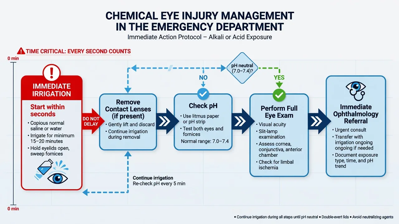
The chemical eye burn is the one diagnosis in which the treatment is started before the history is complete. The alkali burn (lye, cement, ammonia, plaster) is worse than the acid burn because the alkali saponifies the cell membranes and penetrates deeply, while the acid coagulates a protein barrier that limits the penetration. The hydrofluoric burn adds systemic fluoride toxicity.[1][2]
The ocular irrigation, in order
- Irrigate immediately — do not wait for the slit-lamp or the pH paper. Tap water, saline or Hartmann's is acceptable in the first instance; the patient is tilted so the affected eye is dependent, and the fluid is run across the eye from the medial canthus outward.
- Instil topical anaesthetic (amethocaine) and insert the Morgan lens — the irrigation device that sits under the lids and delivers a continuous flow over the cornea and the fornices.
- Evert the eyelids and sweep the fornices with a cotton swab or forceps to remove any retained particulate matter (the cement grain, the plaster fragment) — a retained particle continues to release chemical and prevents the pH from neutralising.
- Run 2 L of saline, Hartmann's or Ringer's through the Morgan lens (the typical first volume; more is given as needed).
- Stop and check the pH with the indicator paper in the inferior fornix, 10 minutes after stopping the irrigation (the lag allows the retained chemical in the tissues to equilibrate).
- Continue irrigation until the pH is 7.0 to 7.2 on two consecutive checks. [1]
The irrigation is driven to a neutral pH, not to a fixed volume. The Dua classification grades the burn on the extent of limbal ischaemia (the limbus is the source of the corneal stem cells, and its ischaemia is the prognostic marker) and the conjunctival involvement, with the worse prognosis in the burn that shows more than half the limbus ischaemic.[1] The complications are the limbal stem-cell deficiency (the corneal opacification), the symblepharon (the adhesion of the lid to the globe), the corneal perforation and the blindness. The patient is referred to ophthalmology once the irrigation is complete and the pH is neutral.
Epistaxis — cautery, anterior and posterior packing
The nosebleed is anterior in 90 per cent (Little's area, the anteroinferior nasal septum where the Kiesselbach's plexus of vessels converges) and posterior in 10 per cent (the sphenopalatine artery, the bleed that runs down the pharynx and is swallowed). The first aid is the sit forward, the pinch of the soft cartilaginous part of the nose (not the bony bridge) for 20 minutes of continuous pressure, and the spit out of the swallowed blood.[11] The patient who continues to bleed after correct first-aid pressure needs the procedure.
The bleeding point is identified with the headlamp, the nasal speculum and the suction, after a topical vasoconstrictor and anaesthetic (cocaine, or co-phenylcaine, or oxymetazoline with lidocaine) on a pledget for 5 to 10 minutes. A visible anterior vessel is cauterised with the silver-nitrate stick applied to the point (not the surrounding mucosa, and never both sides of the septum at one visit — the opposing burns risk a septal perforation).[11] If cautery fails or no vessel is seen, the anterior pack is placed. The Merocel is the polyvinyl-alcohol sponge coated in lubricant and inserted along the floor of the nose, then expanded with saline; the Rapid Rhino is the carboxymethylcellulose-coated balloon that is less traumatic and better tolerated, inserted and inflated with air. Topical tranexamic acid on a pledget or as a spray has evidence for early cessation in the anterior bleed.[10]
The posterior bleed — the patient who swallows blood, continues to bleed despite an anterior pack, or bleeds from both sides — needs a posterior pack. The Foley catheter is the ED option: the catheter is passed into the nasopharynx, the balloon inflated with 10 to 15 mL of water, withdrawn to seat against the choana, and an anterior pack placed in front of it to complete the tamponade, the catheter secured at the nostril. The dedicated posterior balloon (the Brighton or the Storz) serves the same function. The complications are the hypoxia (the posterior pack can obstruct the airway in the elderly and the comorbid, producing the post-nasal-pack oxygen-desaturation), the toxic shock syndrome (rare, debated, but a reason some advocate antibiotic cover for the packed patient), the aspiration, the septal perforation from cautery, and the septal abscess. The coagulation profile and the blood pressure are checked, the anticoagulated patient is assessed for reversal, and the patient with a posterior pack is admitted for the monitoring. [1]
Nasal foreign body — the hook, the Katz catheter and positive pressure
The nasal foreign body is the bead, the pea, the toy part, the food or, dangerously, the button battery in the nose, most often of a child. A button battery is the emergency: the electrical current and the alkali leakage corrode the mucosa and perforate the nasal septum within hours, and the battery is removed immediately, whatever the hour. A unilateral, foul-smelling, bloody nasal discharge in a child is a retained nasal foreign body until proven otherwise.[6]
The extraction is planned after the topical vasoconstrictor and anaesthetic (oxymetazoline with lidocaine) has shrunk the mucosa and improved the view and the space. The techniques, in order of least to most traumatic, are: [1]
Positive pressure (parent kiss)
- The least traumatic first attempt — occlude the unaffected nostril, the parent seals their mouth over the child mouth and gives a short sharp puff
- The puff drives the FB out of the affected nostril; works best for a smooth, anterior FB in a cooperative child
- Variants: the bag-valve-mask or the Katz catheter (the small catheter in the unaffected nostril delivering a puff from the bag)
- Safe, quick, no instrumentation; the first-line for the cooperative child with a smooth object
Hook / right-angle probe
- A right-angle hook (or a bent paperclip in extremis) is passed BEHIND the FB and withdrawn, drawing the FB forward
- For an irregular FB with an edge to catch; needs a clear space behind the object
- Risk: pushing the FB posteriorly into the airway — always control the FB before withdrawing
- Avoid for the smooth round FB that has no edge
Forceps / suction / Foley
- Alligator or Hartman forceps for the FB with a graspable edge; the suction catheter for the soft irregular object
- The Foley: a small Foley passed behind the FB, balloon inflated, withdrawn to deliver the FB forward — useful for the smooth round FB
- Topical vasoconstrictor first to create the space and reduce the trauma
- Reserved for the object that fails the gentler techniques; risk of posterior displacement
The principle is to avoid pushing the object posteriorly into the airway, and to escalate to ENT for the battery that cannot be retrieved in minutes or the deeply impacted object. The smooth round FB resists the forceps and is best retrieved by the positive pressure, the Foley or the suction. After the removal, the nose is inspected for a second object (children insert multiples) and for the septal injury.[6]
Dental avulsion — reimplantation and storage
The avulsed (knocked-out) permanent tooth is the time-critical dental emergency: the sooner the tooth is back in the socket, the better the prognosis, and the best outcome is the replantation within 30 minutes of the injury, with the viability of the periodontal-ligament cells lost after 60 minutes of extraoral dry time.[8][9] A primary (deciduous) tooth is NOT replanted — the replantation ankyloses the root and damages the developing permanent successor.[7]
The avulsed permanent tooth, in order
- Handle by the crown only — never the root, which carries the periodontal-ligament cells on which the reattachment depends.
- If dirty, rinse gently under cold running water for 10 seconds (do not scrub or use antiseptic).
- Reimplant immediately into the socket, orienting the labial surface forward, pressing it firmly into place, and having the patient bite on a gauze or a foil to hold it.
- If reimplantation at the scene is not possible, store the tooth in a transport medium — Hank's balanced salt solution (the best), cold milk, saline, or the patient's own saliva (in the buccal sulcus, in the older cooperative patient). Plain water is avoided — its hypotonicity bursts the periodontal-ligament cells.
- Refer to dental or maxillofacial urgently for the rigid or semi-rigid splint, the antibiotic (doxycycline or penicillin), the tetanus check, and the root-canal planning. [1]
The IADT 2020 guidelines distinguish the mature permanent tooth with a closed apex (reimplant and plan the root canal in 7 to 10 days) from the immature tooth with an open apex (reimplant to preserve the pulp and allow continued root development, with a guarded prognosis). The extraoral dry time over 60 minutes, the dry storage, and the rough handling all predict the inflammatory root resorption that fails the replant.[8][9]
Dry socket — alveolar osteitis, irrigation and dressing
The dry socket (the alveolar osteitis) is the painful post-extraction complication of the loss of the blood clot from the socket, exposing the bone to the oral environment. It presents on the second to fourth day after the extraction with a severe, throbbing pain, a foul odour and taste, and the socket that is empty or contains a breakdown product — but no pus, no swelling, no fever and no lymphadenopathy, because the dry socket is a fibrinolytic, not an infective, process. The pathophysiology is the fibrinolysis of the clot by the tissue activators in the inflamed socket, and the risk factors are the smoking, the oral contraceptive use, the traumatic extraction, the mandibular (especially third-molar) site and the poor post-extraction hygiene.[12]
The management is the gentle irrigation of the socket with warm saline to clear the debris and the breakdown products, and the placement of an obtundent dressing — the zinc-oxide-eugenol dressing (the Alvogyl, the ZOE dressing, the proprietary paste) packed lightly into the socket for the analgesia and the antiseptic effect. The oral analgesia is added (an NSAID, paracetamol, with a stronger agent if needed), the smoking and the rinsing are counselled against for 24 hours, and the patient is reviewed in 48 to 72 hours to replace the dressing until the pain resolves and the socket granulates over. Antibiotics are not indicated in the uncomplicated dry socket; they are reserved for the patient with the systemic infective signs or the immunocompromise.[12]
Local anaesthesia and drug doses
The topical anaesthetic is the shared agent across the cluster. Amethocaine (tetracaine) 1 per cent or oxybuprocaine 0.4 per cent is the topical drop for the cornea and the conjunctiva before the tonometry, the foreign-body removal and the irrigation. Lidocaine 2 per cent gel (or the co-phenylcaine, the lidocaine with the phenylephrine) is the topical agent for the nasal mucosa before the cautery, the packing and the foreign-body extraction, with the oxymetazoline or the cocaine (where available) added for the vasoconstriction. The intra-nasal cocaine 4 per cent (the vasoconstrictor and the anaesthetic, max 1.5 mg/kg) is used in some departments; it is the sympathomimetic that demands the cardiac monitoring in the older and the cardiac patient. The intranasal lidocaine 4 per cent (the pledges or the spray, max 3 mg/kg) is the safer alternative. [1]
The agents and doses of the cluster
Complications and pitfalls
The complications cluster around the missed diagnosis and the procedural error. On the ocular side, the missed open globe (tonometry on a ruptured eye, with the loss of the intraocular contents), the inadequate irrigation of a chemical burn (the retained particle, the alkali that continues to penetrate, the pH not checked 10 minutes after stopping), the residual rust ring left in the cornea, the missed subtarsal foreign body under a non-healing abrasion, and the secondary corneal ulcer from the contaminated FB removal. On the ENT side, the septal perforation from the silver-nitrate cautery on both sides of the septum, the hypoxia and the airway obstruction from the posterior pack, the aspiration of the swallowed blood, the toxic shock (rare) from the retained pack, and the posterior displacement of a nasal foreign body into the airway. On the dental side, the replantation of a primary tooth (the damage to the permanent successor), the replantation after 60 minutes of dry time (the inflammatory root resorption and the failure), the handling by the root (the loss of the periodontal-ligament cells), and the antibiotic treatment of the dry socket (the wrong diagnosis). The recurring pitfalls are to delay the irrigation for the history in the chemical burn, to applanate the suspected open globe, to leave the button battery in the nose overnight, and to dismiss the foul-smelling unilateral nasal discharge in a child as a common cold.[2][6][8]
Post-procedure care and disposition
The disposition follows the diagnosis. The corneal abrasion and the post-FB removal patient are discharged with the topical antibiotic, the analgesia, the tetanus check and the 24- to 48-hour review or the ophthalmology referral if not healed. The chemical eye injury is referred to ophthalmology after the pH-neutral irrigation, and the severe burn (the Dua grade with the limbal ischaemia) is admitted. The acute angle-closure patient is admitted or transferred for the urgent ophthalmology definitive treatment after the medical pressure-lowering. The epistaxis patient with a controlled anterior bleed is discharged with the pack advice, the avoidance of nose-blowing for 48 hours, and the pack or the review as scheduled; the patient with a posterior pack is admitted for the monitoring of the hypoxia, the bleeding control and the coagulation or the anticoagulant reversal. The nasal FB patient is discharged after the removal and the septal check. The avulsed-tooth patient is referred to dental or maxillofacial urgently for the splinting and the follow-up. The dry-socket patient is reviewed at 48 to 72 hours for the dressing change until the socket granulates.[2][8][12]
Special populations
The child is the population of the nasal foreign body and the dental avulsion, and the procedural sedation or the ketamine 1 to 2 mg/kg intravenously (refer to the procedural-sedation topic) is the option for the uncooperative child with the impacted object. The anticoagulated patient is the population of the refractory epistaxis: the topical tranexamic acid, the packing, the reversal of the warfarin or the DOAC per the anticoagulated-patient protocol, and the admission for the posterior pack. The elderly and the comorbid patient with the posterior nasal pack is the population at risk of the post-pack hypoxia, the aspiration and the cardiac stress, and is admitted for the monitoring. The contact-lens wearer with the corneal stain is the population at risk of the Pseudomonas ulcer, and is covered with the fluoroquinolone and never patched. The pregnant patient is managed with the agents that are safe in pregnancy (the topical anaesthetic, the anterior packing, the avoidance of the cocaine and the cautious use of the systemic agents), and the dental and the ENT referral is arranged.[6][8][10]
Evidence and regional guidelines
The contemporary ocular-burn framework is the Dua classification (Dua 2001) that replaced the Roper-Hall system by weighting the limbal ischaemia over the corbal involvement, and the chemical-burn management reviews (Ramponi 2017) that codified the immediate-irrigation-before-assessment principle and the pH-neutral endpoint.[1][2] The rust-ring removal evidence is the rabbit-model comparison (Liston 1991) that established the electric burr as at least as effective as the needle.[3] The corneal-abrasion patching evidence (Patterson 1996) is the early trial that began the move away from the routine patching.[4] The acute angle-closure guideline synthesis (Chan 2025) codified the medical pressure-lowering ladder and the definitive laser iridotomy.[5] The nasal and aural foreign-body review (Lane Wilson 2025) summarised the positive-pressure, the hook, the forceps and the Foley techniques and the button-battery emergency.[6] The dental-trauma guidelines are the International Association of Dental Traumatology 2020 series — the overview (Fouad 2020), the fractures and luxations (Bourguignon 2020) and the luxated and avulsed permanent teeth (Levin 2020) — which codified the 30-minute replantation window, the storage media (the Hank's, the milk, the saline, the saliva) and the primary-tooth non-replantation rule.[7][8][9] The epistaxis evidence is the topical-tranexamic-acid meta-analysis (Fatahi 2026) and the first-aid knowledge assessment (Boldes 2024) that confirmed the correct pinch technique and the topical tranexamic-acid benefit.[10][11] The dry-socket framework is summarised in the alveolar-osteitis narrative review (Siriwattanadom 2026), which codified the irrigation-and-dressing management and the non-antibiotic principle in the uncomplicated case.[12]
ANZ practice note. In the Australian and New Zealand ED, the Rapid Rhino carboxymethylcellulose-coated balloon is the preferred anterior pack for the trained operator, with the Merocel as the alternative. The Morgan lens is stocked in every resuscitation area for the chemical eye irrigation. The cocaine 4 per cent for the nasal mucosa is available in many ANZ EDs under the controlled-drug protocol, with the oxymetazoline-and-lidocaine alternative where cocaine is not held. The dental avulsion is referred to the on-call maxillofacial or dental service, and the topical fluoride or the doxycycline rinse is used per the local protocol. The ACEM G18 policy on the procedural sedation governs the paediatric sedation for the difficult nasal foreign body. [1]
SAQs
SAQ — Acute angle-closure glaucoma
10 minutes · 10 marks
A 68-year-old woman with hyperopia presents to the emergency department at 22:00 with four hours of severe right eye pain, blurred vision, haloes around lights and two episodes of vomiting. She describes a similar but milder episode one week earlier while watching television in a dark room. Examination shows a right eye that is red and firm to gentle palpation, a fixed mid-dilated oval pupil of 5 mm, a hazy (oedematous) cornea and an intraocular pressure of 58 mmHg by Tono-Pen. The left eye is unaffected. Visual acuity is hand movements on the right and 6/9 on the left. The globe is intact on Seidel testing.
SAQ — Epistaxis management in the anticoagulated patient
10 minutes · 10 marks
A 76-year-old man presents to the emergency department with continuous bleeding from the right nostril for 90 minutes despite firm pinching of the soft part of the nose for the last 20 minutes. Blood is running down his posterior pharynx and he is spitting out bright red clots. He takes warfarin 5 mg daily for atrial fibrillation and perindopril for hypertension. On examination his heart rate is 96 beats per minute, blood pressure 156 over 92, he is anxious but haemodynamically stable, and active bleeding continues from the right anterior septum with posterior dripping visible on oropharyngeal inspection.
Exam pearls
- Chemical eye burn — irrigate first, ask questions after, and check the pH 10 minutes after stopping, irrigating until 7.0 to 7.2 on two checks; alkali (lye, cement, ammonia) is worse than acid.
- Open globe suspected — no pressure on the eye, no tonometry; the Seidel test (fluorescein leak), the shield, the antiemetic, the antibiotic and the urgent ophthalmology.
- Acute angle closure — the painful red eye with the fixed mid-dilated pupil, the hazy cornea and the haloes; lower the pressure with the acetazolamide 500 mg IV, the topical timolol and apraclonidine, the pilocarpine 2 per cent after the pressure falls, the mannitol if refractory; the laser iridotomy is definitive.
- Rust ring — the Algerbrush at the slit-lamp, within 24 to 48 hours, the residual ring left to organise for the second debridement.
- Epistaxis — first aid is the sit-forward, the soft-nose pinch for 20 minutes, the spit out; the silver nitrate to the visible vessel (never both septal sides); the anterior pack (Merocel, Rapid Rhino) if cautery fails; the posterior pack (Foley) and the admission for the posterior bleed.
- Nasal FB — the button battery is the emergency; positive pressure (the parent kiss) for the smooth anterior FB, the hook for the irregular FB, the Foley or the suction for the smooth round FB; never push the object into the airway.
- Avulsed permanent tooth — handle by the crown, replant within 30 minutes, store in milk or saline (never plain water), never replant a primary tooth.
- Dry socket — the day-2-to-4 severe pain with the foul odour and the empty socket, no systemic signs; irrigate and dress (the zinc-oxide-eugenol), no antibiotics. [1]
Red flags
[1]References
- [1]Dua HS, King AJ, Joseph A. A new classification of ocular surface burns Br J Ophthalmol, 2001.PMID 11673310
- [2]Ramponi DR. Chemical Burns of the Eye Adv Emerg Nurs J, 2017.PMID 28759511
- [3]Liston RL, Olson RJ, Mamalis N, et al. A comparison of rust-ring removal methods in a rabbit model: small-gauge hypodermic needle versus electric drill Ann Ophthalmol, 1991.PMID 2012370
- [4]Patterson J, Fetzer D, Krall J, et al. Eye patch treatment for the pain of corneal abrasion South Med J, 1996.PMID 8578357
- [5]Chan PP, Wong MH, Husain R. Controversies, consensuses, and guidelines for acute primary angle closure attack (APACA) by the Asia-Pacific Glaucoma Society (APGS) and the Academy of Asia-Pacific Professors of Ophthalmology (AAPPO) Asia Pac J Ophthalmol (Phila), 2025.PMID 40615047
- [6]Lane Wilson J, Ruda BK, Nelson J. Foreign Bodies in the Ear, Nose, and Throat Am Fam Physician, 2025.PMID 40736491
- [7]Bourguignon C, Cohenca N, Lauridsen E, et al. International Association of Dental Traumatology guidelines for the management of traumatic dental injuries: 1. Fractures and luxations Dent Traumatol, 2020.PMID 32475015
- [8]Levin L, Day PF, Hicks L, et al. International Association of Dental Traumatology guidelines for the management of traumatic dental injuries: General introduction Dent Traumatol, 2020.PMID 32472740
- [9]Fouad AF, Abbott PV, Tsilingaridis G, et al. International Association of Dental Traumatology guidelines for the management of traumatic dental injuries: 2. Avulsion of permanent teeth Dent Traumatol, 2020.PMID 32460393
- [10]Fatahi M, Hosseini A, Pishgahi A, et al. A meta-analysis on the efficacy of topical tranexamic acid for epistaxis: Does the method of administration affect the success rate? Australas Emerg Care, 2026.PMID 42297643
- [11]Boldes T, Friedlander AH, Zakeri-Shishvan M, et al. Epistaxis first-aid: a multi-center knowledge assessment study among medical workers Eur Arch Otorhinolaryngol, 2024.PMID 38748311
- [12]Siriwattanadom S, Sittitavornwong S, Paemuang S, et al. An Update on Herbal Remedies for Treatment of Alveolar Osteitis: A Narrative Review Eur J Dent, 2026.PMID 41558540