EM · Procedural & diagnostic ED skills
Foreign body removal in the emergency department
Also known as Foreign body removal · Soft tissue foreign body · Corneal foreign body · Aural foreign body · Nasal foreign body · Button battery removal · Rust ring removal · Splinter removal
Foreign body removal in the ED across the four anatomical compartments — the soft tissue FB (the wood splinter, the glass shard, the metal shaving, the needle), the ocular FB (the metallic rust ring, the conjunctival FB under the upper lid), the aural FB (the bead, the live insect, the cotton-bud tip, the button battery), and the nasal FB (where the button battery is a time-critical emergency). The technique spine — local anaesthetic, a sterile field, the right instrument (forceps, hook, needle), and a systematic wound exploration — is shared, but each compartment has its own equipment, its own pitfalls, and its own escalation trigger. The imaging logic is radio-opaque versus radiolucent: an X-ray for the metal and the glass, a high-frequency ultrasound for the wood and the plastic. The post-removal care is tetanus assessment, wound toilet, and antibiotics only for the contaminated, the bite, and the plantar puncture. The differential on every case is retained versus resolved — and the worst error is to discharge a patient with a residual fragment and a developing infection. ACEM-primary, globally tagged.
On this page & tools
Your progress
Saved locally on this device.
Target exams
Red flags
Related topics
- Wound assessment and management
- Wound closure and suturing techniques
- Local anaesthesia and topical agents
- Foreign body ingestion and aspiration — coins, button batteries, magnets and the choking child
- Procedural sedation in the emergency department
- Paediatric trauma — the modified approach
- Fracture and dislocation reduction in the ED
Foreign body removal is a bread-and-butter emergency department skill that hides three examiner traps. The first is the compartment: soft tissue, ocular, aural, and nasal FBs share a technique spine — anaesthesia, a good light, a sterile field, the right instrument, and a systematic exploration — but each has its own equipment, its own escalation trigger, and its own pitfall. The second is the material: metal and glass are radio-opaque and announce themselves on a plain film, but wood and plastic are radiolucent and will be missed on an X-ray, declared instead as a late necrotising infection. The third is the one true emergency: the button battery, whose electrolysis at the negative pole generates hydroxide and liquefies tissue within hours, is the foreign body the Fellowship candidate must never observe, never delay on, and never mistake for a coin. The bar is that a candidate who has read only this topic could remove the common FBs in an OSCE station, defend the imaging and the post-removal care in a viva, and name the four scenarios that mandate an immediate referral rather than an ED attempt.[1][5]
Definition and indications
A foreign body is any object of exogenous origin that lodges in a tissue or body cavity and is not meant to be there. The indications for removal are the pain, the functional disturbance (a foreign sensation in the eye, an obstructed ear or nose), the infection risk (the contaminated splinter, the plantar puncture), the toxicity (the button battery, the alkaline disk), the chemical injury (the iron rust ring that stains the cornea), and the retained-object-inoculation risk of any contaminated wound. The indication is strengthened when the object is sharp, organic, or a battery, and weakened — but rarely dropped — when the object is inert, small, deep, and asymptomatic, where a conservative leave-and-review strategy is occasionally defensible. [1]
The Fellowship decision point is not whether to remove but where to remove and with whom. The ED scope is the FB that is superficial, visible, accessible, and in a cooperative or sedatable patient. The four compartments and their ED-removable FBs are the soft tissue (the splinter, the superficial glass shard, the metal shaving), the ocular surface (the conjunctival FB, the superficial corneal FB and its rust ring), the external ear canal (the bead, the cotton-bud tip, the dried insect — never a button battery), and the nasal cavity (the bead, the foam, the food — with the button battery as the cannot-miss emergency). Anything deeper — an intraocular FB, a button battery in the oesophagus or the stomach, an FB embedded in a tendon or a joint, a deeply impacted aural FB, or an FB in an uncooperative child who cannot be safely sedated — is referred.[1]
Contraindications — the foreign body that is NOT removed in the ED
The contraindications are the situations in which an ED removal attempt will harm the patient — by pushing the object deeper, by damaging a critical structure, by delaying definitive treatment, or by anaesthetising an uncooperative child unsafely. The Fellowship candidate must name these out loud before reaching for the forceps. [1]
Relevant anatomy and landmarks
The four compartments have distinct anatomy that governs both the technique and the complications. In the soft tissue, a superficial FB lies in the dermis or the subcutaneous fat, and the relevant anatomy is the local neurovascular bundle and the tendon — a glass shard at the wrist, a needle near the digital nerve, a splinter in the palm all sit adjacent to structures a forceps or a hook can damage. The ocular surface comprises the conjunctiva (with the fornices where a FB hides, especially under the upper lid) and the cornea (the epithelium, Bowman's layer, and the stroma — a superficial FB sits in the epithelium and a rust ring forms where iron oxidises into the Bowman layer). The external ear canal is an S-shape: to straighten the adult canal, pull the pinna posterosuperiorly; to straighten the paediatric canal, pull the pinna posteroinferiorly. The tympanic membrane is at the medial end, and a forceps advanced blindly risks a perforation. The nasal cavity has the septum medially, the inferior turbinate laterally, and the floor below; a button battery lodged against the septum erodes through it within hours.[5][7]
The single anatomical pitfall that costs marks is the upper lid eversion. A patient with a foreign-body sensation and a fluorescein-staining linear vertical corneal abrasion (the "rail track") has a retained subtarsal FB under the upper lid until proven otherwise — the lid must be everted (with a cotton bud or a wire swab as a fulcrum) and the fornix swept before the patient is discharged, or the abrasion will recur with every blink. [1]
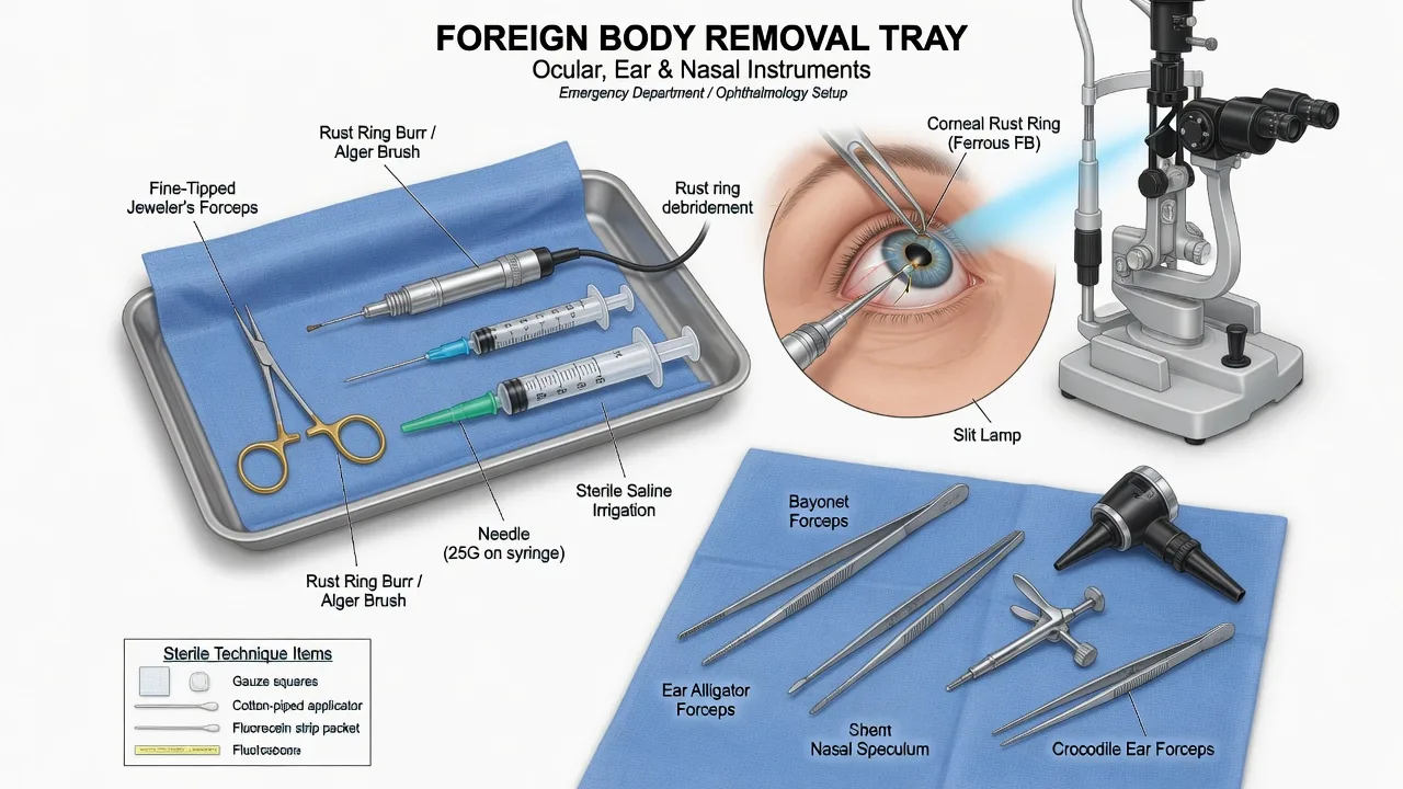
Equipment
The FB tray varies by compartment, but the shared kit is a bright focused light (a headlamp or a slit lamp), magnification (loupes or the slit lamp), sterile gloves and a sterile field, local anaesthetic, and sharp container disposal. The compartment-specific instruments are listed below. [1]
The equipment by compartment
Patient preparation and consent
Preparation is the same four steps for every FB. Position and immobilise — the adult is seated comfortably with the head stabilised against a headrest; the child is held upright on a parent's lap with the parent's hand across the forehead and the other across the arms (the "bear hug"), or in a papoose board if available. Consent — the procedure, the instruments, the possible need for more than one attempt, the fallback of referral, and the rare complications (canal laceration, perforation, infection, a retained fragment). Anaesthesia — never attempt a painful removal on an unsedated patient; the movement causes the iatrogenic injury. Light and exposure — the headlamp or slit lamp aimed directly, the speculum placed, the assistant holding the patient still. [1]
For the child, procedural sedation is often the better part of valour — ketamine 1 to 2 mg/kg intravenously or 4 to 5 mg/kg intramuscularly provides a dissociated, still, breathing patient in whom an aural or nasal FB can be removed safely on the first attempt, sparing the child the trauma of a repeated forceps attempt and the canal trauma that results. Refer to the procedural-sedation topic for the full monitoring standard. [1]
Local anaesthetic and topical doses
The anaesthesia is tailored to the compartment. For the soft tissue, infiltrate lidocaine 1 per cent at a maximum of 3 mg/kg plain (about 20 mL in the 70 kg adult) or 7 mg/kg with adrenaline (about 50 mL), buffered 9:1 with sodium bicarbonate to halve injection pain, through a 25G needle at the field around the FB — never into the FB itself, which distorts the anatomy and pushes it deeper. Adrenaline-containing solution is never injected into an end-artery territory (the finger, the toe, the nose tip, the pinna, the penis). For the ocular surface, instil amethocaine (tetracaine) 0.5 to 1 per cent drops — one drop into the lower fornix, repeated once after a minute; the cornea is anaesthetised within 30 seconds. For the aural canal and the nasal mucosa, soak a cotton pledget in lidocaine 4 per cent with adrenaline 1:1000 and leave it in the canal or the nostril for 5 to 10 minutes for topical anaesthesia and vasoconstriction — this shrinks the mucosa and reduces the bleeding that otherwise obscures the field. LET gel (lidocaine-epinephrine-tetracaine) provides needle-free anaesthesia for the paediatric soft-tissue FB. [1]
Stepwise technique — soft tissue foreign body
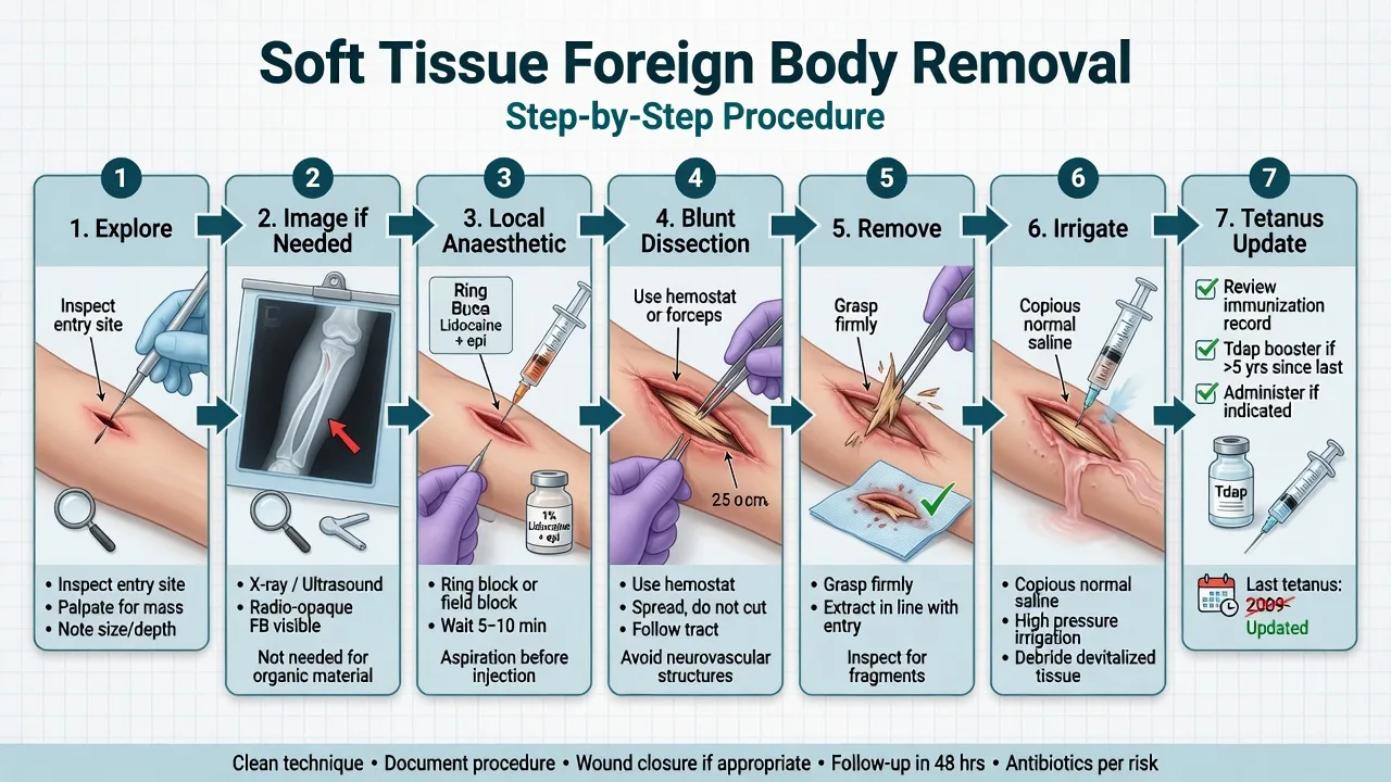
The soft tissue FB is approached as a mini-surgical exploration. After anaesthesia and a sterile field, the technique runs to a fixed sequence. [1]
The soft tissue FB removal, in order
- Locate and image — for the radio-opaque (metal, often glass), a plain X-ray in two projections with a surface marker confirms the location and the depth; for the radiolucent (wood, plastic), a high-frequency linear ultrasound identifies the echogenic object with a posterior acoustic shadow. Do not explore blind.
- Anaesthetise the field — lidocaine 1 per cent infiltrated around (not into) the FB, with a margin; wait 5 minutes for onset.
- Incise to expose — extend the wound along the long axis if needed, or make a small incision over the ultrasound-localised object with a number 11 blade; expose the FB visually.
- Dissect around the object — use the fine forceps to spread the tissue gently, defining the FB's orientation; identify the neurovascular bundle and the tendon in the field before any traction.
- Deliver the object — pass a blunt probe or a right-angle hook behind the deep end of the FB, lever it towards the surface, and lift it out with the forceps. A 25G needle used as a lever beneath one end is the trick for an embedded splinter.
- Irrigate and explore — copious saline irrigation of the cavity; re-examine for a second FB (glass shards, wood fragments), devitalised tissue, and a tendon or nerve injury.
- Close or dress — close the clean wound primarily; leave the bite, the puncture, and the heavily contaminated wound open to heal by secondary intention (refer to wound assessment and closure topics). [1]
The structural errors are to explore blind (the iatrogenic nerve injury), to fail to image a radiolucent FB (the missed wood that returns as necrotising fasciitis), and to close a contaminated wound over a missed fragment (the trapped inoculum).[2][8]
Stepwise technique — ocular foreign body and the rust ring
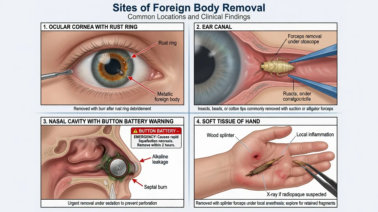
The ocular FB is the one compartment where the slit lamp is the operator's hands — it magnifies the FB, it allows a steady approach with a needle, and it confirms the post-removal fluorescein stain. [1]
The ocular FB removal, in order
- Examine and stain — fluorescein strip, cobalt blue light; identify the punctate stain, the rust ring, or the negative-staining defect of a removed FB.
- Evert the upper lid — sweep the upper fornix with a cotton-tipped applicator or a wire swab; remove any subtarsal FB. A vertical linear corneal abrasion is the rail-track sign of a retained subtarsal FB.
- Anaesthetise — amethocaine 1 per cent, one drop, repeat once; wait 30 seconds.
- Remove the corneal FB — at the slit lamp, with the patient's head on the chin rest and the forehead against the bar, ask the patient to fix on a distant target. Approach the FB with a 25G hypodermic needle on a tuberculin syringe held tangentially to the cornea (never perpendicular); lift the FB off the epithelium with the bevel.
- Remove the rust ring — if iron is present, an orange-brown rust ring remains in the Bowman layer; remove it with a sterile battery-operated rust-ring burr at low speed, or a needle, leaving a small clean defect. A residual rust ring that is deeply embedded can be left to declare itself and removed at the 24- to 48-hour review (it lifts as the epithelium migrates beneath it).
- Irrigate — saline wash to remove any loose particulate matter.
- Treat and review — topical chloramphenicol 1 per cent ointment four times daily for 5 to 7 days; prescribe a cycloplegic (cyclopentolate 1 per cent) if there is an associated iritis. Review at 24 to 48 hours to confirm epithelialisation; the defect heals within 48 to 72 hours. Counsel on the red flags — worsening pain, reduced vision, photophobia — and exclude an open globe if the mechanism was high-velocity. [1]
The post-removal antibiotic for the simple corneal abrasion is debated — the Cochrane review found no clear evidence that topical antibiotics improve healing over placebo for uncomplicated abrasions, but chloramphenicol remains standard practice to prevent bacterial superinfection of the epithelial defect, and is mandatory after a rust-ring removal and for any contact-lens-related abrasion (which needs an anti-pseudomonal cover such as a fluoroquinolone).[3][4] A patient who wears contact lenses is told to discard the lens and not to reinsert it until the defect has healed and been reviewed.
Stepwise technique — aural foreign body
The aural FB is the paediatric favourite, and the technique depends entirely on the shape and the nature of the object. The single rule that governs the whole compartment is the smooth-spherical rule: a bead, a bean, a marble, or a cotton-bud tip cannot be grasped with forceps — forceps push it deeper against the tympanic membrane, and the attempt converts a removable FB into an impacted one needing a general anaesthetic. [1]
Irregular FB (insect, cotton-bud tip, paper, wax plug)
- Grasp with the alligator (crocodile) forceps under direct vision through the speculum
- A live insect is drowned first — instil mineral oil, olive oil, or lidocaine 2 per cent to kill it, then remove the dead insect with forceps or irrigation
- Never pull a struggling insect — it abrades the canal and the tympanic membrane
- Irrigate afterwards to clear debris
Smooth spherical FB (bead, bean, marble)
- Do NOT use forceps — they slip and push it deeper. Pass a right-angle hook (Jobson Horne) behind the FB and lever it out
- Suction with a wide-bore Frazier catheter (Schuknecht) can engage a smooth FB
- Cyanoacrylate on an applicator stick, held to the FB for 60 seconds, bonds and allows the FB to be drawn out — the elegant trick for the impacted bead
- Irrigation (room-temperature saline) works for an organic FB but is contraindicated if the tympanic membrane is perforated or if the FB is a seed/bean that will swell on water contact
Button battery in the ear
- Time-critical emergency — alkaline liquefactive necrosis of the canal and the tympanic membrane within hours
- Remove immediately under direct vision with forceps or suction — never irrigate blindly
- After removal, irrigate the canal with saline to remove the alkaline residue
- Refer to ENT — assess for tympanic membrane perforation, facial nerve palsy, and hearing loss; review for delayed complications
The discriminators the examiner rewards: kill the insect before you pull; never forceps a smooth FB (use a hook, suction, or glue); never irrigate a swollen seed or a perforated TM; and the button battery is removed within the hour, not observed.[5][6]
Stepwise technique — nasal foreign body
The nasal FB is almost always a paediatric presentation, and the button battery is the cannot-miss emergency. The technique splits into the positive-pressure technique for the cooperative child and the instrumental technique for the uncooperative or the impacted FB. [1]
The nasal FB removal, in order
- Position and anaesthetise — sit the child upright; if the FB is accessible, soak a pledget in lidocaine 4 per cent with adrenaline 1:1000 and leave it in the nostril for 5 to 10 minutes to anaesthetise the mucosa and shrink the turbinate. Inspect with the nasal speculum and the headlamp.
- Positive-pressure (the parent's breath) — for the cooperative child with a single FB in the anterior nares, ask the parent to deliver a firm puff of air into the child's mouth while occluding the unaffected nostril (the "parent's kiss" or mouth-to-mouth). The positive pressure expels the FB in a high proportion of cases, with no instrumentation and no trauma.
- Hook behind — pass a right-angle hook (or a Katz extractor, a catheter with a small balloon inflated behind the FB) behind the object and draw it forward and out.
- Forceps — for an irregular FB (foam, food, paper) grasp with the alligator forceps under direct vision; for a smooth FB use the hook or the balloon technique.
- Suction — the wide-bore suction engages a soft or irregular FB.
- After removal — inspect both nostrils for a second FB; check the septum for a battery injury; if a battery was present, irrigate the cavity with saline and refer to ENT for a delayed-perforation review. [1]
The button battery in the nose is the single most dangerous nasal FB. The mechanism is the electrolysis of tissue fluid at the negative pole, which generates hydroxide ions and produces an alkaline (not acidic) liquefactive necrosis of the nasal septum and the turbinates within hours of insertion. A battery left in the nose for over 24 hours causes a septal perforation in a high proportion of cases; the long-term outcomes include saddle-nose deformity, chronic sinonasal disease, and growth disturbances.[7] The candidate must name the mechanism — electrolysis, alkaline necrosis — and the urgency — removal within hours, never observed.
Imaging — radio-opaque versus radiolucent
The imaging decision rests on the material. Metal is radio-opaque and is seen on a plain X-ray in two projections; glass is variably radio-opaque (most soda-lime and leaded glass is visible, but some is not); wood and plastic are radiolucent and are missed on a plain film. The Carneiro Radiographics review established the multimodality approach: a plain film for the metal and the glass, a high-frequency linear ultrasound (10 to 15 MHz) for the wood and the plastic (which appear as an echogenic linear object with a posterior acoustic shadow and an anechoic halo if chronic), and a CT for the orbital, the intraocular, or the deeply embedded FB.[1][9] A CT is also the modality of choice for the suspected wooden orbital FB — the wood is hypodense on CT and can be mistaken for air (a classic pitfall); an MRI is contraindicated if any metal is suspected, and wood on MRI is hypointense on T2.
Metal (and most glass)
- Radio-opaque; visible on a plain X-ray in two orthogonal projections
- Use a surface marker (a paperclip) to localise the depth and the trajectory
- Always image before exploring — the two-view X-ray confirms the location, the depth, and the absence of a second FB
- Glass is variably radio-opaque — most are seen, but a high index of suspicion warrants an ultrasound if the X-ray is negative and the history is of glass
Wood and plastic (radiolucent)
- NOT visible on a plain X-ray — wood is radiolucent and is the missed-FB classic
- Image with a high-frequency linear ultrasound (10 to 15 MHz): the FB is echogenic with a posterior acoustic shadow
- On CT the wood is hypodense and mimics air — a retained orbital wooden FB is repeatedly missed because the operator reads the hypodensity as surgical emphysema
- MRI is contraindicated if metal is possible; on MRI the wood is hypointense on T2-weighted imaging
The intraocular or orbital FB
- CT is the modality of choice — localises the object, identifies the globe injury, and excludes an intraocular FB
- B-scan ultrasound if the globe is intact and the FB is not metal
- NEVER perform MRI if a metallic FB is possible — a ferrous object will torque in the scanner and blind the patient
- Plain X-ray retains a screening role for the high-velocity metallic injury before the definitive CT
The discriminating question on every retained FB is: is this object radio-opaque or radiolucent? If radio-opaque, image with an X-ray; if radiolucent, image with an ultrasound (or a CT for the deep or the orbital).[1][9] The candidate who orders a plain X-ray for a suspected wooden splinter and discharges a negative result has made the textbook error — the wood is invisible, the splinter remains, and the patient returns with a necrotising soft-tissue infection.[2]
Differential diagnosis — retained foreign body versus resolved
The differential on every FB presentation is whether the object is still there. The history is the first clue — a patient who felt something enter and can still feel it usually has it; a patient who felt something enter and now feels nothing may have expelled it (the coughed-out or sneezed-out FB) or may have a residual fragment. The signs that argue for a retained FB are the persistent pain, the localised infection (a cellulitis, an abscess, a draining sinus), the recurrent or a non-healing wound, and the functional disturbance (a foreign-body sensation in the eye, an obstructed nose, a blocked ear). The signs that argue for resolution are the recovered object (the parent brings the bead in a tissue), the resolved symptoms, and the normal examination. [1]
Retained foreign body
- Persistent or worsening pain at the entry site; a foreign-body sensation in the eye
- A localised infection — a cellulitis, an abscess, a draining sinus — that is slow to heal or recurrent
- A linear vertical corneal abrasion (the rail-track sign) argues for a retained subtarsal FB
- Imaging confirms — an X-ray for metal/glass, an ultrasound for wood/plastic; never discharge a suspected FB on a negative history alone
Resolved / expelled FB
- The object was recovered (parent brings it); symptoms resolved after the event
- A normal examination with no fluorescein uptake, no canal laceration, no sinus tract
- A confident history and a normal exam support discharge with a safety-net
- Always examine for a second FB — glass shards and wood fragments are commonly multiple
Cellulitis or abscess (with or without FB)
- A spreading erythema, warmth, swelling, and systemic signs argue for an infection — but a retained FB is the cause until excluded
- Image the area before assuming a primary cellulitis — the underlying retained FB drives the infection
- Manage with antibiotics AND a search for the FB; an abscess needs drainage and an exploration for the source
- A necrotising soft-tissue infection with crepitus or systemic toxicity mandates urgent surgical exploration and broad-spectrum antibiotics
Allergic or toxic reaction
- A localised reaction to the material (a metal allergy, a chemical irritant) can mimic a retained FB
- The history of a known allergen and the absence of an entry wound distinguish it
- Treat with antihistamines and topical steroids; exclude a retained FB first
- A chemical burn (an alkaline disk, a battery leak) needs copious irrigation and an injury assessment
The discriminating question on every presentation is: has this FB resolved, or is a fragment retained? The default is to assume retention until imaging excludes it — a missed retained FB is the most common source of the late return and the litigation.[2][8]
Post-removal care — tetanus, wound toilet, antibiotics
After the FB is removed, the post-care is the same four steps. Tetanus prophylaxis is assessed at every FB removal — the wound type (a clean minor wound versus a contaminated, a puncture, or a tetanus-prone wound) and the vaccination history (the number of documented doses and the time since the last dose). A patient with fewer than three documented doses, or a contaminated wound with over five years since the last dose, receives a tetanus-diphtheria-acellular-pertussis (dTPa) booster; a patient with an unknown or incomplete primary course and a tetanus-prone wound also receives tetanus immunoglobulin 250 IU intramuscularly. Wound toilet — copious saline irrigation, debridement of devitalised tissue, and a non-adherent dressing. Antibiotics are NOT routine for the clean removed FB; they are reserved for the contaminated wound (the bite, the plantar puncture, the heavily soiled splinter), the immunocompromised host, the prosthetic-valve or joint patient, and the established infection. The plantar puncture through a shoe sole is treated with ciprofloxacin 500 mg orally twice daily for 7 days to cover Pseudomonas; the mammalian bite is treated with amoxicillin-clavulanate 875/125 mg orally twice daily.[8]
Discharge and safety-net — the patient is discharged with advice to return for the spreading redness, the increasing pain, the fever, the numbness or weakness (a nerve injury), and the reduced vision (after an ocular FB). The ophthalmology review is at 24 to 48 hours for the corneal FB and the rust ring; the wound review is at 48 hours for the contaminated wound left open; the button-battery patient is reviewed by ENT at 1 week for the delayed perforation.[7]
Complications — procedure-related and disease-related
The complications split into those caused by the procedure and those caused by the missed or the retained FB. The procedure-related complications are the canal laceration (the bleeder that obscures the field after a forceps slip), the tympanic membrane perforation (the forceps advanced blindly, or the irrigation under pressure through a perforated TM), the nasal septal injury (the forceps tearing the mucosa), the corneal abrasion from the needle or the burr (the iatrogenic defect larger than the original FB), and the retained rust ring or fragment (the incomplete removal). The disease-related complications are the infection (the cellulitis, the abscess, the necrotising fasciitis of the missed wood), the alkaline necrosis of the button battery (the septal perforation, the facial nerve palsy, the canal stenosis), the intraocular injury (the endophthalmitis, the retinal detachment, the siderosis from a retained iron FB), and the delayed presentation of a retained FB as a chronic draining sinus or a non-healing wound.[2][7]
Pitfalls and practical tips
The pitfalls are the inverse of the structure. Failing to image the radiolucent FB — the missed wood that returns as necrotising fasciitis. Ordering an X-ray for a wooden splinter and discharging the negative — the textbook error. Reading a hypodense orbital lesion on CT as surgical emphysema — the missed orbital wood. Forceps on a smooth spherical aural FB — the impacted bead that needs a general anaesthetic. Pulling a live insect from the ear — the abraded canal and the perforated TM. Missing a subtarsal FB — the vertical corneal rail-track abrasion that recurs with every blink. Closing a contaminated wound over a missed fragment — the trapped inoculum. Failing to recognise the button-battery halo sign on the X-ray — the delayed necrosis. Attempting an intraocular FB removal in the ED — the globe that is destroyed. The repeated failed attempt in an uncooperative child — the canal that bleeds, the FB that impacts, the general anaesthetic that was avoidable. [1]
The practical tips are the opposite: image before you explore (the two-view X-ray or the high-frequency ultrasound); kill the insect before you pull (oil or lidocaine drops); hook behind a smooth FB, never forceps; evert the upper lid on every ocular FB; recognise the button-battery halo sign and remove within the hour; never remove an intraocular FB in the ED (shield and refer); never irrigate a perforated TM or a swelling seed; refer the deep, the impacted, the uncooperative-child, and the neurovascular-adjacent FB; and apply the two-attempt rule — stop and refer after two failed attempts, before the canal or the mucosa is traumatised.[1][5][6]
Special populations
The child is the commonest FB patient and the one most at risk of an iatrogenic injury from a repeated attempt. The principle is the single best attempt under optimal conditions — a good light, an assistant, topical anaesthesia, and procedural sedation (ketamine 1 to 2 mg/kg intravenously) for the uncooperative child with an aural or nasal FB. The positive-pressure (parent's kiss) technique succeeds for many anterior nasal FBs without any instrumentation.[6] The diabetic and immunocompromised patient has a lower threshold for antibiotic prophylaxis and a 48-hour review after a contaminated FB. The anticoagulated patient bleeds more from a canal laceration or a mucosal tear — meticulous haemostasis and pressure replace dose modification. The contact-lens wearer with a corneal abrasion after FB removal needs an anti-pseudomonal topical (a fluoroquinolone), is told to discard the lens, and is reviewed at 24 to 48 hours to exclude a bacterial keratitis. The occupational or high-velocity injury (the hammering-metal worker with a suspected intraocular FB) is imaged with a CT before any manipulation; the threshold to refer to ophthalmology is low.
Evidence and the regional guidelines
The contemporary framework rests on five evidence streams. The imaging evidence — the Carneiro Radiographics review — established the multimodality approach: a plain X-ray for the metal and the glass, a high-frequency ultrasound for the wood and the plastic, and a CT for the orbital and the deep.[1] The point-of-care-ultrasound evidence — the Osborne study of wooden-FB detection by medics — confirmed the high-frequency linear probe as the bedside tool for the radiolucent soft-tissue FB.[9] The corneal-FB evidence — Xu's review of corneal-FB management and the Cochrane review of antibiotic prophylaxis for the corneal abrasion — established the slit-lamp-and-needle technique and the chloramphenicol post-care, with the caveat that the simple abrasion may not strictly need a topical antibiotic.[3][4] The aural and nasal-FB evidence — the Ponnuvelu management algorithm and the Morris epidemiology study of nasal and aural FB removal — codified the technique-by-shape approach and the positive-pressure method.[5][6] The button-battery evidence — the Heilig study of long-term outcomes after nasal button-battery injuries — established the alkaline-liquefactive-necrosis mechanism, the urgency of removal within hours, and the high rate of delayed complications including septal perforation and saddle-nose deformity.[7] The plantar-puncture evidence — the Chachad review — established the Pseudomonas cover for the through-shoe-sole puncture and the high rate of retained FB in this injury.[8] The missed-FB evidence — the Chaulagain case report of a missed wooden FB causing necrotising fasciitis — is the cautionary example of the radiolucent-FB trap.[2]
ANZ practice note. In Australasian EDs the FB removal follows the compartment-specific technique: the slit-lamp-and-needle for the ocular FB with chloramphenicol post-care, the headlamp-and-speculum with forceps/hook/suction for the aural FB (with procedural sedation for the uncooperative child), and the positive-pressure or hook technique for the nasal FB. The button battery in the ear, the nose, or the oesophagus is a time-critical emergency removed within the hour and referred to ENT for the delayed-perforation review. Imaging follows the material rule — an X-ray for metal and glass, a high-frequency ultrasound (POCUS) for wood and plastic, and a CT for the orbital and the deep. Tetanus prophylaxis is documented at every removal. The intraocular FB is shielded and referred; the uncooperative child after two failed attempts is referred to ENT for a general anaesthetic. ACEM procedural standards and the Royal Australasian College of Surgeons' guidance on paediatric FB removal frame the local practice. [1]
Exam pearls
- The button battery is the emergency — electrolysis at the negative pole generates hydroxide and liquefies the tissue within hours; the halo (double-ring) sign on X-ray distinguishes it from a coin; remove within the hour, never observe.
- Radiolucent FB = ultrasound, not X-ray — wood and plastic are missed on a plain film; a high-frequency linear probe finds the echogenic object with a posterior acoustic shadow; a missed wooden FB returns as necrotising fasciitis.
- Never forceps a smooth spherical aural FB — use a right-angle hook behind it, a wide-bore suction, or cyanoacrylate on an applicator stick; forceps push it deeper and impact it.
- Kill the insect before you pull — instil oil or lidocaine drops to drown a live insect in the ear, then remove the dead insect; a struggling insect abrades the canal and the tympanic membrane.
- Evert the upper lid on every ocular FB — a vertical linear corneal abrasion is the rail-track sign of a retained subtarsal FB; the fluorescein stain is the diagnostic test.
- The two-attempt rule — stop and refer after two failed attempts in an uncooperative child; the third attempt traumatises the canal and impacts the FB.
- Shield the intraocular FB — never remove, never apply pressure; keep nil by mouth, give ceftriaxone 1 g intravenously, and refer to ophthalmology for theatre. [1]
SAQ — Wooden splinter in the plantar foot of a diabetic patient
10 minutes · 10 marks
A 54-year-old insulin-dependent diabetic presents to the ED 48 hours after stepping on a wooden splinter while walking barefoot in the garden. He has not sought medical attention. There is a 5 mm puncture wound on the sole with surrounding erythema, mild swelling and tenderness; the entry site is crusted. He is afebrile, HR 92, BP 138/82, SpO2 98 per cent on room air. The plantar mid-foot is warm and tender to palpation but he can weight-bear. There is no purulent discharge. He last had a tetanus booster 12 years ago.
SAQ — Suspected button battery in the nose of a four-year-old
10 minutes · 10 marks
A four-year-old boy is brought to your ED because his mother saw him put something into his right nostril 90 minutes ago. He is well, afebrile, RR 22, SpO2 98 per cent, no stridor. The right nostril has clear rhinorrhoea and no visible object on anterior inspection. His mother brings an empty button-battery package from a small electronic toy, but is unsure whether the battery is missing because he has inserted it or because she lost it.
Red flags
[1]References
- [1]Carneiro BC, Neves JR, Oliveira AL, et al. Multimodality Imaging of Foreign Bodies: New Insights into Old Challenges Radiographics, 2020.PMID 33136481
- [2]Chaulagain U, Simkhada R, Sharma P, et al. Missed Wooden Perineum Foreign Body Leading to Extensive Necrotizing Soft Tissue Infection: A Case Report Clin Case Rep, 2026.PMID 41550391
- [3]Xu P, Yu T, Li X, Wang Y. Managing corneal foreign body injuries in a primary eye care setting Clin Exp Optom, 2026.PMID 40174878
- [4]Ng SM, O'Connell N, Bhardwaj G, et al. Antibiotic prophylaxis for corneal abrasion Cochrane Database Syst Rev, 2025.PMID 41017778
- [5]Ponnuvelu K, Charles R, Rajan S, Senthilnathan S. Intriguing aural foreign body and algorithm of management of foreign body BMJ Case Rep, 2021.PMID 34400422
- [6]Morris S, Liberman G, O'Dwyer M, Cottier D. Will children ever learn? Removal of nasal and aural foreign bodies: a study of hospital episode statistics Ann R Coll Surg Engl, 2018.PMID 29968507
- [7]Heilig Y, Strychowsky J, Nguyen A, et al. Long-term outcomes following nasal button battery foreign body injuries in children: a 10-year retrospective analysis of 45 patients Int J Pediatr Otorhinolaryngol, 2026.PMID 41985339
- [8]Chachad S, Bhatt M. Management of plantar puncture wounds in children Clin Pediatr (Phila), 2004.PMID 15094944
- [9]Osborne K, Boone D, Wild B, et al. Special Forces Medics Ability to Identify Wooden Foreign Bodies by Point-of-Care Ultrasound J Spec Oper Med, 2025.PMID 39621007