EM · Perforated viscus
Perforated viscus
Also known as Perforated viscus · Perforated peptic ulcer · Perforated appendix · Perforated diverticulum · Pneumoperitoneum · Generalised peritonitis
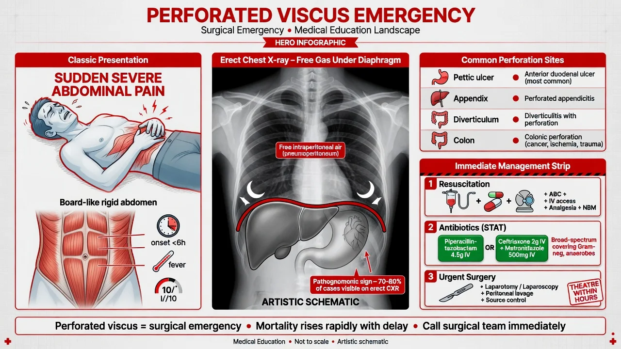
Perforated viscus — the full-thickness breach of a hollow abdominal organ releasing gas and contents into the peritoneal cavity, the perforated peptic ulcer (the sudden severe epigastric pain, the rigid abdomen, the free gas under the diaphragm on the erect CXR), the perforated appendix (the localised right lower quadrant peritonitis), the perforated diverticulum (the Hartmann procedure), the perforated colon (the colorectal cancer, the colitis). The management is the NBM, the NG tube, the IV fluid resuscitation, the broad-spectrum antibiotics (the piperacillin-tazobactam 4.5 g IV or the ceftriaxone 2 g IV plus the metronidazole 500 mg IV), the morphine 5 mg IV, the urgent surgical referral and the laparotomy for the generalised peritonitis. The erect CXR (the free gas under the diaphragm, the sensitivity about 70 per cent), the CT (the definitive test — the free gas, the free fluid, the site of the perforation). The differential — the acute pancreatitis, the mesenteric ischaemia, the ruptured AAA, the inferior MI. ACEM-primary, globally tagged.
On this page & tools
Your progress
Saved locally on this device.
5 MCQs with explanations
Target exams
Red flags
A perforated viscus is the full-thickness breach of a hollow abdominal organ that releases luminal gas and contents into the peritoneal cavity, producing pneumoperitoneum and peritonitis. It is among the most time-critical of the surgical presentations to the emergency department because the peritoneal soiling converts within hours from a chemical to a bacterial, then a faecal, peritonitis, and the mortality climbs steeply with every hour of delay. The Fellowship candidate must, within the first hour, answer four questions: is the patient septic or shocked and what resuscitation does the physiology demand, is there a perforation (and which imaging proves it), where is the perforation (the peptic ulcer, the appendix, the diverticulum, or the colon — because the operation differs for each), and does this patient go to the theatre now or after a CT. The four sites behave differently — the perforated peptic ulcer leaks acidic gastric juice and causes sudden severe epigastric pain with a rigid abdomen; the perforated appendix leaks pus into the right iliac fossa; the perforated diverticulum leaks faeces into the pelvis and demands a Hartmann procedure; the perforated colon leaks from a cancer or an ischaemic or inflamed wall and produces the septic, distended abdomen. The candidate who reaches for the laparotomy in every perforation, or who delays the antibiotics and the fluid resuscitation for the imaging, fails the station.[1][2]
Definition and classification
A perforated viscus is a full-thickness defect in the wall of a hollow abdominal organ (the stomach, the duodenum, the small bowel, the appendix or the colon) that communicates with the peritoneal cavity. The consequence is pneumoperitoneum (free intraperitoneal gas) and peritonitis (inflammation of the peritoneum by the leaked contents). A contained perforation leaks into a walled-off area — a sealed peptic ulcer behind the liver, or a pericolic abscess around a diverticulum — and may be comparatively stable; a free perforation floods the peritoneal cavity and produces generalised peritonitis and septic shock.[1]
The classification that governs management is by anatomic site, because each site has its own cause, its own operation and its own prognosis: [1]
| The site | The common cause | The leaked contents | The typical operation |
|---|---|---|---|
| Peptic ulcer (stomach or duodenum) | NSAIDs, H. pylori, stress | Acidic gastric juice, bile | Omental (Graham) patch, laparoscopic or open |
| Appendix | Untreated acute appendicitis | Purulent fluid, faecolith | Laparoscopic appendicectomy |
| Diverticulum (sigmoid) | Acute diverticulitis | Faeces, pus | Hartmann procedure (Hinchey III to IV) |
| Colon | Colorectal cancer, ischaemic or ulcerative colitis | Faeces | Resection — Hartmann or subtotal colectomy |
The four perforation sites
Epidemiology and risk
Perforated peptic ulcer remains common despite the decline in uncomplicated ulcer disease brought by proton-pump inhibitors and H. pylori eradication, because the population exposed to non-steroidal anti-inflammatory drugs (NSAIDs) and low-dose aspirin has grown. The risk factors are NSAID and aspirin use, H. pylori infection, smoking, alcohol, stress ulceration in the critically ill, and Zollinger-Ellison syndrome. The peak age is the sixth and seventh decades, and the patient often has no prior ulcer history — the perforation is the first presentation. Perforated appendicitis is the natural history of untreated acute appendicitis and is commonest in the young adult and the elderly, in whom delayed presentation drives the perforation rate above 60 per cent. Perforated diverticulitis complicates about 1 to 2 per cent of acute diverticulitis admissions and is commonest in the older patient with known diverticular disease. Perforated colon is dominated by colorectal cancer (often a left-sided obstructing lesion that ischaemically perforates upstream at the caecum, where the wall tension is greatest) and by ischaemic colitis and inflammatory bowel disease in the acutely ill patient.[1]
[1]Pathophysiology — from chemical to faecal peritonitis
The perforation releases luminal contents into the peritoneal cavity, and the peritoneal response evolves in two phases. The first is chemical peritonitis: gastric acid (pH near 1), bile and pancreatic juice pour over the peritoneum, producing an immediate, dramatic inflammation with a massive fluid shift into the peritoneal space — the "third space" — and a sudden, severe pain. This is why the perforated peptic ulcer presents as a bolt from the blue, the patient able to name the exact minute the pain began. The second phase is bacterial peritonitis, beginning within hours as the gut organisms (Streptococcus, Enterobacteriaceae, Bacteroides, anaerobes) seed the peritoneum; the peritonitis becomes purulent, the patient becomes septic, and the once-localised soiling becomes generalised. A colonic perforation skips straight to faecal peritonitis, the most lethal form, because the bacterial load is several orders of magnitude higher than in the upper gut. [1]
[1]The systemic response is driven by the cytokine storm of severe sepsis: capillary leak, hypovolaemia, vasodilation, myocardial depression, acute kidney injury, acute respiratory distress syndrome and disseminated intravascular coagulation. The patient with generalised peritonitis is, in physiological terms, a septic-shock patient with a surgically correctable source, and the resuscitation runs in parallel with — never in sequence before — the imaging and the surgical referral.[2]
Clinical presentation
The perforated peptic ulcer presents with sudden, severe epigastric pain that the patient can time to the minute, often after a meal or in the small hours. The pain rapidly becomes generalised as the gastric contents spread through the peritoneal cavity. The patient lies still, breathing shallowly (movement worsens the pain), with a rigid, board-like abdomen, guarding, rebound tenderness and absent bowel sounds (the paralytic ileus of the peritoneal inflammation). Loss of hepatic dullness on percussion is a classic sign — the free gas has risen over the liver and replaced the normal dull note with a resonant one. A history of dyspepsia, NSAID or aspirin use, or known ulcer disease supports the diagnosis but is often absent. [1]
The perforated appendix presents with the natural history of acute appendicitis that has ruptured — typically one to three days of periumbilical pain that localised to the right iliac fossa, then a transient improvement as the appendiceal pressure was relieved by the rupture, followed by worsening right iliac fossa peritonitis, fever and a rising inflammatory response. The picture is one of localised peritonitis unless the appendiceal stump has leaked freely, when it becomes generalised. [1]
The perforated diverticulum presents in the older patient with known diverticular disease, with left lower quadrant pain, fever, a rigid or tender abdomen and signs of sepsis; a Hinchey III to IV perforation presents in septic shock with generalised peritonitis. The perforated colon presents with sepsis, abdominal distension, and peritonitis; a caecal perforation upstream of an obstructing left-sided cancer presents with sudden severe pain and free gas. [1]
[1]Differential diagnosis
The sudden severe abdominal pain of a perforation overlaps with several lethal mimics, and the first task is to exclude a pancreatic inflammatory catastrophe, a vascular emergency and an inferior myocardial infarction before attributing the picture to a perforation — though resuscitation and imaging run in parallel rather than in sequence. [1]
Perforated peptic ulcer
- Sudden severe epigastric pain becoming generalised; rigid board-like abdomen
- NSAID, aspirin, or known ulcer history; the patient can time the onset
- Free gas under the diaphragm on the erect CXR or CT; lipase normal or mildly raised
- NBM, NG tube, IV fluids, broad-spectrum antibiotics, PPI, urgent surgery — omental patch
Acute pancreatitis
- Severe epigastric pain radiating straight through to the back, vomiting, SIRS
- Lipase at least 3 times the upper limit of normal; gallstones or alcohol
- CT shows peripancreatic fat stranding ± necrosis; no free gas unless a posterior duodenal perforation
- Fluids, analgesia, early enteral feeding; NO prophylactic antibiotics
Mesenteric ischaemia
- Severe pain out of proportion to the examination; atrial fibrillation or vascular disease
- Metabolic acidosis, raised lactate, bloody diarrhoea; lipase normal
- CT angiography shows the mesenteric occlusion; pneumatosis or portal venous gas late
- Urgent revascularisation or embolectomy; laparotomy for necrotic bowel
Ruptured abdominal aortic aneurysm
- Sudden back or abdominal pain, syncope, hypotension in the older male smoker
- Pulsatile mass; bedside ultrasound confirms the aneurysm
- Lipase normal; free gas absent — but a retroperitoneal leak can mimic
- Unstable — straight to theatre, not CT; activate the massive transfusion protocol
Inferior myocardial infarction
- Epigastric pain, nausea, diaphoresis; may mimic a perforation or pancreatitis
- ECG shows inferior ST elevation (II, III, aVF); troponin rises
- Lipase normal; no free gas on imaging
- Always do an ECG in epigastric pain; dual antiplatelet and reperfusion therapy
Ruptured ectopic pregnancy
- A woman of reproductive age with sudden lower abdominal pain and syncope
- Beta-hCG positive; bedside ultrasound shows free fluid and an empty uterus
- Lipase normal; no free gas — the free fluid is blood
- Beta-hCG in every woman with abdominal pain; emergency laparoscopy
The other considerations include acute cholecystitis (right upper quadrant pain, Murphy sign, gallstones on ultrasound), ureteric colic (loin-to-groin pain, haematuria, no peritonism), diabetic ketoacidosis presenting with abdominal pain and a metabolic acidosis, and sickle-cell crisis in the relevant population. The cardinal rule is that a rigid abdomen with free gas is a perforation regardless of the differential, and the surgical referral is not delayed by the workup of the mimics.[1]
Bedside assessment
The assessment begins with ABCDE and runs in parallel with the imaging and the surgical referral. The airway and breathing — the patient with peritonitis is hypovolaemic, hypoxic and at risk of aspiration from the paralytic ileus; give high-flow oxygen, have suction ready, and attach monitoring. The circulation — tachycardia, hypotension, cool peripheries, prolonged capillary refill and oliguria flag the third-space losses and the septic shock; establish two large-bore cannulae, take bloods including a lactate and a venous gas, and begin the fluid resuscitation. The disability — drowsiness is hypovolaemia, sepsis or acidosis. The exposure — a full abdominal examination, including the groins for a hernia, the costophrenic angles for the referred pain of free gas, and a digital rectal examination. [1]
The history establishes the onset (the patient who can time the minute of onset favours a perforation), the NSAID, aspirin and PPI use, the known ulcer, diverticular disease, or colorectal cancer, the change in bowel habit and bleeding per rectum (malignancy), the comorbidities (the cardiac, the renal and the respiratory disease govern the resuscitation and the anaesthetic risk), the medications (the anticoagulants complicate the surgery), and the allergies. Screen explicitly for the signs of septic shock — the confusion, the mottled skin, the cold peripheries — because finding them changes the disposition from the CT scanner to the theatre. [1]
Investigations
The investigations confirm the perforation, localise the site, grade the severity and exclude the mimics. [1]
The erect chest X-ray is the traditional first-line test for free gas. With the patient upright for at least 5 to 10 minutes (or a left lateral decubitus film in the patient who cannot sit), free gas rises to collect under the right hemidiaphragm in about 70 per cent of perforations, and under the left in a smaller proportion. The sensitivity is limited — the erect CXR misses about 30 per cent of perforations, and a supine film is far less sensitive. A normal erect chest X-ray does NOT exclude a perforation. The CT is far more sensitive and is now the definitive test.[3][4]
[1]The CT of the abdomen and pelvis with intravenous contrast is the definitive investigation. It confirms the free intraperitoneal gas (often tracking to the upper abdomen and around the falciform ligament), demonstrates the free fluid, localises the site of the perforation by the focal bowel wall thickening, the adjacent inflammatory stranding and the extraluminal gas pocket, and grades the Hinchey stage for a diverticular perforation. The CT also excludes the mimics — the peripancreatic inflammation of pancreatitis, the mesenteric occlusion of ischaemia, the aneurysm — and is the investigation of choice in the haemodynamically stable patient.[2]
The classic CT and radiographic signs of pneumoperitoneum include: [1]
| The sign | The appearance | The meaning |
|---|---|---|
| Rigler sign | Both sides (inner and outer) of the bowel wall visible | Free gas outlining the bowel on both sides |
| Football sign | A large oval lucency over the abdomen | A large-volume pneumoperitoneum |
| Falciform ligament sign | The falciform ligament outlined by gas | Free gas in the upper abdomen |
| Hyperlucency of the liver | A dark liver on the supine film | Gas interposed between the anterior abdominal wall and the liver |
| Cupola sign | An inverted-cup gas collection under the diaphragm | Free gas trapped under the central tendon |
The blood panel includes the full blood count (leukocytosis and neutrophilia of the peritonitis), the urea and electrolytes (dehydration raises the urea; the acute kidney injury of the septic shock), the lactate (the rising lactate is the perfusion failure — the single most useful blood test for the severity), the C-reactive protein, the liver function tests, the amylase or lipase (to exclude the pancreatitis — but remember a posterior duodenal perforation can raise the amylase), the group and save or crossmatch, the coagulation, and the venous or arterial blood gas (the pH, the base deficit, the lactate). The ECG and a troponin exclude an inferior myocardial infarction; the beta-hCG is mandatory in every woman of reproductive age. A point-of-care ultrasound (eFAST) may show free fluid and, in skilled hands, the enhancement of the peritoneal stripe by free gas, but it does not replace the CT.[3]
The Hinchey classification — grading the perforated diverticulum
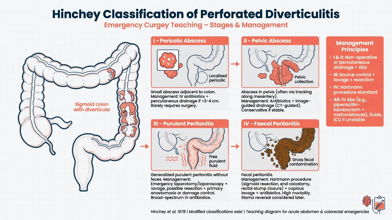
The Hinchey classification grades the severity of the perforated diverticulum and governs the operation, from the localised abscess to the faecal peritonitis: [1]
Hinchey classification of perforated diverticulitis
Immediate management — resuscitation
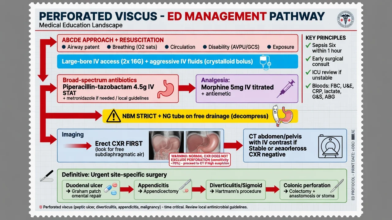
The management of the perforated viscus is concurrent, not sequential: resuscitation, antibiotics, analgesia and the surgical referral run in parallel with the imaging. The candidate who sends the patient to the CT scanner before establishing the intravenous access and giving the antibiotics fails the station.[1][2]
The ABCDE and the intravenous access — two large-bore cannulae, the monitoring (the pulse, the blood pressure, the oxygen saturation, the urine output via the catheter). The fluid resuscitation — give a bolus of 0.9 per cent saline 500 mL to 1 litre (or a balanced crystalloid such as Hartmann) over 15 to 30 minutes in the septic patient, then titrate to a mean arterial pressure over 65 mmHg, a urine output of 0.5 mL per kilogram per hour, and a falling lactate. Reassess frequently — the over-resuscitation causes pulmonary oedema in the elderly comorbid patient, and the under-resuscitation permits the septic shock to deepen. [1]
The analgesia — morphine 5 mg intravenously, titrated (0.1 mg per kilogram, the adult 5 to 10 mg, repeated at 5 to 10 minute intervals to the pain), is the standard opioid and does not mask the physical signs or delay the diagnosis — the systematic reviews support the generous analgesia. The ondansetron 4 mg intravenously for the nausea and the vomiting. The paracetamol 1 g intravenously for the baseline analgesia and the fever. Avoid the NSAIDs — the patient may already have an NSAID-induced ulcer, and the NSAIDs worsen the acute kidney injury of the sepsis. [1]
The nasogastric tube — a large-bore NG tube on the free drainage, with the intermittent aspiration, decompresses the stomach, reduces the further soiling of the peritoneal cavity, relieves the vomiting, and reduces the aspiration risk. The urinary catheter for the urine-output monitoring. The NBM (nil by mouth) — the patient takes nothing by mouth. [1]
The broad-spectrum antibiotics — piperacillin-tazobactam 4.5 g intravenously every 8 hours is the standard first-line regimen, covering the enteric Gram-negatives and the anaerobes; the alternative is ceftriaxone 2 g intravenously once daily plus metronidazole 500 mg intravenously every 8 hours. Give the first dose within the first hour of the recognition of the sepsis — the source control is surgical, but the antibiotics reduce the bacterial load before the operation. For the penicillin-allergic or the septic-shock patient, meropenem 1 g intravenously every 8 hours or the combination of ciprofloxacin 400 mg intravenously twice daily plus metronidazole 500 mg intravenously every 8 hours is the escalation.[1]
The proton-pump inhibitor — pantoprazole 80 mg intravenously as a bolus, then 8 mg per hour by infusion (or the equivalent omeprazole or esomeprazole) — is given for the perforated peptic ulcer to reduce the further acidic soiling and to aid the sealing; the WSES guidelines support the perioperative PPI.[1]
The drug doses for the perforated viscus
Definitive management — by site
The definitive management is the surgical source control, and the operation differs by the site and the severity. The general principles — the peritoneal lavage, the repair or the resection of the perforation, and the drainage — are common to all; the technique is site-specific.[1][2]
The perforated peptic ulcer
The perforated peptic ulcer is managed by the omental (Graham) patch — a patch of the greater omentum sutured over the perforation — combined with a thorough peritoneal lavage. The contemporary WSES and Cesena guidelines favour a laparoscopic-first approach in the haemodynamically stable patient without the extensive adhesions, with the conversion to the open if the perforation cannot be visualised or controlled.[1][2] A gastroduodenoscopy at the time of the surgery or soon after biopsies the ulcer (to exclude the malignancy in the gastric ulcer and to test for H. pylori) and guides the long-term management — the H. pylori eradication and the continuation or the step-down of the PPI. The simple closure is favoured over the definitive acid-reducing procedure (the vagotomy, now largely historical) in the modern era of the PPI. The non-operative management — the NBM, the NG tube, the antibiotics and the PPI, with the serial imaging — is reserved for the contained perforation in the high-risk surgical candidate, and requires the close monitoring for the deterioration.
Model answer — when to operate laparoscopically and when to convert to open
The perforated appendix
The perforated appendix is managed by the laparoscopic appendicectomy with the peritoneal lavage of the pus. The laparoscopic approach is preferred in the perforated appendicitis for the lower wound-infection rate and the better visualisation of the pelvis. A post-operative abscess may require the image-guided drainage. The interval appendicectomy is largely abandoned — the recurrent appendicitis after a conservatively managed appendix mass is uncommon. [1]
The perforated diverticulum
The perforated diverticulum is managed by the Hinchey stage. The Hinchey I to II (the localised or distant abscess) is managed with the antibiotics and the image-guided drainage, often without the immediate surgery. The Hinchey III (the purulent peritonitis) and the Hinchey IV (the faecal peritonitis) require the emergency laparotomy — the Hartmann procedure (the sigmoid resection with the end colostomy and the rectal stump) for the faecal peritonitis and the unstable patient, and the primary anastomosis with or without a defunctioning ileostomy for the selected, stable Hinchey III patient. The Hartmann avoids the anastomotic leak in the grossly contaminated abdomen and the patient in shock; the penalty is the stoma and the planned reversal. The laparoscopic lavage and drainage (without the resection) for the Hinchey III purulent peritonitis was promoted by the LADIES trials but the evidence is contested and it is not standard care.[2]
The perforated colon
The perforated colon is managed by the resection. The obstructing left-sided colorectal cancer that perforates the caecum is managed by the subtotal colectomy or the Hartmann; the ischaemic or the colitic perforation is managed by the resection of the affected segment with the stoma. The primary anastomosis is reserved for the stable, well-perfused patient with the minimal contamination. The faecal peritonitis of the colonic perforation carries the highest mortality of the four sites.[1]
Subtypes and scenarios
The contained perforated peptic ulcer — the perforation that has sealed behind the liver or against the gallbladder — presents with a less dramatic picture, may have a small pocket of free gas on the CT, and may be managed non-operatively in the high-risk surgical candidate with the antibiotics, the PPI and the close monitoring. The perforation in the elderly NSAID user is the classic scenario — the muted pain, the delayed presentation, the sepsis on arrival, and the high mortality. The perforation in the intensive-care patient is the stress-related mucosal disease — the upper-GI bleed or the perforation in the critically ill, prevented by the acid suppression. [1]
The perforated appendicitis in the elderly presents late, often as a small-bowel obstruction or a generalised peritonitis, and the perforation rate is above 60 per cent — have a low threshold for the CT. The perforated diverticulitis in the patient with known diverticular disease presents with the left lower quadrant pain, the fever and the sepsis; the Hinchey stage on the CT governs the operation. The stercoral perforation of the constipated or the institutionalised patient is a faecal peritonitis at the rectosigmoid — a high-mortality scenario.[2]
Complications and pitfalls
The complications of the perforation itself are the septic shock and the multi-organ failure, the intra-abdominal abscess, the wound infection and dehiscence, the paralytic ileus, the adhesions and the subsequent obstruction, and the anastomotic leak (after a primary anastomosis). The complications of the surgery include the bleeding, the enterotomy (the inadvertent), the stoma complications (the retraction, the necrosis, the parastomal hernia), and the incisional hernia. The complications of the non-operative management are the missed generalised peritonitis, the delayed surgery, and the death. [1]
[1]The pitfalls in the management are the failure to recognise the perforation (the sudden pain, the rigid abdomen, the free gas), the over-reliance on the normal erect CXR (the 30 per cent miss rate), the delay of the antibiotics and the fluids for the imaging, the failure to refer to the surgery early, the non-operative management of a free perforation, and the failure to exclude the lethal mimics — the inferior MI, the mesenteric ischaemia, the ruptured aneurysm and the pancreatitis.[4]
Prognosis and disposition
The mortality of the perforated peptic ulcer is about 10 to 30 per cent, driven by the delayed presentation, the comorbidity and the shock on arrival; the mortality doubles for every 24 hours of the delay to the surgery. The mortality of the Hinchey III perforated diverticulum is about 5 to 15 per cent, and the Hinchey IV (the faecal peritonitis) up to 30 per cent. The mortality of the colonic perforation is the highest of the four sites, often above 30 per cent, because of the older age, the malignancy and the faecal load. The POSSUM and the P-POSSUM scores predict the perioperative mortality and guide the surgical decision in the high-risk patient. [1]
The disposition: every patient with a confirmed perforation is admitted under the surgical team and goes to the theatre for the source control, with the resuscitation and the antibiotics running. The patient in the septic shock goes to the theatre emergently, not to the CT. The contained perforation managed non-operatively is admitted for the close monitoring with the serial examinations, the lactate and the imaging. The intensive-care admission is for the septic shock, the multi-organ failure, or the significant comorbidity. The patient is discharged once the sepsis has resolved, the source has been controlled, and the stoma (if formed) is functioning, with the plan for the H. pylori eradication, the PPI, and the stoma reversal in the appropriate interval. [1]
Special populations
The elderly perforate with muted pain, no rigidity, a delayed presentation and a high mortality — have a low threshold for the CT, give the antibiotics early, and involve the surgery and the intensive care from the start. The anticoagulated patient (the warfarin, the DOAC) complicates the surgery — reverse the anticoagulation where appropriate (the vitamin K, the prothrombin complex concentrate for the warfarin; the specific antidotes for the DOAC) and balance the bleeding risk against the urgency of the source control. The immunosuppressed (the steroid, the transplant, the chemotherapy) present atypically and tolerate the sepsis poorly. The pregnant patient — the appendix and the gallbladder are the common sources, the imaging is the ultrasound first, the MRI second, the CT with the counselling if the diagnosis cannot be made otherwise, and the surgery is not delayed by the pregnancy. The paediatric perforation is usually the appendicitis — the perforation rate is high in the under-5 child, and the management is the laparoscopic appendicectomy with the antibiotics.[1]
Evidence and regional guidelines
The contemporary framework rests on the World Society of Emergency Surgery (WSES) guidelines for the perforated peptic ulcer (the Tarasconi 2020 guidelines) and the Cesena guidelines on the laparoscopic-first approach to the general surgery emergencies (the Sermonesi 2023 consensus).[1][2] The practice-defining points are the early broad-spectrum antibiotics (the piperacillin-tazobactam 4.5 g IV first line), the perioperative PPI for the peptic ulcer, the laparoscopic-first approach for the stable patient, the omental patch for the peptic ulcer, the Hartmann for the faecal peritonitis, and the CT as the definitive imaging where the erect CXR is normal. The imaging evidence confirms the limited sensitivity of the erect CXR (about 70 per cent) and the superiority of the CT for the site and the severity.[3][4]
ANZ practice note. The Royal Australasian College of Surgeons and the ACEM endorse the early surgical referral, the broad-spectrum antibiotics within the first hour, and the CT-first imaging for the haemodynamically stable patient. The perforated peptic ulcer is managed by the laparoscopic omental patch in the capable centre, with the open conversion for the unstable or the difficult case. The perforated diverticulum follows the Hinchey-based approach with the Hartmann procedure for the faecal peritonitis. The surgical registrar and the consultant are the senior decision-makers; the disposition and the timing of the surgery are the joint decisions. [1]
Model answer — the ED workup of the suspected perforated viscus
Exam practice
SAQ — Perforated peptic ulcer with generalized peritonitis
12 minutes · 10 marks
A 64-year-old man on ibuprofen for osteoarthritis and a former heavy smoker presents with sudden severe epigastric pain that began four hours ago and is now generalised. He lies completely still on the trolley. T 38.4, HR 124, BP 98/60, RR 26, SpO2 96 per cent on room air. The abdomen is rigid and board-like with absent bowel sounds and loss of hepatic dullness. WCC 18, lactate 3.2. An erect chest radiograph shows free gas under both hemidiaphragms.
SAQ — Perforated diverticulum with faecal peritonitis
12 minutes · 10 marks
A 74-year-old woman with known diverticular disease presents with sudden severe lower abdominal pain and a rigid abdomen. She is confused and peripherally shut down. T 38.8, HR 138, BP 82/48 on a fluid bolus, RR 30, lactate 5.6. CT abdomen shows free gas and free fluid with a perforated sigmoid diverticulum and generalized faecal contamination.
Exam pearls
- The four perforation sites are the peptic ulcer (the sudden epigastric pain, the omental patch), the appendix (the right iliac fossa, the appendicectomy), the diverticulum (the Hinchey, the Hartmann), and the colon (the cancer or the colitis, the resection).
- The erect CXR detects free gas in only about 70 per cent — a normal film never excludes a perforation; go to the CT.
- The resuscitation, the antibiotics, the analgesia and the surgical referral run in parallel, not in sequence — do not send the patient to the CT before the IV access and the first antibiotic dose.
- The first-line antibiotic is the piperacillin-tazobactam 4.5 g IV; the alternative is the ceftriaxone 2 g IV plus the metronidazole 500 mg IV.
- The Hinchey classification grades the perforated diverticulum — the Hartmann for the faecal peritonitis (the Hinchey IV).
- Exclude the lethal mimics — the pancreatitis (the lipase), the mesenteric ischaemia (the lactate, the CT angiography), the ruptured AAA (the ultrasound), and the inferior MI (the ECG).
- The patient who can time the minute of the onset, who lies still with the rigid abdomen and the absent bowel sounds, and who has the loss of the hepatic dullness, has the perforated peptic ulcer. [1]
Red flags
[1]References
- [1]Tarasconi A, Coccolini F, Biffl WL, et al. Perforated and bleeding peptic ulcer: WSES guidelines World J Emerg Surg, 2020.PMID 31921329
- [2]Sermonesi G, Ceresoli M, Marrazza A, et al. Cesena guidelines: WSES consensus statement on laparoscopic-first approach to general surgery emergencies and abdominal trauma World J Emerg Surg, 2023.PMID 38066631
- [3]Pinto A, Miele C, Tinto A, et al. Spectrum of Signs of Pneumoperitoneum Semin Ultrasound CT MR, 2016.PMID 26827732
- [4]Chiu YH, Chen JD, Tiu CM, et al. Reappraisal of radiographic signs of pneumoperitoneum at emergency department Am J Emerg Med, 2009.PMID 19328377