EM · Paediatric emergencies
Neonatal emergencies (the sick neonate in the emergency department)
Also known as Sick neonate · Neonatal collapse · Duct-dependent congenital heart disease · Neonatal sepsis · Congenital adrenal hyperplasia · Salt-wasting crisis · Necrotising enterocolitis · Neonatal seizures · Hypoxic ischaemic encephalopathy · Prostaglandin E1 infusion · Neonatal cardiac emergency
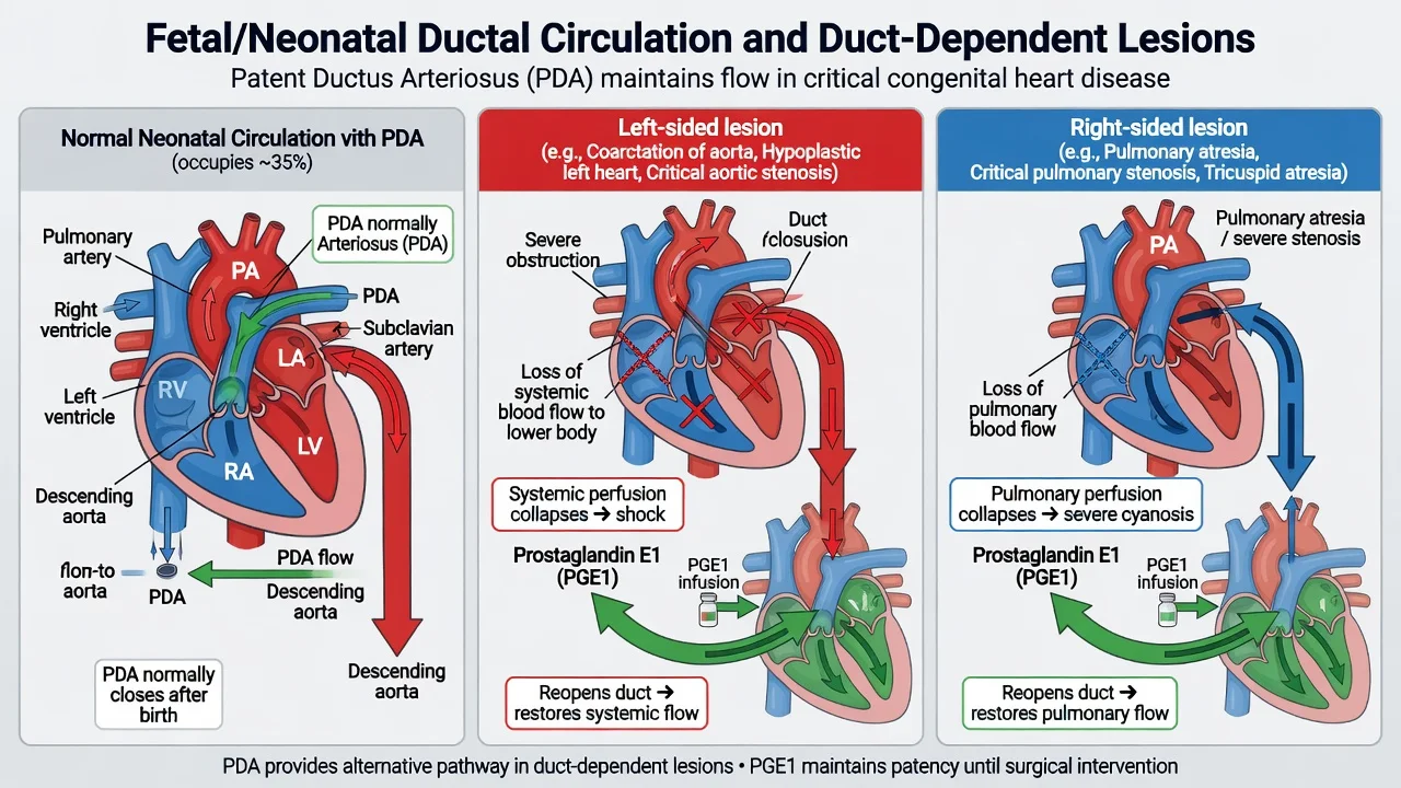
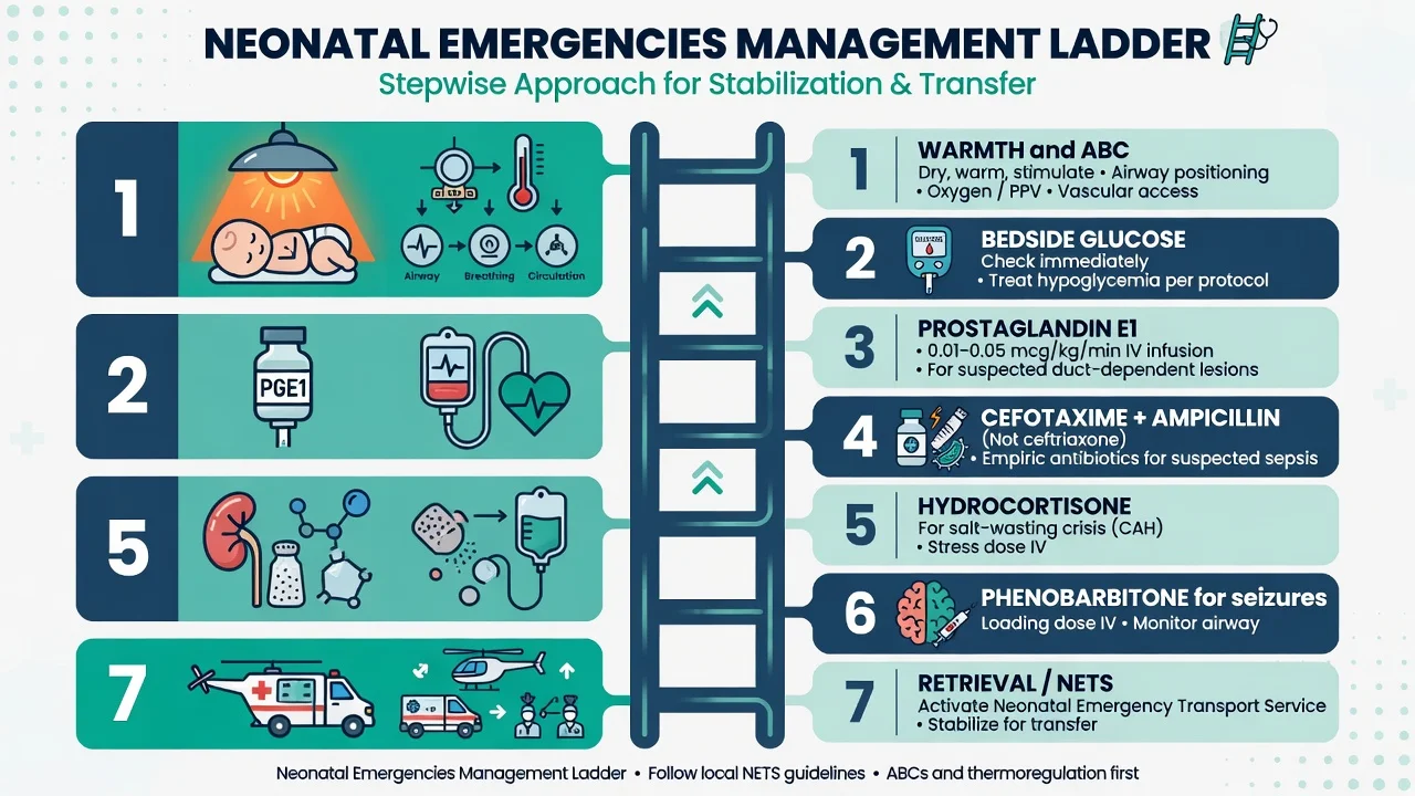
Neonatal emergencies are the life-threatening presentations of the first 28 days of life — the well-then-sick neonate who deteriorates as a ductus closes, an infection declares itself, a metabolic pathway fails, or an adrenal enzyme deficit unmasks. The four reversible, time-critical diagnoses are the duct-dependent congenital heart lesion (the hypoplastic left heart, the coarctation, the pulmonary atresia, the tricuspid atresia — collapse at 24 to 72 hours as the ductus arteriosus constricts, reversed by prostaglandin E1 0.01 to 0.05 mcg per kilogram per minute to keep the duct open), the neonatal sepsis (the group B streptococcus, the E. coli, the Listeria — full septic workup of the blood culture, the lumbar puncture and the suprapubic aspirate, then cefotaxime 50 mg per kilogram IV plus ampicillin 50 mg per kilogram IV, with aciclovir 20 mg per kilogram IV added if HSV is suspected), the congenital adrenal hyperplasia salt-wasting crisis (vomiting and dehydration with hyponatraemia and hyperkalaemia at one to two weeks, reversed by hydrocortisone 25 mg IV), and the neonatal seizure (the hypoglycaemia, the hypocalcaemia, the meningitis). The Fellowship candidate must recognise the sick neonate, start the specific therapy on suspicion rather than waiting for transfer, and pre-empt the side effects — the prostaglandin apnoea above all.
On this page & tools
Your progress
Saved locally on this device.
5 MCQs with explanations
Target exams
Red flags
Related topics
- Paediatric fever and serious bacterial illness (the febrile child in the emergency department)
- Paediatric trauma — the modified approach
- Meningitis and encephalitis (emergency department diagnosis and management)
- Status epilepticus
- Fluid resuscitation in the emergency department
- DKA, HHS and hypoglycaemia
- Adrenal crisis (Addisonian crisis)
- Cardiogenic shock in the emergency department
Neonatal emergencies are the life-threatening presentations of the first 28 days of life. The archetype is the well-then-sick neonate — an infant who fed normally for days, then over hours becomes mottled, grunting, hypothermic, and refuses feeds. Four reversible, time-critical diagnoses account for most of the danger: the duct-dependent congenital heart lesion that collapses as the ductus arteriosus closes, the neonatal sepsis of the first days to weeks, the congenital adrenal hyperplasia salt-wasting crisis of the second week, and the neonatal seizure driven by a metabolic or infectious cause.[1][2] Every one of these is fully reversible if the specific therapy is started on suspicion, and lethal within hours if it is not. The Fellowship candidate's task is to recognise the sick neonate from age-specific signs that do not resemble the adult presentation, to run resuscitation and the diagnostic workup in parallel, and to give the prostaglandin, the antibiotic, or the steroid before the transfer to the tertiary centre, because the neonate may not survive the journey without them.
Definition and classification
A neonatal emergency is any acute, life-threatening presentation in an infant under 28 days. The emergency department presentation is rarely a single disease with a clear history; it is an undifferentiated sick neonate, and the classification is therefore by the four time-critical, reversible groups that must be actively excluded in parallel rather than sequenced one at a time. [1]
The four groups are: (i) duct-dependent congenital heart disease, in which the systemic or pulmonary circulation depends on a patent ductus arteriosus and fails as it constricts at 24 to 72 hours; (ii) neonatal sepsis, divided into early-onset (within 72 hours of birth, acquired intrapartum from maternal colonisation) and late-onset (after 72 hours, often nosocomial or community-acquired); (iii) congenital adrenal hyperplasia salt-wasting crisis, the cortisol and aldosterone deficiency of 21-hydroxylase failure that declares itself at one to three weeks as the maternal salt and glucocorticoid reserve is exhausted; and (iv) the neonatal seizure, in which a metabolic cause (hypoglycaemia, hypocalcaemia, hypomagnesaemia, hyponatraemia) or an infective cause (meningitis, encephalitis) drives abnormal neuronal discharge in a brain with a low seizure threshold. Two surgical or metabolic mimics — pyloric stenosis and the inborn errors of metabolism — must be distinguished because their management diverges sharply from the first four.[2][3]
Epidemiology and risk factors
Critical congenital heart disease affects roughly 1 to 3 per 1000 live births, and around a third of critical lesions are duct-dependent — meaning that without the ductus they cannot sustain life beyond the first days. The lesions that present as neonatal collapse are the hypoplastic left heart syndrome, the coarctation of the aorta, the critical aortic stenosis, the interrupted aortic arch, the pulmonary atresia, the severe tetralogy of Fallot, and the tricuspid atresia.[1]
Neonatal sepsis has an early-onset incidence of approximately 0.5 to 1 per 1000 live births in the antibiotic-prophylaxis era. The organisms of early-onset sepsis are the group B streptococcus, Escherichia coli, and Listeria monocytogenes; late-onset sepsis adds the coagulase-negative staphylococci (especially in the line-bearing premature infant), the Gram-negatives, and Staphylococcus aureus.[2] The risk factors are maternal colonisation with group B streptococcus, prolonged rupture of the membranes beyond 18 hours, maternal intrapartum fever, chorioamnionitis, and prematurity — but a sick neonate without any of these still has sepsis until proven otherwise.
Congenital adrenal hyperplasia from 21-hydroxylase deficiency has an incidence of roughly 1 in 15,000 live births in its classic salt-wasting form; it presents at one to three weeks as the maternal salt and glucocorticoid supply falls away. The inborn errors of metabolism are individually rare (1 in 10,000 to 1 in 100,000) but collectively common enough that any neonate with recurrent hypoglycaemia, a metabolic acidosis, or an altered conscious level after a feed must be screened for them. [1]
Pathophysiology
The duct-dependent lesion fails for a single reason: the foetal circulation assumed a patent ductus arteriosus, and the postnatal constriction of that duct at 24 to 72 hours removes either the systemic output (in the left-sided obstructive lesions) or the pulmonary output (in the right-sided obstructive lesions). In hypoplastic left heart syndrome and coarctation, the systemic circulation depends on the right ventricle pumping through the duct into the aorta; when the duct closes, systemic perfusion fails and the infant shocks with metabolic acidosis. In pulmonary atresia and tricuspid atresia, the pulmonary circulation depends on the duct; when it closes, the infant cyanoses profoundly. Prostaglandin E1 (alprostadil) relaxes the ductal smooth muscle and reopens or maintains the duct, restoring the parallel circulation as a bridge to definitive surgery or catheter intervention.[1]
The neonatal immune system is immature: complement levels are low, neutrophil function is impaired, and the immunoglobulin repertoire is undeveloped. Transplacental maternal immunoglobulin G provides partial cover but is insufficient against a heavy intrapartum bacterial load. Group B streptococcus and E. coli translocate across the immature gut and respiratory mucosa, producing an overwhelming bacteraemia, pneumonia, or meningitis that is rapidly progressive. [1]
In congenital adrenal hyperplasia, the deficiency of 21-hydroxylase blocks the conversion of progesterone to aldosterone and cortisol. Aldosterone deficit removes the renal sodium-retaining and potassium-excreting drive, producing salt-wasting, volume depletion, and a hyperkalaemia that can be lethal; cortisol deficit removes vascular tone and hepatic gluconeogenesis, producing shock and hypoglycaemia; and the shunting of steroid precursors into the androgen pathway produces the virilisation that is the visible clue in the female infant.[3]
[1]Clinical presentation
The sick neonate does not present with the adult vocabulary of pain or fever. The presentation is the well-then-sick story: a previously normal feeder who over hours becomes lethargic, refuses the breast or bottle, feels cold to the mother, and is brought in mottled, grunting, or floppy. The cardinal signs are non-specific: poor feeding, lethargy or irritability, hypothermia (a neonate more often cools than mounts a fever with sepsis), grunting or tachypnoea (the respiratory compensation for a metabolic acidosis, or the work of a duct-dependent pulmonary lesion), apnoea, and a changed colour — pallor, mottling, or central cyanosis.[1][2]
The duct-dependent cyanotic lesion presents with central cyanosis that does not correct with oxygen, a quiet or absent murmur (the obstructive right-sided lesions often have no murmur), and a heaving right ventricle. The duct-dependent systemic lesion (coarctation, hypoplastic left heart) presents with shock, weak or absent femoral pulses, a differential in the four-limb blood pressures (the arms higher than the legs), and hepatomegaly from backward failure. Differential cyanosis — pink hands with blue feet, or the reverse — points to a specific duct-dependent anatomy. [1]
The salt-wasting neonate presents at one to three weeks with projectile or persistent vomiting, dehydration, weight loss, and the biochemistry of aldosterone and cortisol deficit: hyponatraemia, hyperkalaemia, hypoglycaemia, and a metabolic acidosis. The ambiguous genitalia of the virilised female infant (clitoromegaly, labial fusion) is the visible clue, but male infants with CAH have normal genitalia and are easily missed.[3]
The neonatal seizure is subtle: rather than the grand mal convulsion of the older child, the neonate shows focal clonic movements, bicycling of the legs, lip-smacking, tonic deviation of the eyes, or apnoea. A seizure in the first 72 hours raises hypoxic ischaemic encephalopathy, meningitis, and the metabolic causes; a seizure after the first week raises hypocalcaemia, hypoglycaemia, meningitis, and the inborn errors. [1]
Differential diagnosis
The differential of the sick neonate is narrow but high-stakes, and the distinction is made at the bedside by the pattern of the cyanosis, the biochemistry, and the age. The five diagnoses below are the ones an examiner expects a candidate to distinguish with a sentence each. [1]
Duct-dependent CHD
- Collapse at 24 to 72 hours, central cyanosis that does not correct with oxygen, weak femoral pulses, differential four-limb BP, hepatomegaly; a soft or absent murmur
- Metabolic acidosis from poor perfusion; chest X-ray may show pulmonary oligaemia or oedema; pre- and post-ductal SpO₂ gap
- Prostaglandin E1 0.01 to 0.05 mcg/kg/min IV immediately, pre-empt the apnoea, transfer for surgery or catheter intervention
- The single most urgent drug is the prostaglandin, not the antibiotic — though both may be needed if sepsis is not excluded
Neonatal sepsis
- Lethargy, poor feeding, hypothermia or fever, grunting, apnoea; risk factors of maternal GBS, prolonged rupture, chorioamnionitis, prematurity
- Raised or normal CRP (early), positive blood culture, CSF pleocytosis, urine from a suprapubic aspirate; metabolic acidosis
- Cefotaxime 50 mg/kg IV plus ampicillin 50 mg/kg IV, plus aciclovir 20 mg/kg IV if HSV suspected; fluid boluses cautiously
- The septic neonate is more often cold than hot — never triage on the absence of fever
Congenital adrenal hyperplasia
- Vomiting and dehydration at one to three weeks, ambiguous genitalia in the female infant, weight loss, lethargy; the male infant looks normal externally
- Hyponatraemia with HYPERkalaemia, hypoglycaemia, metabolic acidosis; raised 17-hydroxyprogesterone (treat empirically, confirm later)
- Hydrocortisone 25 mg IV immediately, 0.9% saline boluses, glucose for hypoglycaemia; fludrocortisone once stable
- The hyperkalaemia is the biochemical fingerprint — no other mimic of sepsis in this age group gives a high potassium with a low sodium
Pyloric stenosis
- Projectile non-bilious vomiting at three to six weeks, hungry infant, visible peristalsis, palpable olive-shaped pyloric mass in the epigastrium
- HYPOchloraemic HYPOkalaemic METABOLIC ALKALOSIS (the only metabolic alkalosis in paediatrics); ultrasound confirms a hypertrophied pylorus
- Fluid and chloride correction first (never operate on the alkalotic infant), then pyloromyotomy
- The biochemistry is the mirror image of sepsis and CAH — alkalosis, not acidosis
Inborn errors of metabolism
- Recurrent or persistent hypoglycaemia, encephalopathy, vomiting after a protein feed, a family history of neonatal death; often after the first feed of protein
- Metabolic acidosis with an ANION gap, hyperammonaemia, ketones or the absence of expected ketones; ammonia and lactate sent urgently
- Stop the protein feed, give 10% dextrose, treat the hyperammonaemia; urgent metabolic and genetics referral
- The pattern recurs with feeds and the ammonia is the discriminator from sepsis
Investigations and diagnostic targets
The investigations run in parallel with the resuscitation and the first specific drug. A venous or capillary blood gas is the single most useful test: it gives the pH, the metabolic acidosis that marks poor perfusion or sepsis, the lactate, and the glucose at the bedside within a minute. A bedside glucose is checked immediately in every neonate and rechecked after any dextrose — hypoglycaemia is common, reversible, and a leading seizure precipitant. Urea and electrolytes with calcium, magnesium, and a corrected sodium define the metabolic pattern: the hyponatraemia with hyperkalaemia of CAH, the hypocalcaemia of the late-neonatal seizure, and the deranged sodium of the inborn error. A full blood count, CRP, and coagulation are sent; blood cultures are drawn before the first antibiotic but never delay it. [1]
The full septic workup of the neonate is the blood culture, the lumbar puncture (CSF cell count, protein, glucose, culture, and a PCR for herpes simplex if vesicles, maternal history, or an unexplained encephalopathy are present), and the urine obtained by suprapubic aspirate or a sterile in-out catheter — a bag urine is never acceptable in the neonate because the contamination rate is too high. The lumbar puncture is deferred if the neonate is unstable, and the antibiotics are given empirically with the LP performed once the perfusion is restored.[2] An ammonia and a lactate are added whenever an inborn error is plausible. A chest radiograph assesses the heart size, the pulmonary vasculature (oligaemia in the right-sided obstructive lesions, oedema in the left-sided failure), and the lung fields for pneumonia. Four-limb blood pressures and pre- and post-ductal saturations are recorded to expose the coarctation and the differential cyanosis. An ECG and a bedside echocardiogram (where available) complete the cardiac assessment, but the prostaglandin is never delayed for the echo — the clinical suspicion is enough to start it.[1]
The sick neonate at a glance
Immediate management and resuscitation
The resuscitation is adapted to the neonate: the airway is positioned with the head in the neutral or slightly sniffing position (the neonate's large occiput flexes the neck on a flat surface); high-flow oxygen or CPAP is applied for respiratory distress, and intubation is undertaken early in respiratory failure or before a prostaglandin infusion that is likely to cause apnoea. Access is the first practical challenge — two peripheral cannulae are sought, but an intraosseous needle (anteromedial tibia) or an umbilical venous line is placed without hesitation if the neonate is collapsed and a peripheral line cannot be secured within 60 seconds. Cardiac monitoring is attached from the outset. [1]
Fluid resuscitation is given in 10 mL per kilogram boluses of 0.9 per cent saline, repeated to clinical end-points of perfusion, capillary refill, and conscious level — but with a critical caveat: in the duct-dependent cardiac lesion and any cardiogenic shock, the boluses are reduced to 5 mL per kilogram aliquots because the failing heart cannot tolerate the volume, and aggressive saline can precipitate pulmonary oedema. Hypoglycaemia is treated immediately with 2 mL per kilogram of 10 per cent dextrose intravenously and rechecked; hypocalcaemia with calcium gluconate 10 per cent at 1 mL per kilogram under cardiac monitoring; hypomagnesaemia with magnesium sulphate 50 mg per kilogram. The neonate is warmed actively — hypothermia worsens the acidosis and the respiratory failure. [1]
[1]The first 60 minutes of the collapsed neonate
Recognise the sick neonate — poor feeding, lethargy, hypothermia, grunting, mottling, central cyanosis, weak femoral pulses. Open and position the airway; apply high-flow oxygen or CPAP, and intubate early if respiratory failure or if a prostaglandin infusion is about to be started. Secure access — two peripherals, or an intraosseous/umbilical line within 60 seconds if collapsed. Send the venous gas, glucose, urea and electrolytes with calcium and magnesium, full blood count, CRP, coagulation, lactate, ammonia, and blood cultures. Give glucose 2 mL/kg of 10 per cent if hypoglycaemic. If a duct-dependent lesion is suspected (cyanosis not correcting with oxygen, or shock with weak femorals), start prostaglandin E1 0.01 to 0.05 mcg per kilogram per minute intravenously immediately. If sepsis is suspected or not excluded, begin cefotaxime 50 mg per kilogram IV plus ampicillin 50 mg per kilogram IV, adding aciclovir 20 mg per kilogram IV if HSV is plausible. If hyponatraemia with hyperkalaemia and hypoglycaemia is present, give hydrocortisone 25 mg IV with saline and glucose for the salt-wasting crisis. Perform the lumbar puncture once the neonate is stable. Contact the paediatric retrieval service (NETS in ANZ) and the tertiary cardiology or metabolic service early — the specific therapy given in the first hour determines the outcome. [1]
Definitive management — prostaglandin, antibiotics, steroid and seizure control
The definitive therapy depends on the working diagnosis, but in the undifferentiated neonate more than one treatment is often begun in parallel and refined as the results return. [1]
Prostaglandin E1 (alprostadil) is the bridge therapy for the duct-dependent lesion. The dose is 0.01 to 0.05 microgram per kilogram per minute intravenously, started on clinical suspicion and titrated to the response (an improving pH, lactate, perfusion, and saturation). The older teaching of a higher initiating dose (0.1 microgram per kilogram per minute) is reserved for the complete arrest or profound collapse, because the side effects are dose-dependent. The prostaglandin side effects are the dose-limiting danger: apnoea (in up to a quarter of infants on a therapeutic infusion), hypotension, fever, flushing, and hyperglycaemia. The apnoea is predictable and pre-empted by elective intubation and ventilation before transfer, which is the standard of care for any neonate on a prostaglandin infusion being moved between hospitals. The prostaglandin is continued until the definitive surgical or catheter intervention — it is a bridge, not a cure.[1]
Neonatal sepsis is treated with cefotaxime 50 mg per kilogram IV plus ampicillin 50 mg per kilogram IV. Cefotaxime is chosen over ceftriaxone in the neonate because ceftriaxone displaces bilirubin from albumin (risking kernicterus) and causes biliary sludging. Ampicillin covers Listeria, which the cephalosporins do not. Aciclovir 20 mg per kilogram IV every eight hours is added whenever herpes simplex is suspected — a maternal history of genital HSV, vesicles on the neonate, a vesicular rash, hepatitis, or an unexplained encephalopathy with a CSF pleocytosis — because the untreated disease is devastating and the treatment is safe. The duration is guided by the culture and the clinical course: 7 to 10 days for an uncomplicated bacteraemia, 14 to 21 days for meningitis, 21 days for HSV encephalitis.[2]
Congenital adrenal hyperplasia salt-wasting crisis is treated with hydrocortisone 25 mg intravenously as a stat dose (the equivalent of roughly 10 to 25 mg per square metre), followed by ongoing hydrocortisone at 25 to 100 mg per square metre per day in divided doses, with 0.9 per cent saline boluses for the volume depletion and intravenous glucose for the hypoglycaemia. The hyperkalaemia usually corrects as the aldosterone activity is restored by the high-dose hydrocortisone (which has mineralocorticoid activity at these doses). Fludrocortisone is added once the infant is stable and enteral feeding is re-established, because its onset is in hours to days and the intravenous hydrocortisone already provides mineralocorticoid cover in the acute phase.[3]
The neonatal seizure is treated by correcting the underlying metabolic cause first: glucose 2 mL per kilogram of 10 per cent for hypoglycaemia, calcium gluconate 10 per cent at 1 mL per kilogram for hypocalcaemia, magnesium sulphate 50 mg per kilogram for hypomagnesaemia, and 3 per cent saline carefully for symptomatic hyponatraemia. If the seizures persist or the cause is structural or infectious (HIE, meningitis), phenobarbitone 20 mg per kilogram intravenously is the first-line anticonvulsant in the neonate, with a further 5 to 10 mg per kilogram if needed; midazolam 0.1 mg per kilogram IV or levetiracetam is added for refractory seizures. The choice of phenobarbitone first-line is a neonate-specific convention that does not apply to the older child or adult. [1]
Drug doses in the neonatal emergency
The septic workup — blood, CSF and urine
The full septic workup of the neonate is the blood culture, the lumbar puncture, and the urine obtained by suprapubic aspirate or sterile in-out catheter, supported by the CRP and the full blood count. The blood culture is drawn before the first antibiotic but never delays it; a single adequate volume is preferable to none. The lumbar puncture is performed once the neonate is cardiovascularly stable — it is deferred in the collapsed or coagulopathic infant, the antibiotics given empirically, and the LP done after resuscitation. The CSF is sent for cell count, differential, protein, glucose, Gram stain, culture, and a herpes simplex PCR when indicated. The urine is never collected by a bag in the neonate — the contamination rate approaches 50 per cent and renders the culture uninterpretable; a suprapubic aspirate (any growth of a single organism is significant) or a sterile in-out catheter (more than 10,000 colony-forming units per millilitre of a single organism) is the standard.[2]
The interpretation of the neonatal CSF is harder than in the older child because the normal values overlap with the abnormal: a CSF white cell count up to 20 to 30 per microlitre, a protein up to 1.0 to 1.5 g per litre, and a glucose below half the plasma value can all be normal in the first weeks of life. The decision to treat is therefore made on the clinical state, the Gram stain, and the trend, not on a single number. [1]
Complications and pitfalls
The recurring pitfalls in the neonatal emergency are the inverse of the protocol. The first is treating the duct-dependent lesion as sepsis alone and never starting the prostaglandin — a cyanotic or shocked neonate may have both, and the prostaglandin is harmless if the lesion is not duct-dependent, whereas its omission is lethal if it is. The second is the prostaglandin apnoea that catches the unprepared team: the infusion is started, the neonate improves, and then arrests because nobody readied the intubation. The third is using ceftriaxone in the neonate, displacing bilirubin and precipitating kernicterus. The fourth is missing the salt-waster because the male infant has normal genitalia and the vomiting was attributed to reflux — the hyperkalaemia is the tell, and any hyponatraemia with a high potassium in a two-week-old is CAH until proven otherwise.[3] The fifth is not checking a glucose in every neonate, missing a reversible hypoglycaemic brain injury. The sixth is performing an LP in an unstable neonate, causing a respiratory arrest on the procedure trolley — the LP is deferred and the antibiotics given.
The complications of the disease itself are the cardiogenic shock and acidosis of the duct-dependent lesion, the disseminated intravascular coagulation and meningitis of the septic neonate, the arrhythmia of the hyperkalaemic salt-waster, and the hypoglycaemic or hypoxic brain injury of the seizing neonate. Each is preventable by the prompt specific therapy.[3]
Prognosis and disposition
Every sick neonate is admitted to a neonatal or paediatric intensive care unit at a tertiary centre; the duct-dependent lesion is transferred for surgical or catheter intervention, the septic neonate for ongoing antibiotic and supportive care, the salt-waster for endocrine stabilisation, and the seizing neonate for neurology and metabolic workup. The outcome is determined by the timeliness of the first specific therapy: a duct-dependent lesion reversed with prostaglandin before cardiovascular collapse has an excellent surgical outlook, a septic neonate given the antibiotic within the hour does far better than one in whom it was delayed, and the salt-waster responds to hydrocortisone within hours. The retrieval is initiated early — in ANZ through NETS (the Newborn and Paediatric Emergency Transport Service) or the equivalent state service — and the neonate is transferred on a prostaglandin infusion, intubated and ventilated, with a trained retrieval team. [1]
Special populations
The premature neonate has an immature immune system, surfactant deficiency, and a fragile cerebral vasculature at risk of intraventricular haemorrhage; the septic threshold is lower and the respiratory failure more rapid. The surgical neonate raises necrotising enterocolitis (abdominal distension, blood in the stool, pneumatosis intestinalis on the abdominal radiograph) and the congenital duct-dependent malformations. The infant of a diabetic mother is at risk of hypoglycaemia, polycythaemia, and the cardiac and caudal malformations. The mother on long-term steroids, anticonvulsants, or with chorioamnionitis transmits the risk of neonatal adrenal suppression, vitamin K deficiency bleeding, and early-onset sepsis respectively. [1]
Duct-dependent congenital heart disease — the lesion-by-lesion deep dive
The duct-dependent lesion is the single most time-critical reversible diagnosis in the neonate, because the specific therapy — prostaglandin E1 — works in minutes, costs nothing to start, and is harmless if the working diagnosis is wrong. The Fellowship candidate must know not only that the duct closes at 24 to 72 hours, but which circulation fails in which lesion, because the bedside pattern (the cyanosis versus the shock, the murmur versus the silent chest, the pre- versus post-ductal saturation gap) narrows the anatomy before the echo.[4]
The duct-dependent lesions divide into two physiological families. The right-sided obstructive lesions — pulmonary atresia, critical pulmonary stenosis, severe tetralogy of Fallot, tricuspid atresia, Ebstein anomaly — fail the pulmonary circulation: when the duct closes, deoxygenated blood cannot reach the lungs and the neonate cyanoses profoundly with a quiet chest and pulmonary oligaemia on the radiograph. The left-sided obstructive lesions — hypoplastic left heart syndrome, coarctation of the aorta, interrupted aortic arch, critical aortic stenosis — fail the systemic circulation: when the duct closes, the left ventricle cannot deliver cardiac output to the body and the neonate shocks with weak pulses, hepatomegaly, metabolic acidosis, and often pulmonary oedema. The transposition complexes sit across the divide: in d-transposition of the great arteries, the parallel circulations only mix at the duct or the atrial level, and the neonate cyanoses as the duct closes unless an atrial or ventricular septal defect allows mixing. [1]
Hypoplastic left heart
- The left ventricle, mitral and aortic valves are underdeveloped; systemic output depends entirely on the right ventricle pumping through the duct into the aorta
- Shock, mottling, weak pulses globally, hepatomegaly, metabolic acidosis as the duct closes at 24 to 72 hours; a soft systolic murmur or none
- Prostaglandin E1 to keep the duct open; the systemic circulation is now right-ventricle-dependent — avoid anything that drops the pulmonary vascular resistance and steals flow from the body
- The staged Norwood palliation, not a corrective repair, is the surgical path
Coarctation of the aorta
- A discrete narrowing at the isthmus, often with a bicuspid aortic valve; the legs are perfused through the duct until it closes
- Shock with weak or absent femoral pulses, a differential four-limb blood pressure (arms more than 10 to 20 mmHg above the legs), differential cyanosis (pink hands, blue feet) while the duct is open
- Prostaglandin E1 to reopen the duct and restore lower-body flow; treat the cardiogenic shock with 5 mL/kg aliquots, not the standard 10 to 20 mL/kg
- Four-limb blood pressures and pulses are the bedside fingerprint — never examine a neonate without feeling all four pulses
d-Transposition
- The aorta arises from the right ventricle and the pulmonary artery from the left; the two circulations run in parallel and only survive if they mix
- Profound cyanosis from birth that does not correct with oxygen, a loud single second heart sound, and sometimes a harsh murmur of a ventricular septal defect
- Prostaglandin E1 to keep the duct open as a mixing site; an urgent balloon atrial septostomy (Rashkind) if the atrial communication is too small to allow mixing
- The hyperoxia test does NOT improve the saturation — the blood cannot reach the lungs however much oxygen is given
Pulmonary atresia
- The pulmonary valve is imperforate; pulmonary blood flow depends entirely on the duct and on collateral vessels
- Deep cyanosis, a quiet chest, a normal-sized heart on the radiograph with pulmonary oligaemia, and often a prominent right-to-left ductal shunt
- Prostaglandin E1 is the lifeline — the cyanosis will not improve until the duct is open and pulmonary flow is restored
- Definitive management depends on the right ventricle size — catheter or surgical, single or two-ventricle repair
Tricuspid atresia
- The tricuspid valve is absent; the right atrium drains only through an atrial septal defect to the left heart, and pulmonary flow depends on a VSD or the duct
- Cyanosis, a single first heart sound, and a holosystolic murmur if a VSD is present; the ECG shows a superior-leftward axis and left ventricular hypertrophy
- Prostaglandin E1 if pulmonary flow is duct-dependent; the ECG axis is the discriminator from other cyanotic lesions
- A single-ventricle (Fontan) pathway is the long-term surgical strategy
Initiating and managing the prostaglandin E1 infusion — the ED sequence
1 — Recognise the duct-dependent pattern
Central cyanosis that does not correct with oxygen, OR shock with weak femoral pulses and a differential blood pressure, in a neonate at 24 to 72 hours of life. A pre-/post-ductal saturation gap of more than 10 per cent supports the diagnosis. Do NOT wait for the echocardiogram.
2 — Draw up and start the infusion
Alprostadil (prostaglandin E1) at 0.01 to 0.05 microgram per kilogram per minute intravenously, ideally via a central or good peripheral line with a controlled infusion. Titrate to the clinical response: a rising saturation, a falling lactate, a warming periphery, a strengthening pulse. Reserve the higher initiating dose (0.1 mcg/kg/min) for the complete arrest.
3 — Pre-empt the apnoea
Up to a quarter of neonates on a therapeutic infusion develop dose-dependent apnoea, plus hypotension, fever and flushing. Electively intubate and ventilate ANY neonate on a prostaglandin infusion who is being transferred, and have the intubation equipment ready at the bedside from the moment the infusion starts. A respiratory arrest in an unprepared team is a preventable death.
4 — Manage the side effects
Treat the hypotension with cautious volume (5 mL/kg saline aliquots) and an inotrope ( adrenaline or dobutamine) if it persists; control the fever with active cooling if it is marked. Reassess the perfusion, the acidosis and the saturation every 15 minutes and titrate the prostaglandin down to the lowest effective dose once the circulation is stable.
5 — Arrange the definitive transfer
Contact the paediatric retrieval service (NETS in ANZ) and the tertiary cardiac centre early. The neonate transfers on the prostaglandin infusion, intubated and ventilated, with a trained retrieval team and full monitoring. The prostaglandin runs until the surgical or catheter intervention (a balloon septostomy, a stent, or the first-stage surgical palliation).
The duct-dependent lesion at a glance
Neonatal seizures — the cause-driven deep dive
The neonatal seizure is unlike the seizure of the older child or the adult. The immature brain discharges in subtle, fragmentary patterns — focal clonic movements of a limb, bicycling or pedalling of the legs, lip-smacking, tonic deviation of the eyes, or apnoea as the sole manifestation — because the cortical synapses are not yet organised enough to generalise. The Fellowship candidate must recognise these subtleties, because the FIRST question in the seizing neonate is never "which anticonvulsant?" but "what is the cause?" — and the leading causes are metabolic, infectious, and hypoxic, each with a specific reversible therapy that takes precedence over the drug ladder. [1]
The causes stratify by the timing. A seizure in the first 24 to 72 hours raises hypoxic ischaemic encephalopathy (the leading cause in the term infant), meningitis and encephalitis, the inborn errors of metabolism, drug withdrawal, and the structural brain malformations. A seizure after the first week raises hypocalcaemia, hypoglycaemia, hypomagnesaemia, late-onset meningitis, and the inborn errors. The bedside glucose, the calcium, the magnesium, the sodium, and a septic workup are sent before or alongside the anticonvulsant. The landmark evidence that therapeutic hypothermia improves outcome in moderate to severe HIE — the only neuroprotective intervention — comes from the TOBY and the NICHD whole-body cooling trials, which underpin the 6-hour cooling window and the urgent cooling-centre referral.[6][7]
Hypoxic ischaemic encephalopathy
- The leading cause of neonatal seizures in the term infant; follows a perinatal asphyxia event (low Apgars, cord acidosis, resuscitation)
- Seizures in the first 24 to 48 hours, a depressed conscious level, multi-organ failure (renal, hepatic, cardiac), and an abnormal background on the amplitude-integrated EEG
- Treat the seizures; offer THERAPEUTIC HYPOTHERMIA within 6 hours of the insult if the criteria are met (moderate to severe HIE in a term infant) — refer urgently
- The seizure is the marker of the brain injury; the hypothermia is the only neuroprotective therapy that improves outcome
Metabolic causes
- Hypoglycaemia (glucose below 2.6 mmol/L), hypocalcaemia, hypomagnesaemia, hyponatraemia — each fully reversible in minutes
- Recurrent or refractory seizures, often with a family history or an inborn error; an anion-gap metabolic acidosis or a hyperammonaemia points to the metabolic pathway
- Treat the specific deficit: 2 mL/kg of 10 per cent dextrose for hypoglycaemia; 1 mL/kg of 10 per cent calcium gluconate for hypocalcaemia; 50 mg/kg magnesium sulphate for hypomagnesaemia
- A seizure that does not respond to phenobarbitone may be hypocalcaemic or hypomagnesaemic — always re-check the electrolytes
Infective causes
- Meningitis (group B streptococcus, E. coli, Listeria) and herpes simplex encephalitis; the seizure accompanies the septic or encephalopathic picture
- A full septic workup including a lumbar puncture and a CSF herpes simplex PCR; a CSF pleocytosis with a low glucose and a high protein
- Cefotaxime plus ampicillin, with aciclovir 20 mg/kg IV every 8 hours added if HSV is plausible; the anticonvulsant is adjunctive
- HSV encephalitis is devastating if untreated — a vesicular rash, a maternal history, or a hepatitis all lower the threshold to add aciclovir
Structural and vascular
- Intracranial haemorrhage (intraventricular in the premature, subdural or subarachnoid in the term), cerebral malformations, cortical dysplasia, and the rare stroke
- Focal seizures, an asymmetric examination, a bulging fontanelle, or an apnoeic spell; the imaging (cranial ultrasound then MRI) defines the lesion
- Treat the seizures and the raised intracranial pressure if present; the prognosis depends on the lesion and its extent
- A focal seizure in a term neonate is a stroke or a malformation until the imaging proves otherwise
The neonatal seizure management ladder — cause first, drug second
0 to 2 minutes — recognise and protect
Confirm the seizure from the clinical picture (a neonate rarely generalises — look for the focal clonic, the bicycling, the apnoea). Protect the airway, position, give oxygen, attach monitoring. Check the bedside glucose immediately.
2 to 5 minutes — treat the reversible cause
Give 2 mL/kg of 10 per cent dextrose IV if the glucose is low (below 2.6 mmol/L). Send the venous gas, the urea and electrolytes, the calcium, the magnesium, the ammonia, the lactate, the blood cultures, and a full blood count. Correct hypocalcaemia with 1 mL/kg of 10 per cent calcium gluconate under cardiac monitoring, and hypomagnesaemia with 50 mg/kg magnesium sulphate.
5 to 10 minutes — first-line anticonvulsant
Phenobarbitone 20 mg per kilogram intravenously over 20 minutes is the first-line drug in the neonate — a neonate-specific convention that does NOT apply to the older child or adult. A further 5 to 10 mg/kg may be given if seizures persist, up to a total of 40 mg/kg.
10 to 20 minutes — second-line
If seizures continue, add midazolam 0.1 mg/kg IV (or a continuous infusion), or levetiracetam (increasingly used second-line). Phenytoin or fosphenytoin 20 mg/kg IV is an older alternative. Intubate if the airway or the breathing is threatened by the drugs or the ongoing seizure.
Ongoing — the parallel workup and the neuroprotective referral
Perform the lumbar puncture once stable; send the CSF for cells, protein, glucose, culture and a herpes simplex PCR. If HIE is the cause and the infant is within 6 hours of the insult, refer urgently to a cooling centre for therapeutic hypothermia — the only intervention proven to improve neurodevelopmental outcome. Arrange the continuous EEG and the neurology and metabolic review.
TOBY — moderate hypothermia for perinatal asphyxial encephalopathy (NEJM 2009)
New England Journal of Medicine
PMID 19797281
Key finding
A multicentre randomised trial of 325 term infants with moderate to severe perinatal asphyxial encephalopathy, comparing whole-body cooling to 33.5 degrees for 72 hours against standard care. Cooling reduced the combined outcome of death or severe neurodevelopmental disability at 18 months from 51 percent to 41 percent, and increased the rate of survival without neurological abnormality.
Practice change
Therapeutic hypothermia within 6 hours of birth is the standard of care for moderate to severe HIE in the term infant — the only intervention proven to improve neurodevelopmental outcome, and the reason a seizing neonate with a perinatal asphyxia history must be referred to a cooling centre urgently.
NICHD whole-body hypothermia trial (NEJM 2005)
New England Journal of Medicine
PMID 16221780
Key finding
A randomised trial of 208 term infants with moderate to severe encephalopathy, comparing whole-body cooling to 33.5 degrees within 6 hours of birth against standard care. Cooling reduced the composite of death or moderate-to-severe disability at 18 to 22 months from 62 percent to 44 percent, establishing the efficacy and the safety of the intervention.
Practice change
The companion trial to TOBY that established the 6-hour therapeutic window and the cooling protocol now used worldwide. Any neonate presenting to the ED with a history of perinatal asphyxia, an abnormal conscious level, or seizures within the window is referred to a cooling centre without delay.
Necrotising enterocolitis — the surgical neonate
Necrotising enterocolitis (NEC) is the most common gastrointestinal emergency of the neonate, and the one a Fellowship candidate must not miss because the early signs overlap with benign prematurity and the late course is catastrophic. The disease is an ischaemic-inflammatory necrosis of the intestinal wall, most often the terminal ileum and the proximal colon, that progresses from mucosal injury to full-thickness gangrene and perforation over hours to days. The risk is concentrated in the premature, low-birth-weight infant (over 90 per cent of cases are in infants born before 37 weeks), but NEC also occurs in the term infant, where it is associated with congenital heart disease, perinatal asphyxia, polycythaemia, and hypoplastic left heart syndrome in particular.[5]
The presentation is the feed-intolerant premature infant: increasing gastric residuals or vomiting, abdominal distension and tenderness, blood in the stool (frank or occult), lethargy, apnoea, and temperature instability. The disease may declare itself subtly as a deteriorating preterm who simply "does not look right", or overtly as a discoloured, rigid abdomen with abdominal wall erythema and a palpable mass. The Bell staging stratifies the severity and guides the management: stage I (suspected — feed intolerance, mild distension), stage II (definite — pneumoperitoneum on imaging, abdominal tenderness, occult or frank blood), and stage III (advanced — shock, peritonitis, perforation). [1]
NEC (suspected, Bell I to II)
- Premature infant, feed intolerance, increasing gastric residuals, abdominal distension, occult or frank blood in the stool, apnoea and lethargy
- Pneumatosis intestinalis (gas in the bowel wall) is the pathognomonic radiograph sign; portal venous gas and a fixed dilated loop are advanced
- Make nil by mouth, decompress with a large-bore nasogastric tube, start broad-spectrum antibiotics (e.g. ampicillin, gentamicin, metronidazole), fluid resuscitate, and observe serial abdominal radiographs
- The decision to operate is for the advanced stage with perforation or clinical deterioration — most suspected cases settle on medical management
NEC (advanced, Bell III)
- Shock, peritonitis, a discoloured or rigid abdominal wall, a palpable abdominal mass, metabolic acidosis, disseminated intravascular coagulation
- Pneumoperitoneum on the cross-table lateral or left-lateral decubitus radiograph signals bowel perforation — a surgical emergency
- Aggressive fluid and inotrope resuscitation, correction of the coagulopathy and acidosis, broad-spectrum antibiotics, and an urgent surgical review for laparotomy or drainage
- The mortality of perforated NEC is 20 to 30 per cent; survivors face short-bowel syndrome and neurodevelopmental impairment
Spontaneous intestinal perforation
- A focal perforation, often in the terminal ileum, in the very-low-birth-weight infant in the first week; distinct from NEC in its focality and its lack of pneumatosis
- A sudden pneumoperitoneum with minimal systemic upset early in the course; the infant may look surprisingly well for the radiograph
- NPO, antibiotics, and a surgical review; many isolated perforations are managed by primary peritoneal drainage without laparotomy
- The absence of pneumatosis and the focal nature distinguish it from NEC, and the prognosis is generally better
Volvulus / malrotation
- A midgut volvulus from malrotation presents with bilious vomiting (the cardinal sign in any infant) and a sudden abdominal catastrophe with ischaemic gut
- Bilious vomiting in a neonate is a surgical emergency until proven otherwise; an upper GI contrast study shows the abnormal duodenojejunal flexure
- Urgent surgical referral for a Ladd procedure; the gut can infarct within hours — the window is narrow
- Bilious vomiting is NEVER "reflux" in a neonate — it is malrotation with volvulus until the contrast study proves otherwise
The ED management of suspected necrotising enterocolitis
1 — Recognise the feed-intolerant premature infant
Increasing gastric residuals, vomiting, abdominal distension, blood in the stool, and systemic signs (apnoea, lethargy, temperature instability). Any premature infant who deteriorates after feeds has NEC until proven otherwise.
2 — Resuscitate and decompress
Make the infant nil by mouth immediately. Pass a large-bore nasogastric tube for free drainage and intermittent aspiration to decompress the bowel. Secure IV access; give fluid boluses (10 mL/kg normal saline) for shock and correct the metabolic acidosis.
3 — Investigate
A plain abdominal radiograph (anteroposterior plus a cross-table lateral or left-lateral decubitus) looking for pneumatosis intestinalis, portal venous gas, a fixed dilated loop, or free gas. Send a venous gas, full blood count, coagulation, CRP, urea and electrolytes, and blood cultures.
4 — Start broad-spectrum antibiotics
Empiric cover with ampicillin plus gentamicin plus metronidazole (or a local equivalent such as piperacillin-tazobactam plus gentamicin) to cover the Gram-negatives, the enterococci, and the anaerobes. Treat for 7 to 14 days depending on the severity and the culture.
5 — Surgical review and disposition
Involve the paediatric surgical team early — the decision to operate rests on the perforation, the clinical deterioration, or a fixed loop that progresses. Stable suspected NEC is managed medically with serial examinations and radiographs. Transfer to a neonatal surgical centre (NETS retrieval in ANZ) for any infant needing operative intervention.
Neonatal sepsis — the antibiotic regimen deep dive
The empiric antibiotic regimen for neonatal sepsis is one of the most frequently examined facts, because the wrong choice (ceftriaxone) is harmful and the right choice depends on the age, the likely organism, and the local resistance pattern. The Fellowship candidate must know the three accepted regimens, the organism each covers, and the reason ceftriaxone is avoided.[2]
The organisms of early-onset sepsis (within 72 hours of birth, acquired intrapartum) are the group B streptococcus (the single most common), Escherichia coli, and Listeria monocytogenes. The organisms of late-onset sepsis (after 72 hours) add the coagulase-negative staphylococci (especially in the line-bearing infant), the Gram-negatives, and Staphylococcus aureus. Herpes simplex is considered separately because its therapy (aciclovir) does not overlap with the bacterial regimens. [1]
Benzylpenicillin + gentamicin
- A classic regimen for suspected early-onset sepsis: benzylpenicillin covers the group B streptococcus and the Listeria, gentamicin covers the E. coli and the other Gram-negatives and provides synergy
- Used widely in the United Kingdom (per NICE NG195) and in many ANZ centres as the first-line for early-onset sepsis
- Dose: benzylpenicillin 25 mg/kg IV 12-hourly (the first week) or 8-hourly (thereafter); gentamicin 5 to 6 mg/kg IV once daily with trough monitoring
- The synergy of the penicillin and the aminoglycoside against GBS and enterococci is the rationale; once-daily gentamicin is nephrotoxicity-sparing
Ampicillin + gentamicin
- The North American standard for suspected early-onset sepsis: ampicillin covers the Listeria (which the cephalosporins do not) and the GBS and enterococci, gentamicin covers the Gram-negatives and provides synergy
- Widely used across North America and many ANZ units; ampicillin has marginally broader cover than benzylpenicillin against some Gram-negatives and Listeria
- Dose: ampicillin 50 mg/kg IV 12-hourly (the first week) or 6-hourly (thereafter); gentamicin 5 to 6 mg/kg IV once daily
- The Listeria cover is the key point — cephalosporins miss Listeria, and the untreated neonatal listerial meningitis is fatal
Cefotaxime + ampicillin
- A regimen that adds the third-generation cephalosporin for broader Gram-negative cover and for meningitis penetration; cefotaxime achieves reliable CSF levels
- Used when meningitis is suspected or when a cephalosporin is preferred; cefotaxime is chosen OVER ceftriaxone in the neonate
- Dose: cefotaxime 50 mg/kg IV 6- to 12-hourly depending on the age; ampicillin 50 mg/kg IV as above
- Cefotaxime (not ceftriaxone) — ceftriaxone displaces bilirubin (kernicterus) and causes biliary sludging in the neonate
Add aciclovir if HSV
- Herpes simplex encephalitis and disseminated HSV are devastating if untreated; add aciclovir 20 mg/kg IV every 8 hours whenever HSV is plausible
- Triggers: a maternal history of genital HSV, vesicles on the neonate, a vesicular rash, an unexplained hepatitis or coagulopathy, or an encephalopathy with a CSF pleocytosis
- Send a CSF and a surface HSV PCR; treat empirically while the results are pending — the cost of waiting is the brain
- Duration 21 days for HSV encephalitis, with a suppressive course thereafter
The neonatal sepsis antibiotic doses
Evidence and regional guidelines
The contemporary framework draws on the NICE guideline NG195 on neonatal infection (2021, updated), which sets the antibiotic strategy for early- and late-onset neonatal infection and the indications for investigation; the RCPCH sepsis guidance and the NICE NG51 sepsis framework, which place the sick neonate in the highest-risk band for prompt antibiotics; the Endocrine Society congenital adrenal hyperplasia guideline (Speiser and colleagues), which sets the hydrocortisone and fludrocortisone strategy; and the Cochrane reviews of intrapartum antibiotic prophylaxis for group B streptococcus. The Kuok and colleagues case series (2025) documents the real-world emergency department presentation of cardiac-related neonatal collapse and reinforces the prompt prostaglandin strategy.[1] The De Rose and colleagues review (2024) summarises the occult serious bacterial infection in neonates and infants under three months, including the limits of clinical assessment.[2] The Canlas case series (2019) illustrates the salt-wasting crisis presenting with arrhythmia, underscoring that the hyperkalaemia is not merely biochemical but acutely dangerous.[3]
ANZ practice note. In Australia and New Zealand, the retrieval of the sick neonate is coordinated through NETS (Newborn and Paediatric Emergency Transport Service) or the equivalent state-based paediatric retrieval service, with a specialist neonatal or paediatric retrieval team. The duct-dependent lesion is stabilised with prostaglandin E1 0.01 to 0.05 microgram per kilogram per minute, the neonate electively intubated and ventilated for the transfer, and routed to a tertiary paediatric cardiac centre. Neonatal sepsis is managed with cefotaxime plus ampicillin per the local antibiotic stewardship policy (ceftriaxone is avoided in the neonate); benzylpenicillin plus gentamicin is an alternative regimen used in some centres for suspected early-onset sepsis. The CAH salt-waster is managed jointly with paediatric endocrinology. The Australian Health Minister's Advisory Council and the New Zealand equivalent coordinate the newborn screening for 21-hydroxyprogesterone, but the salt-wasting crisis may present before the screen result is back. [1]
SAQ — respiratory distress syndrome in a late-preterm neonate in the ED
10 minutes · 10 marks
A 5-hour-old baby boy born at 34 weeks gestation by an unplanned home delivery is brought to the emergency department with increasing work of breathing. He is grunting, has subcostal and intercostal recession with nasal flaring, respiratory rate 72, SpO2 84 per cent on room air improving to 92 per cent in 30 per cent oxygen, and central cyanosis when oxygen is removed. Temperature 36.2 degrees Celsius, heart rate 160, blood pressure 60/35, capillary glucose 2.6 mmol per litre, no murmur. He is alert and vigorous between episodes of apnoea. The chest film shows a diffuse ground-glass appearance with air bronchograms and low lung volumes.
SAQ — persistent pulmonary hypertension of the newborn in a term neonate with refractory hypoxaemia
10 minutes · 10 marks
A 12-hour-old term neonate, born by emergency caesarean for fetal distress after meconium-stained liquor, presents to the emergency department with severe respiratory distress and cyanosis. On 100 per cent oxygen via a neonatal mask he remains centrally cyanosed, SpO2 68 per cent, respiratory rate 80 with marked recession and grunting, heart rate 170, blood pressure 55/30. Pre-ductal SpO2 is 88 per cent and post-ductal SpO2 is 65 per cent. He is intubated and ventilated but the saturation barely improves to 75 per cent on a ventilator set to FiO2 1.0. The chest film shows patchy bilateral infiltrates with a normal cardiac silhouette. Bedside echocardiography is pending.
Exam pearls
- Prostaglandin E1 0.01 to 0.05 mcg/kg/min IV — start on suspicion, never wait for the echo; the apnoea is the dose-limiting side effect, so ready the intubation.
- Cefotaxime 50 mg/kg IV plus ampicillin 50 mg/kg IV for neonatal sepsis — ceftriaxone is avoided because of bilirubin displacement and biliary sludging; add aciclovir 20 mg/kg if HSV is suspected.
- The septic neonate is more often hypothermic than febrile — never triage on the absence of a fever.
- Hydrocortisone 25 mg IV immediately for the salt-wasting crisis — any two-week-old with vomiting, dehydration, hyponatraemia and hyperkalaemia is CAH until proven otherwise; the male infant has normal genitalia and is easily missed.
- Check a bedside glucose in every sick neonate — hypoglycaemia is common, reversible in a minute, and a leading cause of the neonatal seizure.
- Phenobarbitone 20 mg/kg IV is first-line for the neonatal seizure — a neonate-specific convention that does not apply to the older child or adult.
- Pyloric stenosis is the only metabolic alkalosis in paediatrics — hypochloraemic, hypokalaemic, and at three to six weeks with projectile non-bilious vomiting.
- The full septic workup is the blood culture, the lumbar puncture, and a suprapubic aspirate — never a bag urine in the neonate.
- Pre- and post-ductal saturations and four-limb blood pressures expose the duct-dependent lesion — a gap of more than 10 per cent in saturation or 10 mmHg in pressure between the limbs is significant.
- Intubate before transfer if the neonate is on a prostaglandin infusion — the apnoea is predictable, and a respiratory arrest in transit is preventable.
- The duct-dependent lesion divides into right-sided obstructive (cyanosis, quiet chest, pulmonary oligaemia) and left-sided obstructive (shock, weak femorals, hepatomegaly) — the prostaglandin E1 is the bridge for both.
- The hyperoxia test: a PaO2 that does not rise above 50 to 60 mmHg in 100 per cent oxygen suggests a cyanotic shunt; the test is an aid, not a gatekeeper — start the prostaglandin on the clinical picture.
- Hypoplastic left heart and coarctation are systemic-duct-dependent — avoid anything that drops the pulmonary vascular resistance and steals flow from the body; resuscitate the cardiogenic shock with 5 mL/kg aliquots, not the standard 10 to 20 mL/kg.
- d-Transposition presents with profound cyanosis from birth that does not correct with oxygen — the blood cannot reach the lungs however much oxygen is given; a balloon atrial septostomy (Rashkind) is needed if the atrial mixing is inadequate.
- Neonatal seizures are subtle — focal clonic movements, bicycling of the legs, lip-smacking, tonic eye deviation, or apnoea — because the immature brain cannot generalise; the first question is the cause, not the anticonvulsant.
- Therapeutic hypothermia within 6 hours of birth is the only intervention proven to improve outcome in moderate to severe HIE — a seizing neonate with a perinatal asphyxia history is referred to a cooling centre urgently.
- Necrotising enterocolitis is the feed-intolerant premature infant — increasing gastric residuals, distension, blood in the stool; pneumatosis intestinalis is the pathognomonic radiograph sign and pneumoperitoneum means perforation and surgery.
- Benzylpenicillin plus gentamicin (NICE/UK) and ampicillin plus gentamicin (North America) are equivalent first-line regimens for early-onset sepsis — both cover GBS, E. coli and Listeria; the cephalosporins miss Listeria. [1]
Red flags
[1]References
- [1]Kuok MCI, Cole AD, Ayer J, et al. Cardiac-related neonatal collapse presenting to the emergency department: a retrospective cohort study BMJ Paediatr Open, 2025.PMID 39965867
- [2]De Rose DU, D'Amico G, Tzialla C, et al. Occult Serious Bacterial Infections in Neonates and Infants Up to Three Months of Age with Bronchiolitis: Are Invasive Cultures Required? Antibiotics (Basel), 2024.PMID 39200002
- [3]Canlas JF, Jefferies A, Amed S Congenital adrenal hyperplasia with salt-wasting crisis and arrhythmia: a case study BMJ Case Rep, 2019.PMID 30700462
- [4]Strobel AM, Lu le N The Critically Ill Infant with Congenital Heart Disease Emerg Med Clin North Am, 2015.PMID 26226862
- [5]Hu X, Liang H, Li F, et al. Necrotizing enterocolitis: current understanding of the prevention and management Pediatr Surg Int, 2024.PMID 38196049
- [6]Azzopardi DV, Strohm B, Edwards AD, et al. Moderate hypothermia to treat perinatal asphyxial encephalopathy N Engl J Med, 2009.PMID 19797281
- [7]Shankaran S, Laptook AR, Ehrenkranz RA, et al. Whole-body hypothermia for neonates with hypoxic-ischemic encephalopathy N Engl J Med, 2005.PMID 16221780