EM · Procedural & diagnostic ED skills
Point-of-care ultrasound: aortic, lung and cardiac (the RUSH protocol)
Also known as POCUS · Point-of-care ultrasound · Bedside ultrasound · RUSH protocol · Rapid Ultrasound in Shock · Lung ultrasound · Focused cardiac ultrasound
Bedside point-of-care ultrasound of the aorta, the lung and the heart for the critically ill patient — the aortic measurement (the abdominal aortic aneurysm is an outer-to-outer aortic diameter over 3 cm), the lung patterns (the B-lines for pulmonary oedema, the A-lines for the normal lung or COPD, the absent lung sliding and the lung point for pneumothorax), the cardiac windows (the pericardial effusion, the eyeball left-ventricular function, the right-ventricular dilatation of pulmonary embolism) and the inferior-vena-cava collapsibility for the volume status. Integrated by the RUSH protocol (Rapid Ultrasound in Shock), which maps the pump, the tank and the pipes to answer the shock question at the bedside. ACEM-primary, globally tagged.
On this page & tools
Your progress
Saved locally on this device.
5 MCQs with explanations
Target exams
Red flags
Related topics
- Focused Assessment with Sonography in Trauma (FAST and E-FAST)
- Abdominal aortic aneurysm (ruptured and intact)
- Pneumothorax (including tension pneumothorax)
- Pulmonary embolism (acute, in the emergency department)
- Pericardial tamponade
- Acute decompensated heart failure and cardiogenic pulmonary oedema
- Acute exacerbation of chronic obstructive pulmonary disease
Point-of-care ultrasound (POCUS) of the aorta, the lung and the heart is the bedside skill that lets the emergency physician localise the cause of shock, breathlessness and chest pain within minutes, at the point of care, without moving the patient. Three focused examinations are woven together: the aortic sweep for the abdominal aortic aneurysm, the lung survey for the artefact patterns that separate oedema from pneumothorax and from the normal lung, and the cardiac windows for the effusion, the left-ventricular function and the right ventricle. The RUSH protocol — Rapid Ultrasound in Shock — integrates them into a single resuscitation algorithm that maps the pump, the tank and the pipes, so that the sonographic findings drive the next drug, the next fluid bolus or the next call to theatre.[1][8]
Definition and the principle
The principle is that each examination answers a single, focused question at the bedside, and the answers combine to discriminate the four shock states. The aorta is measured in its longitudinal and transverse planes from the diaphragm to the bifurcation, and an outer-to-outer anteroposterior diameter over 3 centimetres defines the abdominal aortic aneurysm.[6][10] The lung is interrogated through the intercostal spaces for the pleural line and its artefacts: the horizontal A-lines (reverberation artefacts of a dry, air-filled lung, normal or COPD), the vertical B-lines (comet-tail artefacts arising from the pleural line that erase the A-lines and move with sliding, indicating a water-logged interlobular septum and pulmonary oedema), and the absent lung sliding with a lung point that confirms pneumothorax.[2][3][4] The heart is viewed through the parasternal, the apical and the subxiphoid windows to find the pericardial effusion, to eyeball the left-ventricular systolic function, and to detect the right-ventricular dilatation of massive pulmonary embolism.[7][8]
Indications
The aortic, lung and cardiac surveys are indicated whenever a sonographic answer would change the next decision in a critically ill patient, and the Fellowship candidate should reach for the probe early rather than late. The undifferentiated hypotension or shock is the prime indication — the RUSH exam is performed within the first minutes of the resuscitation to separate the hypovolaemic from the cardiogenic, the obstructive and the distributive causes.[1] The acute dyspnoea or respiratory failure triggers the lung and the cardiac survey together: bilateral B-lines flag cardiogenic pulmonary oedema, the A-line pattern with absent sliding flags pneumothorax, and the A-line lung with a dilated right ventricle flags pulmonary embolism — the BLUE protocol's discriminant profiles.[5] The syncopal, the chest-pain or the back-pain patient of the aneurysmal age gets the aortic sweep, as does any pulsatile abdominal mass or the older male smoker with new abdominal pain. The unexplained tachycardia, the peri-arrest patient and the cardiac arrest all warrant the cardiac and the lung survey to find the reversible causes.
Contraindications — when not to delay for the scan
There is no absolute contraindication to the examination itself, which is non-invasive and adds no radiation. The contraindication is to delaying a life-saving intervention for the sake of the scan. The haemodynamically shattered patient with a pulsatile abdominal mass and the classic aneurysm triad goes to theatre without a scan; the patient in refractory ventricular fibrillation gets the defibrillation and the CPR, with the cardiac windows grabbed during the rhythm check; the tension pneumothorax is decompressed on clinical grounds before any probe is placed. A technically limited scan — the morbidly obese, the patient with extensive subcutaneous emphysema, the agitated uncooperative patient, the heavily bandaged chest — is documented as indeterminate rather than falsely read as negative, and the decision is made on the clinical picture and the other windows that do succeed. [1]
Differential diagnosis — the shock states and the sonographic pattern
The Fellowship candidate must hold the four shock states and their sonographic fingerprints at once, because the RUSH exam exists to discriminate them at the bedside. Each row below is a complete sonographic profile — the pump, the tank and the pipes read together. [1]
Hypovolaemic / haemorrhagic
- Hyperkinetic small LV (kissing walls) on the cardiac view
- Flat, collapsing IVC — under 1.5 cm, more than half collapse on sniff
- Dry A-line lungs, often with pneumothorax if traumatic
- Aortic sweep for AAA; FAST for free fluid; the tank is empty
Cardiogenic
- Bilateral B-lines (the water-logged lung) — pulmonary oedema
- Poor LV contractility, dilated LV on eyeball
- Plethoric, fixed IVC — over 2.5 cm, no collapse
- May show regional wall-motion abnormality from ischaemia
Obstructive
- Pericardial effusion with RV free-wall collapse — tamponade
- OR dilated hypokinetic RV with septal bowing — massive PE (McConnell sign)
- Plethoric fixed IVC; A-line lungs (the dry lung of PE)
- Absent lung sliding with lung point — tension pneumothorax
Distributive (septic / anaphylactic)
- Hyperkinetic, normally or reduced filling LV in early septic shock
- Variable IVC — often flat and volume-responsive early
- Lungs may show B-lines if ARDS, A-lines if pure vasodilation
- A normal-appearing pump and tank with warm shock — the diagnosis of exclusion once the others are ruled out
The discriminating power of the protocol comes from reading the patterns together rather than in isolation: a single dilated right ventricle is meaningless without the A-line lung and the plethoric IVC that complete the pulmonary-embolism profile, just as a single effusion is harmless until the right-ventricular free wall buckles. [1]
The aortic window — the abdominal aortic aneurysm
The abdominal aorta is scanned with the curvilinear probe in the longitudinal and the transverse planes, swept from the xiphisternum to the bifurcation, because an aneurysm may lie at any point and a single measurement misses it. The probe is placed in the epigastrium, the depth set to see the full width of the aorta and the vertebra behind it, and the gain adjusted so the vessel lumen is dark. The anteroposterior diameter is measured outer-to-outer (the leading edge of the anterior wall to the leading edge of the posterior wall) at the widest point, in the transverse plane, with the measurement perpendicular to the long axis of the vessel — a tangential cut overestimates the size and overcalls the aneurysm.[10] The sweep captures the suprarenal and the infrarenal aorta and continues onto the common iliac arteries; the proximal extent matters because it determines the surgical and the endovascular options. An aortic diameter over 3 centimetres is an aneurysm, and the diameter that predicts rupture is the large one — the rupture risk rises steeply above 5.5 centimetres in men, the threshold for elective repair established by the screening trials.[6]
In the shocked patient with back or abdominal pain, the aortic sweep is read alongside the FAST: an aneurysm plus a retroperitoneal fluid stripe or free intraperitoneal fluid is rupture until proven otherwise, and the patient goes to theatre or to endovascular repair without a CT. The echoconfirms whether tamponade complicates a dissection. The classic error is the tangential measurement that overcalls a normal aorta, and the second is accepting a single transverse slice that misses a saccular aneurysm at an unsampled level. [1]
The lung windows — A-lines, B-lines and pneumothorax
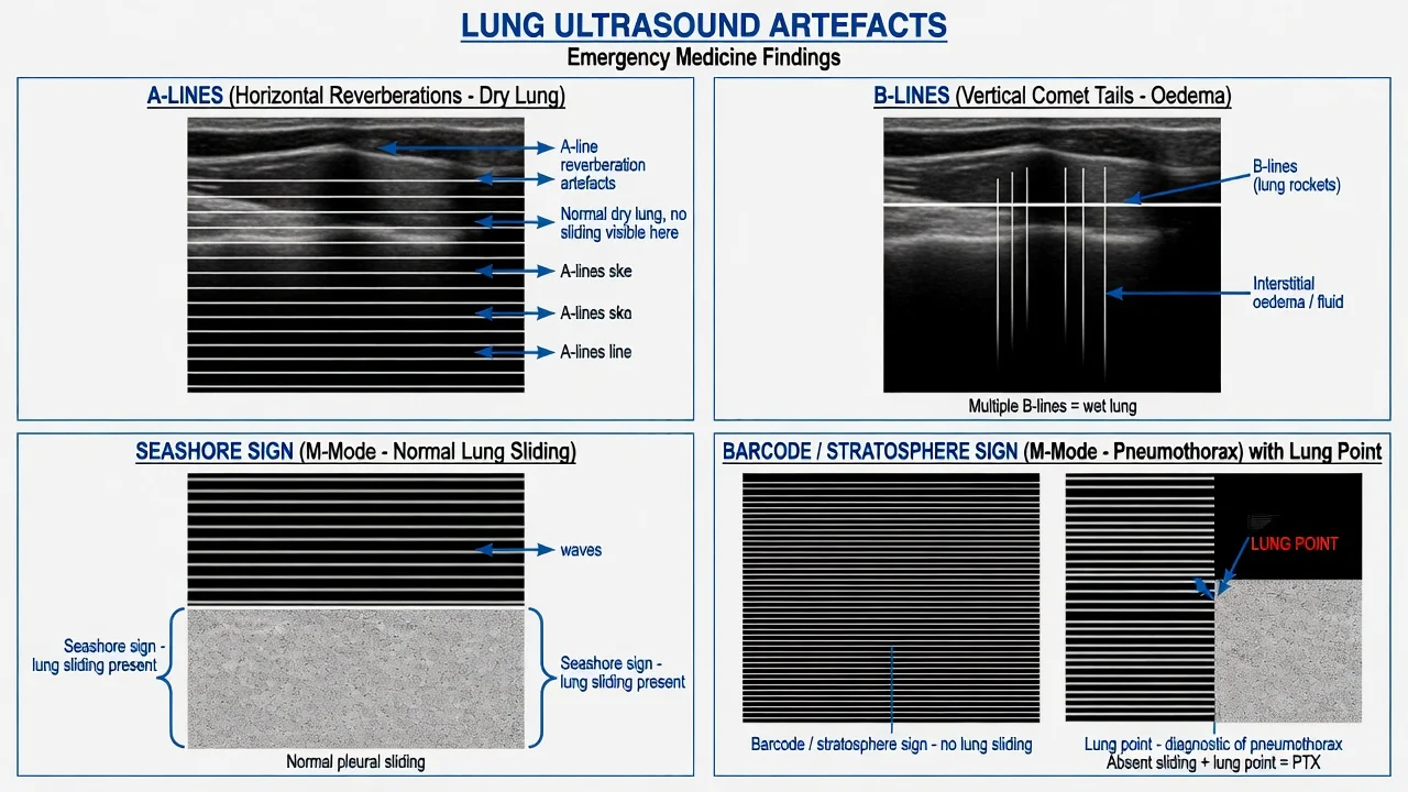
The lung is examined with a high-frequency linear probe (or the phased-array probe) at eight zones — the upper and lower anterior and lateral chest on each side — and the Fellowship candidate should run the survey to a fixed sequence so no zone is missed. The pleural line lies between the two rib shadows (the bat-wing sign), and its movement and its artefacts are the whole of the examination.[2]
Lung sliding is the to-and-fro shimmer of the visceral pleura against the parietal pleura with respiration; on M-mode it produces the seashore sign — a grainy sandy pattern below the pleural line over horizontal wave-like lines above. Its absence abolishes the shimmer and converts M-mode to the barcode or stratosphere sign of parallel horizontal lines throughout. Absent sliding is pneumothorax, but also apnoea, a main-stem intubation, the post-pleurodesis lung, severe bullous disease and ARDS — so sliding is read alongside the artefacts.[2]
The A-line pattern — horizontal reverberation lines deep to the pleural line at regular intervals — is the normal dry lung, also seen in COPD and asthma where the lung is air-filled and the septa dry. The B-lines (also called comet-tail artefacts) are vertical, laser-like, erasing the A-lines, arising from the pleural line and moving with sliding; three or more in a zone indicate the water-logged interlobular septum of pulmonary oedema, and bilateral diffuse B-lines with a hyperdynamic or failing heart on the cardiac view confirm cardiogenic oedema against the A-line alternative.[3] The BLUE protocol harnesses these profiles: bilateral A-lines with a normal left ventricle point to asthma or COPD; bilateral B-lines with a low ejection fraction point to pulmonary oedema; and the A-line lung without B-lines, with a dilated right ventricle, points to pulmonary embolism — the dry-lung, large-heart profile.[5]
Pneumothorax is sought over the anterior chest in the supine patient (where the air rises). The signs, in order of specificity, are the absent lung sliding, the absent B-lines, and the lung point — the boundary on the chest wall where the sliding lung meets the non-sliding pneumothorax, seen as a flickering transition on two-dimensional and a transition on M-mode. The lung point is the only sign near-diagnostic of pneumothorax (specificity approaching 100 per cent), but it is insensitive because it is seen only when the pneumothorax is partial and the probe happens to sit on the margin; a completely collapsed lung has no lung point.[4]
The cardiac windows — effusion, function and the right ventricle
The focused cardiac exam uses four windows — the parasternal long-axis, the parasternal short-axis, the apical four-chamber and the subxiphoid — and the candidate should be able to obtain at least two in any patient. The phased-array probe is used, set to the cardiac preset, with the depth at 15 to 18 centimetres in the adult and the ECG gated where available.[8]
The pericardial effusion appears as an anechoic stripe separating the visceral from the parietal pericardium, circumferential or loculated. The Fellowship candidate must distinguish the effusion that threatens tamponade from the one that does not: right-ventricular free-wall collapse in early diastole and right-atrial collapse in late diastole are the echocardiographic signs of tamponade, and they correlate with the clinical picture of the Beck triad and the pulsus paradoxus. A large effusion that has not yet produced chamber collapse is not tamponade; a small effusion under pressure in the trauma patient may be. Tamponade is always a clinical diagnosis the scan confirms, never a scan diagnosis alone. [1]
Left-ventricular systolic function is estimated by eye — the eyeball ejection fraction — by watching the inward movement of the walls and the change in cavity size through the cycle. Hyperkinetic (the walls nearly kissing), the normal-to-hyperdynamic pump with a small cavity points to hypovolaemia; the poorly contracting dilated ventricle points to cardiogenic failure and explains the B-lines on the lung survey; the regional wall-motion abnormality — a segment that bulges outward in systole — points to the acute coronary syndrome. The eyeball method is less precise than formal quantification but is accurate enough to drive the resuscitation decision.[8]
The right ventricle is examined for the dilatation and the hypokinesis of acute pulmonary embolism. The normal right ventricle is smaller than the left and contracts briskly; in massive embolism it dilates to equal or exceed the left, the interventricular septum bows into the left ventricle in systole (the D-shaped left ventricle in the short-axis view), and the contraction becomes hypokinetic — except, classically, at the apex, which is the McConnell sign of regional right-ventricular dysfunction.[7] A dilated right ventricle with A-line lungs and a plethoric fixed IVC completes the obstructive profile; the candidate reads the whole RUSH pattern, not the single chamber.
The inferior vena cava — the volume status
The inferior vena cava is imaged in the subxiphoid long-axis view, a few centimetres from the right atrium where the vessel is straight, with the diameter measured through the respiratory cycle. A small (under 1.5 centimetre) IVC that collapses by more than half with a sniff implies a low volume state and predicts fluid responsiveness; a plethoric, fixed IVC (over 2.5 centimetres, little or no collapse) implies right-heart strain, tamponade, tension pneumothorax or volume overload. The IVC gives a dynamic, repeatable read on the volume status that complements the static vital signs, and the collapsibility index (the change in diameter with respiration) is the bedside surrogate for fluid responsiveness, though its accuracy is modest and it is read alongside the pump and the lungs rather than in isolation.[9]
Equipment and the machine setup
The three examinations are run from a single cart with a curvilinear probe (the low-frequency 2 to 5 megahertz transducer) for the aorta and the deep cardiac views, a phased-array probe for the intercostal and the subxiphoid cardiac windows, and a high-frequency linear probe (5 to 10 megahertz) for the pleural line and the lung sliding. The depth is set to 15 to 18 centimetres for the aorta and the heart so the target fills the screen, and to 4 to 6 centimetres for the lung so the pleural line sits in the near field. The gain is set so the anechoic structures (the aortic lumen, the effusion) are genuinely black and the parenchyma is mid-grey; too much gain fills free fluid with echoes and hides small collections. The cardiac preset is selected for the heart and the abdominal preset for the aorta; M-mode is added for the lung sliding. A warm gel, a clean probe and a probe cover complete the setup, and the probe marker convention is fixed so the images are reproducible. [1]
Patient preparation and consent
The patient is examined supine, fully exposed, with the head of the bed flat for the aortic and the subxiphoid views and slightly raised for the apical window if tolerated; the left lateral decubitus position brings the heart forward for the apical and the parasternal windows in the harder patient. The probe is placed directly on the skin with coupling gel, and the windows are swept to the optimal image. Consent is implicit in the emergency setting — the scan is non-invasive and adds no radiation — and the team proceeds under the doctrine of necessity, explaining the procedure to the conscious patient as it runs. The scan is integrated into the resuscitation: it is performed by one team member while the airway, the breathing, the access and the haemorrhage control proceed in parallel, and it is interrupted and resumed as the patient demands. [1]
Stepwise technique — the RUSH protocol
The RUSH protocol (Rapid Ultrasound in Shock) runs to a fixed sequence, structured as the pump, the tank and the pipes, so that no window is missed under the pressure of the resuscitation.[1]
The RUSH exam, step by step, in order
- The pump (the heart) — subxiphoid four-chamber for the effusion and the gross function; parasternal long-axis for the LV size, the septum and the aortic root; parasternal short-axis for the septal bowing and the regional function; apical four-chamber if obtainable for the chamber sizes. Read for: the effusion with chamber collapse (tamponade), the dilated hypokinetic RV with septal bowing (PE), the poor LV function (cardiogenic), the hyperkinetic small LV (hypovolaemia).
- The tank (the volume) — the IVC in the subxiphoid long-axis for the diameter and the collapsibility; the lung for the A-line dry lung versus the B-line wet lung; the FAST views for free fluid. Read for: the flat collapsing IVC (empty tank), the plethoric fixed IVC (obstructed or overloaded tank), the bilateral B-lines (the tank overflows into the lung).
- The pipes (the great vessels) — the aortic sweep from the xiphisternum to the bifurcation in transverse and longitudinal, measured outer-to-outer; the FAST for the pericardium and the abdomen; the leg veins for the deep-vein thrombosis where PE is suspected. Read for: the aneurysm (the pipe that bursts), the dissection flap, the DVT that sourced the embolism.
- Synthesise — combine the pump, the tank and the pipes into one of the four shock profiles and act: hypovolaemic (fluid, blood, stop the bleed), cardiogenic (inotrope, diuretic, the cause), obstructive (decompress the pneumothorax, drain the tamponade, lyse the embolism), distributive (fluid, vasopressor, the source). [1]
The structural error is to read each window in isolation — the single effusion called tamponade, the single B-line called oedema — instead of synthesising the pump, the tank and the pipes into the shock profile. The protocol is an algorithm, not a checklist, and the synthesis is the mark. [1]
Drug doses and the resuscitation decisions
The scan does not itself deliver a drug, but every finding drives a defined therapy, and the Fellowship candidate must state the doses the protocol triggers. The flat, collapsing IVC of the hypovolaemic is challenged with a 500 millilitre crystalloid bolus (or 20 millilitres per kilogram in the child) and rescanned; a responsive IVC and a rising pressure confirm volume responsiveness, and the bleeding patient is crossmatched and transfused to a permissive target. The B-line lung of cardiogenic oedema is treated with glyceryl trinitrate (sublingual 300 to 600 micrograms, then an infusion of 10 to 200 micrograms per minute) or with furosemide 40 to 80 milligrams intravenously, and supported with non-invasive ventilation. The dilated right ventricle of massive pulmonary embolism is treated with systemic thrombolysis — alteplase 50 milligrams intravenously over 2 minutes in the arrest, or 100 milligrams over 2 hours in the haemodynamically unstable — or with embolectomy where lytic is contraindicated. The aneurysm with rupture triggers the massive transfusion protocol and the tranexamic acid 1 gram over 10 minutes, on the way to theatre. The tension pneumothorax is decompressed before any scan, and the tamponade is drained. [1]
The drugs the scan triggers
Complications — the interpretive errors
The examination is non-invasive and carries no procedural complication beyond the theoretical probe pressure. The real complications are interpretive — the wrong disposition driven by a misread scan. The tangential aortic measurement overcalls an aneurysm and sends a stable patient to an unnecessary CT; the single unsampled level misses a saccular aneurysm at an unsampled point. The B-line called pneumonia or the A-line called normal when the lung is dry but the patient has PE loses the diagnosis. The effusion called tamponade without chamber collapse, or the small effusion called harmless when it is under pressure in the trauma patient, drives the wrong drainage decision. The dilated right ventricle of cor pulmonale misread as acute embolism sends the patient to needless thrombolysis. Each is prevented by honest reporting, by correlation with the clinical picture and the other windows, and by repeating the scan as the patient changes. [1]
Pitfalls and practical tips
The pitfalls are the inverse of the discipline. Accepting a single window as the answer — the effusion without the chamber, the B-line without the heart, the aorta without the bifurcation. The tangential aortic cut that overestimates the diameter. Forgetting the lung point is the only specific pneumothorax sign — its absence never excludes one, so a high-clinical-suspicion pneumothorax is decompressed on the clinical picture even with a sliding lung. Confusing the IVC with the aorta in the subxiphoid view — the IVC is to the patient's right, enters the right atrium, and collapses with respiration; the aorta is to the left, is pulsatile and does not. Over-reading the eyeball ejection fraction as precise — it is a gross estimate, and the regional wall-motion abnormality, not the number, drives the ischaemia decision. Missing the apical window in the obese or the bandaged patient and declaring the heart unassessable when the subxiphoid or the parasternal window would have answered. The practical tips are the opposite: scan all the windows every time, measure the aorta outer-to-outer at multiple levels, read the pump-tank-pipes together, label indeterminate honestly, decompress the tension pneumothorax clinically, and treat the patient — not the scan. [1]
Post-procedure care and disposition
The disposition hinges on the synthesised shock profile and the response to the triggered therapy. The ruptured aneurysm goes to theatre or to endovascular repair, the massive transfusion running and the vascular surgeon waiting. The tamponade goes to pericardiocentesis or to theatre. The massive pulmonary embolism goes to thrombolysis or to embolectomy, with the critical-care bed ready. The cardiogenic pulmonary oedema responds to the diuretic and the non-invasive ventilation, and is admitted to the monitored bed for the workup of the cause. The hypovolaemic patient who stabilises with fluid is admitted for the source — the bleeding, the sepsis, the loss. The stable patient with an incidental small aneurysm is referred for surveillance, the interval dictated by the diameter. The scan is repeated serially — at any change in physiology, and at the milestones of the resuscitation — because the sonographic picture is dynamic and the single snapshot is never the whole story. [1]
Special populations
The pregnant patient is scanned with the gravid uterus displacing the viscera and the relative hypervolaemia masking the shock until late; the team tilts the uterus left, and the cardiac and the lung windows remain reliable when the aortic view is distorted. The paediatric patient is examined with the same windows at a shallower depth and a smaller probe, and the IVC collapsibility and the lung patterns hold; the volume bolus is weight-based (20 millilitres per kilogram). The elderly patient carries the comorbidity, the anticoagulation and the diminished reserve that lower the threshold to scan, and in whom the B-lines of heart failure are distinguished from the effusion and consolidation of pneumonia by the bilateral profile and the cardiac function. The obese patient is the technically limited scan, honestly labelled indeterminate where the windows fail, and the chronic lung disease patient is the one in whom the A-lines of COPD and asthma are not mistaken for the normal lung, and in whom the B-lines of cor pulmonale are distinguished from the acute oedema by the chronic right-heart profile. [1]
Evidence and regional guidelines
The diagnostic framework is built on the RUSH protocol of Perera and colleagues, which formalised the pump-tank-pipes approach to the undifferentiated shock patient and showed that clinician-performed bedside ultrasound changed the working diagnosis and the management in a substantial proportion of the critically ill.[1] The lung ultrasound evidence rests on the international consensus of Volpicelli and the International Liaison Committee, which standardised the technique, the terminology (A-lines, B-lines, the lung point) and the eight-zone survey, and on the BLUE protocol of Lichtenstein, which demonstrated that the artefact profiles discriminated pulmonary oedema, pneumothorax, COPD or asthma, and pulmonary embolism with high accuracy in acute respiratory failure.[2][5] The B-line to pulmonary artery occlusion pressure correlation of Lichtenstein showed that the diffuse bilateral B-line profile corresponds to the wedge pressure above 18 millimetres of mercury, and the lung point of Lichtenstein was the sign that made the sonographic diagnosis of pneumothorax specific.[3][4] The abdominal aortic aneurysm diameter that defines the disease and the rupture risk was established by the Multicentre Aneurysm Screening Study (MASS), and the emergency-department aortic measurement dates to the early sonography series of Shuman.[6][10] The right-ventricular regional dysfunction of acute pulmonary embolism — the McConnell sign — was defined by McConnell and colleagues, and the clinician-performed focused echo was validated in the emergency department by Mandavia.[7][8] The IVC collapsibility as a fluid-responsiveness surrogate was synthesised in the meta-analysis of Orso and the International Liaison Committee on Lung Ultrasound.[9]
ANZ practice note. POCUS of the aorta, the lung and the heart is a core Fellowship skill of the ACEM training programme, embedded in the primary survey and the resuscitation, and reinforced by the ATLS, the EMST and the ACEM policy on clinician-performed ultrasound. The RUSH protocol and the focused cardiac, lung and aortic surveys are performed by the treating clinician within the first minutes of the resuscitation, repeated serially, and used to drive the fluid, the vasopressor, the thrombolytic and the surgical decisions. The ANZ standard is that every senior emergency trainee can obtain and interpret the four cardiac windows, the aortic sweep, the lung survey and the IVC, and can synthesise them into the shock profile. [1]
Exam pearls
- The pump, the tank and the pipes — the three-line summary of the RUSH protocol, and the structure the examiner wants for any "undifferentiated shock" station.
- AAA is an outer-to-outer diameter over 3 cm — measure at the widest point, in the transverse plane, perpendicular to the long axis; a tangential cut overestimates.
- A-lines are normal or COPD; B-lines are pulmonary oedema; absent sliding with a lung point is pneumothorax — the three-line lung summary.
- The lung point is the only specific pneumothorax sign — its absence never excludes one; its presence confirms it.
- Bilateral B-lines with a failing LV is pulmonary oedema; A-line lungs with a dilated RV is pulmonary embolism — the BLUE protocol discriminator.
- A pericardial effusion with RV free-wall collapse in diastole is tamponade — a clinical diagnosis the scan confirms.
- A dilated hypokinetic RV with septal bowing, sparing the apex, is the McConnell sign of massive PE — read it with the A-line lung and the plethoric IVC.
- A flat, collapsing IVC (under 1.5 cm, more than half collapse on sniff) is the empty tank; a plethoric fixed IVC (over 2.5 cm) is the obstructed or overloaded tank.
- Synthesise the three, do not read a single window — the single effusion is not tamponade, the single B-line is not oedema, the single chamber is not PE. [1]
Exam practice
SAQ — The bedside aortic sweep that diagnoses the ruptured AAA in undifferentiated shock
10 minutes · 10 marks
A 76-year-old male smoker is brought to the resuscitation bay 30 minutes after the sudden onset of tearing central abdominal and lower-back pain at home, followed by a brief syncopal episode. He is pale, diaphoretic and drowsy (GCS 14), BP 82/50, HR 124, SpO2 95 per cent on 15 L oxygen via a non-rebreather mask. The abdomen is rigid with a tender, pulsatile epigastric mass. The team has called the RUSH protocol. Lactate 5.8 mmol/L, haemoglobin 98 g/L. The curvilinear probe is in your hand.
SAQ — Cardiac POCUS discriminating the obstructive from the cardiogenic shock
10 minutes · 10 marks
A 68-year-old woman is brought to the resuscitation bay with sudden onset of severe dyspnoea, presyncope and pleuritic chest pain two weeks after a total knee replacement. She is profoundly hypotensive: BP 78/52, HR 124, RR 32, SpO2 88 per cent on 15 L oxygen, GCS 14, JVP distended to the angle of the jaw at 45 degrees, clear lung fields on auscultation. The ECG shows a sinus tachycardia with an S1Q3T3 pattern. The bedside troponin is mildly elevated at 65 ng/L. You perform the focused cardiac ultrasound as part of the RUSH protocol.
Red flags
[1]References
- [1]Perera P, Mailhot T, Riley D, Mandavia D. The RUSH exam: Rapid Ultrasound in SHock in the evaluation of the critically lll Emerg Med Clin North Am, 2010.PMID 19945597
- [2]Volpicelli G, Elbarbary M, Blaivas M, et al. International evidence-based recommendations for point-of-care lung ultrasound Intensive Care Med, 2012.PMID 22392031
- [3]Lichtenstein DA, Mezière GA, Lagoueyte JF, Biderman P, Goldstein I, Gepner A. A-lines and B-lines: lung ultrasound as a bedside tool for predicting pulmonary artery occlusion pressure in the critically ill Chest, 2009.PMID 19809049
- [4]Lichtenstein D, Mezière G, Biderman P, Gepner A. The lung point: an ultrasound sign specific to pneumothorax Intensive Care Med, 2000.PMID 11126253
- [5]Lichtenstein DA, Mezière GA. Relevance of lung ultrasound in the diagnosis of acute respiratory failure: the BLUE protocol Chest, 2008.PMID 18403664
- [6]Ashton HA, Buxton MJ, Day NE, et al. The Multicentre Aneurysm Screening Study (MASS) into the effect of abdominal aortic aneurysm screening on mortality in men: a randomised controlled trial Lancet, 2002.PMID 12443589
- [7]McConnell MV, Solomon SD, Rayan ME, Come PC, Goldhaber SZ, Lee RT. Regional right ventricular dysfunction detected by echocardiography in acute pulmonary embolism Am J Cardiol, 1996.PMID 8752195
- [8]Mandavia DP, Hoffner RJ, Mahaney K, Henderson SO. Bedside echocardiography by emergency physicians Ann Emerg Med, 2001.PMID 11574793
- [9]Orso D, Paoli S, Piani T, Cilenti FL, Cristiani L, Guglielmo N, Berti L, Gargani L, Volpicelli G, Copetti R, Maccarini D, Barbieri F, Tonerini M, Catena F, Pelusi G, Bertolini G, Nazerian P, International Liaison Committee on Lung Ultrasound (ILC-LUS) for the International Consensus Conference on Pleura and Lung Ultrasound. Accuracy of Ultrasonographic Measurements of Inferior Vena Cava to Determine Fluid Responsiveness: A Systematic Review and Meta-Analysis J Intensive Care Med, 2020.PMID 29343170
- [10]Shuman WP, Hastrup W Jr, Kohler TR, et al. Suspected leaking abdominal aortic aneurysm: use of sonography in the emergency room Radiology, 1988.PMID 3289085