EM · CT interpretation and usage (approach)
Computed tomography — the emergency department interpretation and usage approach
Also known as CT interpretation · CT usage · CT angiography · CT perfusion · pan-scan
The systematic approach to requesting and interpreting computed tomography in the emergency department — the appropriate-versus-inappropriate decision, contrast versus non-contrast phases, CT angiography for pulmonary embolism and aortic dissection, CT perfusion for the stroke penumbra, the early ischaemic stroke signs with the ASPECTS score, subarachnoid haemorrhage sensitivity within six hours, renal colic and appendicitis imaging, the contrast premedication and contrast-induced nephropathy drug doses, and the 2 to 10 mSv radiation dose per scan. ACEM-primary, globally tagged.
On this page & tools
Your progress
Saved locally on this device.
2 MCQs with explanations
Target exams
Red flags
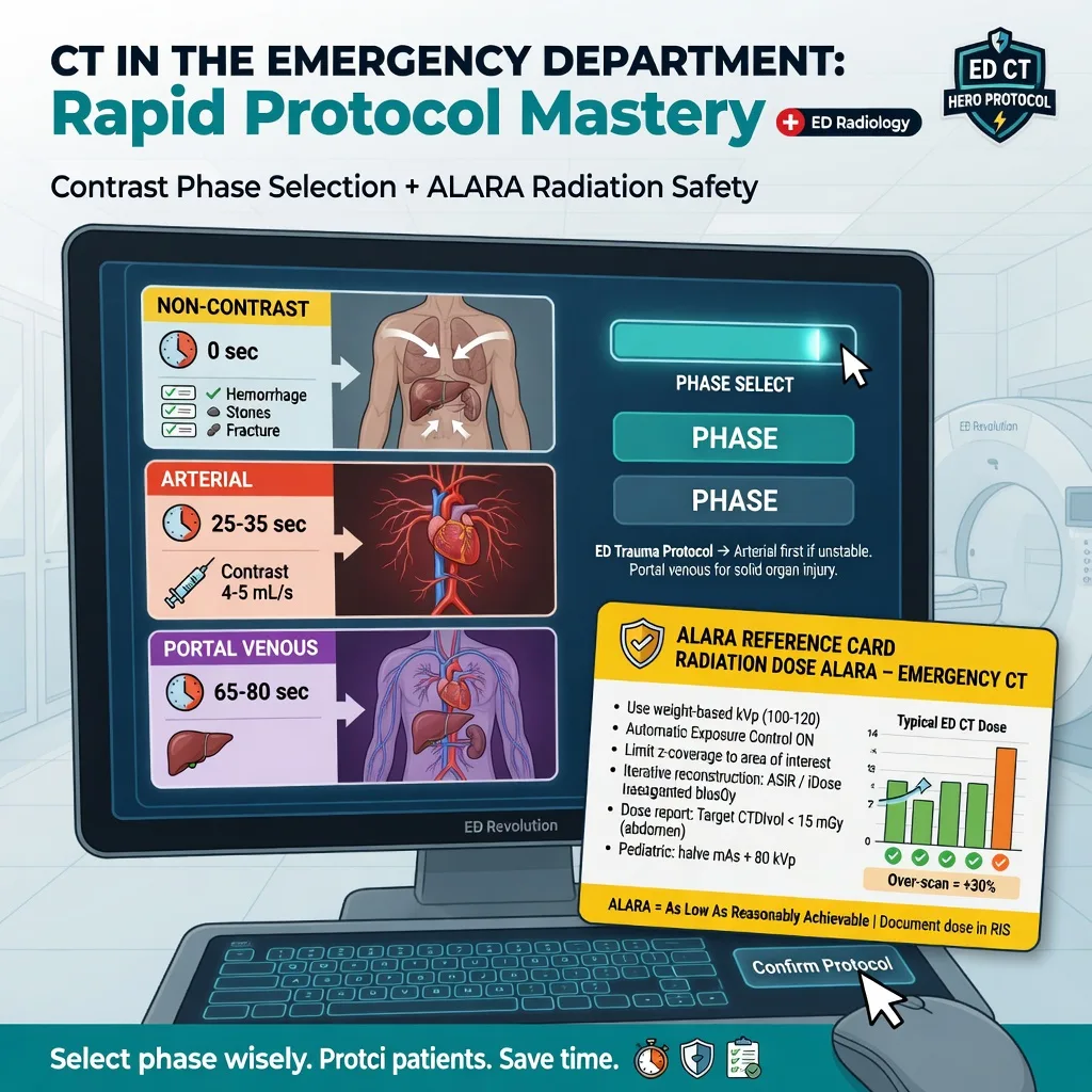
Computed tomography is the workhorse cross-sectional investigation of the emergency department. It is fast, widely available, and it answers time-critical questions — a pulmonary artery filling defect, an intimal flap, a hyperdense middle cerebral artery — that change disposition within minutes. The Fellowship examiner is not testing whether a candidate can recognise a favourite slice; the test is whether a candidate can decide when a CT is justified, choose the right phase, interpret the slice systematically, and weigh the radiation and contrast cost against the clinical yield. This topic is the diagnostic-skill framework, applied across the common ED indications: trauma, stroke, subarachnoid haemorrhage, renal colic, the acute aortic syndrome, appendicitis, pulmonary embolism, and the stroke penumbra. [1]
When and why to use the framework
A CT is justified only when the answer to a defined clinical question will change management, and the brief is governed by three questions applied to every request. First, what is the clinical question? Second, what is the pre-test probability, and will a positive or a negative result change the plan? Third, is there a safer or more informative alternative — bedside ultrasound, plain radiography, or magnetic resonance imaging? Computed tomography is a high-radiation, contrast-requiring, and resource-limited test; a scan performed "to be sure" without a question is the definition of an inappropriate scan. The framework below works through requesting, phase selection, systematic interpretation, and the contrast and radiation cost, in the order in which the clinician encounters them. [1]
The physics that underpins the read
Computed tomography measures the attenuation of X-rays by tissue and assigns each voxel a Hounsfield unit: water is zero, air is approximately negative 1000, and dense bone approaches positive 1000; fat is around negative 60 to negative 100, soft tissue 30 to 60, and acute haemorrhage 50 to 70, which is why fresh blood appears bright on a non-contrast scan. Iodinated contrast agent increases the X-ray attenuation of whatever it fills — the arterial tree in the arterial phase, the organs and venous system in the portal venous phase — and it is the basis of contrast-enhanced CT, CT angiography, and CT perfusion. The radiation dose is measured in millisieverts: a single phase of body CT delivers roughly 2 to 10 mSv, an order of magnitude above a chest radiograph at 0.02 mSv, and that dose carries a small but real stochastic lifetime cancer risk that rises with cumulative exposure, in the young, and in the multiply-scanned patient.[1]
Hounsfield units — the attenuation map every voxel carries
Every voxel on a CT slice carries a Hounsfield unit (HU) — a linear scale of X-ray attenuation anchored to two fixed reference points: distilled water at zero and air at negative 1000. The scale is the reason fresh blood, bone, fat and brain each have a characteristic greyness on the slice, and knowing the canonical values lets the interpreter predict what a lesion should look like before it is found. Dense cortical bone approaches positive 1000; acute haemorrhage and acute clot sit at 60 to 80; iodinated contrast-enhanced blood pool is brighter still, around 100 to 300; brain grey matter 35 to 45; brain white matter 20 to 30; liver and spleen 40 to 60; water and cerebrospinal fluid zero to 10; fat negative 60 to negative 100; and lung air negative 700 to negative 900. The non-contrast head CT works because acute haemorrhage (60 to 80 HU) is intrinsically brighter than brain parenchyma (20 to 45 HU), so an extradural, subdural or subarachnoid bleed declares itself without contrast. [1]
Bright — high HU
- Dense cortical bone: +1000
- Acute haemorrhage/clot: +60 to +80
- Iodinated contrast in vessels: +100 to +300
- Calcification (choroid plexus, calculi): +100 to +400
Grey — mid HU
- Brain grey matter: +35 to +45
- Brain white matter: +20 to +30
- Liver, spleen, soft tissue: +40 to +60
- Muscle: +10 to +40
Dark — low/negative HU
- Water and CSF: 0 to +10
- Fat: −60 to −100
- Lung parenchyma (air): −700 to −900
- Air (bowel gas, pneumothorax): −1000
Windowing — choosing the right window for the question
The human eye resolves only a narrow band of the thousands of greyscale values a CT slice encodes, so the data are mapped to a display window defined by a centre (level) and a width. The window determines which structures pop and which fade — reading a haemorrhage on a bone window, or a fracture on a brain window, is a classic error, and the emergency clinician must consciously select the window for the question. The brain window (centre approximately 35 to 40, width 80 to 100) maximises grey-white differentiation and reveals subtle parenchymal oedema and early ischaemia. The subdural window (a wider, lower window such as centre 50 to 75, width 130 to 180) is tuned to detect a thin extra-axial collection against the skull that the standard brain window can miss. The bone window (centre 400 to 500, width 1500 to 4000) brings out calvarial and skull-base fractures. The lung window (centre negative 600, width 1500) is essential for pneumothorax, pulmonary nodules and ground-glass change. The soft-tissue or mediastinal window (centre 40 to 60, width 350 to 400) shows the aorta, great vessels and mediastinal haematoma. [1]
Brain window
level ~35–40 HU, width ~80–100
- Grey–white differentiation; subtle oedema and early ischaemia
- Use to read the non-contrast head for stroke and SAH
- May MISS a thin subdural against the inner table
Subdural window
level ~50–75 HU, width ~130–180
- Tuned for thin extra-axial blood against the skull
- Wider window reduces the bright bone flare
- Check this whenever a subdural is clinically suspected
Bone window
level ~400–500 HU, width ~1500–4000
- Reveals calvarial and skull-base fractures
- Use in head and facial trauma and for temporal bone
- Pneumocephalus and air-fluid levels also visible
Lung window
level ~−600 HU, width ~1500
- Detects pneumothorax, ground glass, nodules
- Essential on every chest CT and trauma chest
- Lung pathology invisible on the soft-tissue window
Contrast versus non-contrast — the phase decision
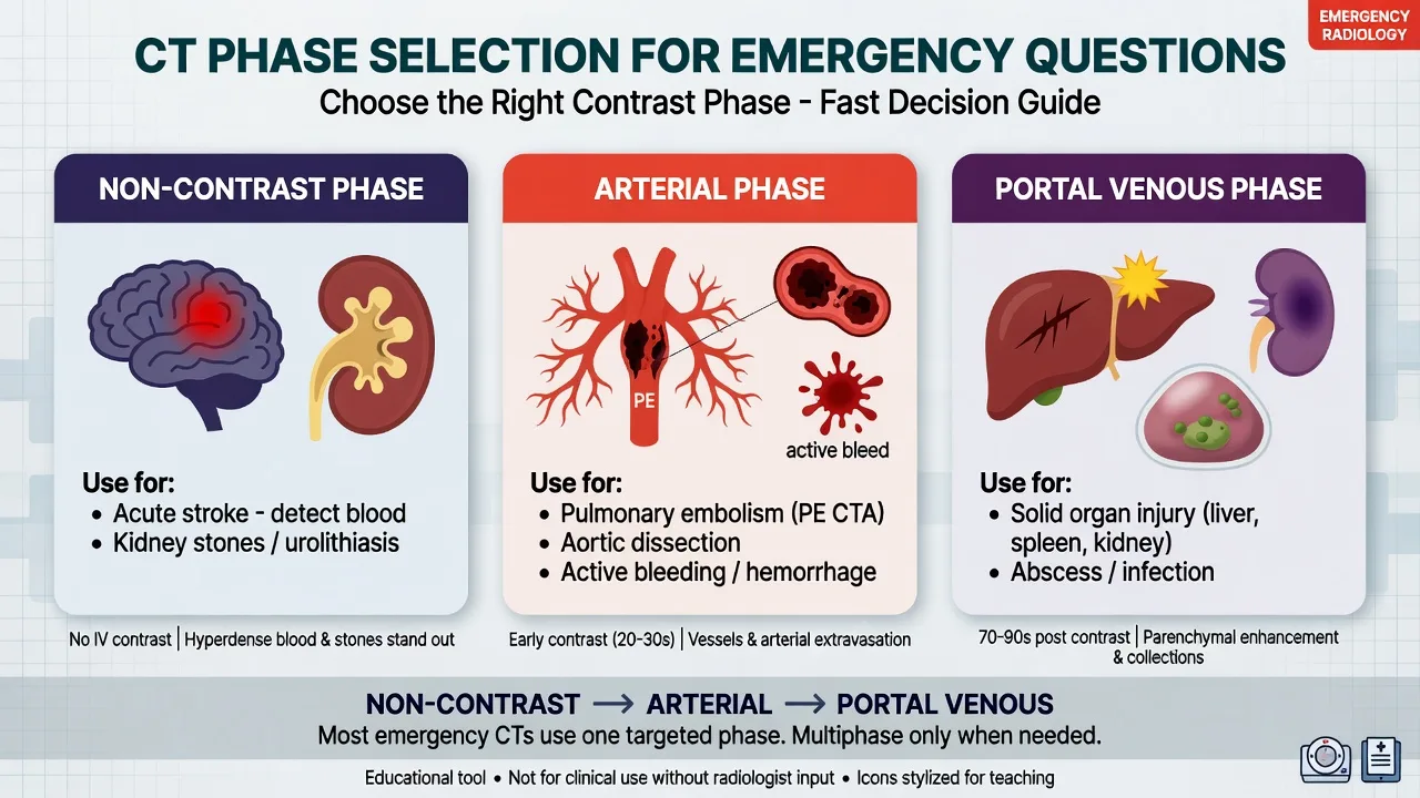
The single most important request decision is whether to give contrast, and the answer is dictated by the question. A non-contrast scan is required whenever the pathology is, or might be, blood or calcification: a non-contrast head CT for haemorrhage, subarachnoid blood, and a hyperdense vessel; a non-contrast abdomen for renal colic and ureteric calculi; and a non-contrast CT to measure an abdominal aortic aneurysm. A contrast-enhanced scan is required when the question is about perfusion, vascular anatomy, or an enhancing lesion: the portal venous phase abdomen for appendicitis, diverticulitis, and intra-abdominal abscess; CT angiography for the aorta, the pulmonary arteries, and the cerebral vessels; and CT perfusion for the stroke core and penumbra. [1]
Trauma — pan-scan versus selective imaging
The defining modern question in trauma imaging is whether to scan everything. Total-body CT, or the "pan-scan", images the head, cervical spine, chest, abdomen and pelvis in a single acquisition, and it was hoped that finding every injury at once would save lives. The REACT-2 randomised controlled trial tested this directly: immediate total-body CT versus conventional imaging with selective CT in blunt trauma patients with compromised vital signs or a severe injury mechanism.[2] It found no difference in in-hospital mortality at the cost of a higher radiation dose, and it did not change the rate of missed injuries. The implication for practice is that the pan-scan is not a default — it is applied to the seriously injured patient in whom the mechanism and the physiology predict polytrauma, while the stable patient with an isolated injury is imaged selectively, guided by clinical assessment and by validated decision rules. The Canadian C-spine rule, for example, safely reduces unnecessary cervical-spine imaging in alert, stable trauma patients by imaging only those with a high-risk factor or absent low-risk criteria.[8]
Stroke — the early ischaemic signs and ASPECTS
Acute ischaemic stroke is the diagnosis in which CT interpretation is most time-critical, because the clock for reperfusion starts at symptom onset and the imaging drives it. The initial scan is a non-contrast head CT, and its first purpose is to exclude haemorrhage — a contraindication to thrombolysis. Once blood is excluded, the scan is read for the early signs of ischaemia, which may be subtle within the first hours: the hyperdense MCA sign, a bright middle cerebral artery from thrombus within it; loss of the insular ribbon, effacement of the cortex over the insula; obscuration of the lentiform nucleus with loss of the grey-white interface in the basal ganglia; and a wedge of cortical sulcal effacement with subtle hypodensity in the affected territory. [1]
The severity is quantified by the Alberta Stroke Program Early CT Score, ASPECTS, a ten-point territorial score applied to two standardised slices (the ganglionic and supraganglionic levels) of the middle cerebral artery territory.[3] A normal territory scores ten and an infarcted territory scores zero; one point is subtracted for each of the ten regions that shows early ischaemic change.
A non-contrast CT that excludes haemorrhage and shows no early change does not exclude ischaemia — the scan is often normal in the first hours, and that is expected. CT angiography is then performed to find a large-vessel occlusion eligible for thrombectomy, and CT perfusion is added when the time window is extended. [1]
Subarachnoid haemorrhage — sensitivity and the lumbar-puncture question
Non-contrast head CT is exquisitely sensitive to subarachnoid blood in the first hours. A systematic review and meta-analysis of CT performed within six hours of headache onset reported a pooled sensitivity of approximately 98 percent, leading several guidelines to accept that a normal CT within six hours, in the right patient, may safely exclude subarachnoid haemorrhage without a lumbar puncture.[4] The sensitivity falls with time as blood is diluted and degrades, so a patient presenting after six hours — or with a story suggesting an earlier sentinel leak — still requires a lumbar puncture to exclude xanthochromia after the CT. The conservative ACEM position in many departments is to perform a lumbar puncture whenever the CT is normal and the index of suspicion persists, particularly after six hours; the scan is read for blood in the basal cisterns, the Sylvian fissure, and the interhemispheric fissure.
CT sensitivity for subarachnoid haemorrhage by time from onset
Systematic CT head interpretation — a repeatable read
The Fellowship viva demands a structured, repeatable head CT read, not a hunt for a favourite sign. A widely taught mnemonic is Blood Can Be Very Bad (Blood, Cisterns, Brain, Ventricles, Bone), but the logic holds regardless of the mnemonic chosen: read in the same order every time, so that a one-millimetre midline shift is never missed for want of a system. Begin by confirming patient and laterality, then work through symmetry and the midline, the ventricles, the sulci and gyri, the basal cisterns, the parenchyma, the blood, and finally the bone on the bone window. [1]
The systematic non-contrast head CT read
Confirm patient identity, scan date and time, and orientation. Check the right-left labelling and confirm the scan is NON-contrast — the first question in a suspected haemorrhage is whether contrast was given, because contrast can masquerade as blood.
Assess symmetry and the midline. Compare the hemispheres side to side and identify the midline markers — the falx, the septum pellucidum and the pineal gland. A shift of the septum pellucidum or pineal of more than a few millimetres is mass effect and signals raised intracranial pressure.
Inspect the ventricles — the frontal horns, temporal horns, third and fourth ventricles. Compression or effacement indicates mass effect; dilatation, particularly of the temporal horns or upward bowing of the corpus callosum, indicates obstructive hydrocephalus from intraventricular blood or a posterior fossa mass.
Examine the sulci and gyri for focal effacement (mass effect or swelling) and for asymmetry indicating oedema or infarct. Sulcal effacement in a vascular territory is an early sign of an evolving infarct; complete effacement on one side suggests a large hemispheric mass.
Check the basal cisterns — perimesencephalic, quadrigeminal and suprasellar. Effacement of the cisterns is a sign of raised intracranial pressure and impending herniation; blood within them is subarachnoid haemorrhage.
Read the parenchyma on the brain window. Compare grey and white matter for a loss of the grey-white interface (early ischaemia), for hypodensity (oedema or completed infarct), and for hyperdensity (acute blood or a hyperdense vessel sign).
Search for blood explicitly and characterise it by shape and location: extradural (lentiform), subdural (crescentic), subarachnoid (in sulci, fissures and cisterns), intraparenchymal (within tissue), and intraventricular (layering in the ventricles).
Switch to the bone window and inspect the calvarium, skull base and sinuses for fractures; then look for pneumocephalus and for fluid in the mastoid air cells or sinuses, which may signal a basilar skull fracture.
Integrate the findings with the clinical picture — a negative scan in the right clinical context (a normal CT after 6 hours of a thunderclap headache) does not exclude the diagnosis and dictates the next test, whether lumbar puncture, CT angiography or perfusion.
The five intracranial haemorrhage patterns
Distinguishing the five patterns of intracranial blood by their shape and location is a viva staple. Each pattern points to a distinct mechanism and urgency, and each is read on the non-contrast brain and subdural windows. The shape of a collection is not arbitrary — it is dictated by which anatomical compartment the blood occupies. [1]
Extradural (epidural)
- Lentiform / biconvex shape; does not cross suture lines
- Arterial (middle meningeal) bleed; look for an overlying temporal bone fracture
- Does NOT cross the falx or tentorium
- Lucid interval; surgical emergency that expands rapidly
Subdural
- Crescentic shape; CROSSES suture lines
- Venous (bridging veins); elderly falls, anticoagulated, alcoholics
- Crosses sutures but does not cross the midline falx
- Acute is bright (+60 to +80 HU); chronic may be iso- or hypodense
Subarachnoid
- Blood within sulci, fissures and basal cisterns
- Thunderclap headache; ruptured aneurysm is the classic cause
- May show a hyperdense vessel (the bearing artery)
- CT ~98% sensitive within 6 h; lumbar puncture if normal after 6 h
Intraparenchymal
- Blood within the brain substance — wedge or rounded focus
- Hypertensive (basal ganglia, thalamus, pons, cerebellum) or traumatic
- Surrounding oedema and mass effect evolve over hours
- Look for extension into the ventricles — a poor prognostic sign
Intraventricular
- Blood layering within the ventricles — a blood–CSF level
- Causes obstructive hydrocephalus with ventricular dilatation
- Usually from extension of an intraparenchymal or subarachnoid bleed
- Poor prognosis; may require external ventricular drainage
CT cervical spine — imaging for clearance
The cervical spine is cleared clinically whenever a validated decision rule permits, and imaged by CT only when it does not. The Canadian C-spine rule applies to alert, stable trauma patients (GCS 15, no distracting injury): imaging is required if any high-risk factor is present (age 65 or over, dangerous mechanism, paraesthesia in the extremities) OR if no low-risk factor allows safe assessment of the neck range of motion.[8] The NEXUS low-risk criteria offer the alternative pathway: imaging is unnecessary only when all five criteria are met — no midline tenderness, no focal neurological deficit, normal level of alertness, no intoxication, and no painful distracting injury.[11] Both rules share the same purpose — to reduce unnecessary imaging — and CT has supplanted plain radiography as the first-line modality in adults because plain films miss a clinically important fraction of injuries, particularly at the craniocervical and cervicothoracic junctions.
Canadian C-spine rule
- Alert and stable trauma patient (GCS 15), no distracting injury
- Image if age ≥65, dangerous mechanism, or extremity paraesthesia
- Otherwise image UNLESS a low-risk factor permits range-of-motion assessment
- Higher sensitivity than NEXUS; more selective
NEXUS low-risk criteria
- No imaging if ALL FIVE are met: no midline tenderness, no focal deficit
- Normal alertness, no intoxication, no painful distracting injury
- Simpler to apply; lower sensitivity (misses some injuries)
- If any criterion is absent, image with CT
Plain radiography
- Largely supplanted by CT in adults
- Misses craniocervical and cervicothoracic junction injuries
- Retained role in paediatrics and resource-limited settings
- Inadequate as a clearance tool in the high-energy adult
NEXUS — National Emergency X-Radiography Utilization Study
New England Journal of Medicine, 2000
Prospective multicentre validation study of 34,069 blunt-trauma patients, testing a five-criterion low-risk rule (no midline tenderness, no focal deficit, normal alertness, no intoxication, no distracting injury) for excluding cervical-spine injury without imaging.
Key finding
The rule was 99.0% sensitive and 12.9% specific for clinically important cervical-spine injury; its application would have reduced imaging by 12.6%. Eight injuries were missed, all but one judged clinically insignificant.
Practice change
Established one of the two standard decision rules that allow cervical-spine clearance without imaging in selected blunt-trauma patients.
Canadian CT Head Rule — Stiell et al.
Lancet, 2001
Prospective cohort of 3121 alert, stable adults with minor head injury (GCS 13–15 with witnessed loss of consciousness, amnesia or confusion), deriving high- and medium-risk criteria predicting the need for CT.
Key finding
Five high-risk factors (signs of skull fracture, vomiting ≥2, age ≥65, dangerous mechanism, GCS under 15 at 2 h) were 100% sensitive for neurosurgical intervention; two medium-risk factors predicted clinically important brain injury. The rule would have reduced CT use.
Practice change
Provided the widely adopted decision rule governing CT use in minor head injury across ANZ, North American and UK practice.
Renal colic — the stone and the hydronephrosis
Non-contrast CT of the abdomen and pelvis is the reference standard for ureteric colic: it identifies the stone directly (as a bright calcific focus), locates it along the ureter, and measures its size, which predicts the likelihood of spontaneous passage. The secondary signs are hydronephrosis and perinephric and periureteric fat stranding from obstruction. A stone under 5 millimetres usually passes; a stone over 10 millimetres usually requires intervention. The non-contrast technique avoids contrast, which would obscure the calcific stone, and modern ultralow-dose protocols reduce the radiation dose substantially in the typically young, recurrently-imaged patient. The alternative of ultrasound first — to find hydronephrosis and avoid radiation, particularly in pregnancy — is preferred where the question is only whether obstruction is present. [1]
Abdominal aortic aneurysm and the acute aortic syndrome
A non-contrast CT measures the aneurysm diameter and shows a retroperitoneal haematoma if rupture has occurred, but the definitive investigation of the suspected acute aortic syndrome is CT angiography. In dissection it demonstrates the intimal flap separating the true and false lumens, defines the Stanford type (A involves the ascending aorta and is a surgical emergency; B is distal to the left subclavian and is managed medically or endovascularly), and identifies malperfusion of branch vessels. A non-contrast CT abdomen for a symptomatic aneurysm reports the maximum transverse diameter; aneurysms at or above 5.5 centimetres in a man, or with rapid growth or a tender presentation, are referred for repair. Bedside ultrasound by the emergency clinician is the faster test for the haemodynamically unstable patient with a suspected ruptured aneurysm and may avert the trip to the scanner. [1]
Appendicitis — the CT signs
Contrast-enhanced CT in the portal venous phase is the most accurate imaging test for acute appendicitis in adults, and a meta-analysis of CT and ultrasound in children and adults confirmed the higher sensitivity of CT.[5] The diagnostic findings are an appendiceal outer diameter over 6 millimetres, wall enhancement with intraluminal contrast, peri-appendiceal fat stranding and fluid, and an appendicolith; a frankly perforated appendix shows a peri-appendiceal abscess or extraluminal gas. Ultrasound is preferred as the first test in children and in young or pregnant patients to avoid radiation, with CT reserved for an equivocal or ultrasound-negative study.
CT abdomen — the systematic trauma and surgical read
A contrast-enhanced CT of the abdomen and pelvis answers the four questions that dominate the ED abdominal presentation: is there free air, is there free fluid, is there fat stranding, and is there solid-organ injury. Free intraperitoneal gas is a perforated viscus — typically a peptic ulcer, the appendix or the colon — and is sought in the least dependent regions (the epigastric subdiaphragmatic space on the slices over the liver). Free fluid tracks to the dependent spaces — Morison's pouch, the pouch of Douglas, and the paracolic gutters — and is distinguished by its Hounsfield value: simple fluid and ascites are near water (0 to 20 HU), while acute haemorrhage is 30 to 70 HU and an unclotted haemoperitoneum or a leaking vascular injury may show a high-attenuation sentinel clot near the bleeding source. Fat stranding — the smudgy increased attenuation of normally dark mesenteric and retroperitoneal fat — is the CT signature of inflammation and points to the diseased segment: appendicitis, diverticulitis, pancreatitis, mesenteric ischaemia, or pyelonephritis. Solid-organ injury is graded by the organ-injury scales of the American Association for the Surgery of Trauma — liver and spleen laceration, subcapsular haematoma, and active contrast extravasation, which is the angiographic sign of ongoing bleeding that may mandate embolisation. [1]
Free gas
- Perforated viscus — peptic ulcer, appendix, colon
- Sought in the non-dependent epigastric subdiaphragmatic space
- Bispecific: check for a thickened or inflammatory wall at the source
- Surgical emergency; the plain erect CXR may also show subdiaphragmatic air
Free fluid
- Tracks to dependent spaces — Morison pouch, pouch of Douglas, paracolic gutters
- Ascites/simple fluid ~0–20 HU; acute blood 30–70 HU
- A sentinel clot near a source localises the bleed
- Active extravasation of contrast = ongoing haemorrhage
Fat stranding
- Smudgy increase in attenuation of mesenteric/retroperitoneal fat
- The CT signature of inflammation — localises the diseased segment
- Appendicitis, diverticulitis, pancreatitis, mesenteric ischaemia
- May precede a visible abscess or free gas
Solid-organ injury
- Liver and spleen laceration, subcapsular and intraparenchymal haematoma
- AAST grades I–VI; grade V–VI splenic injury has high non-operative failure
- Active contrast extravasation = ongoing bleeding — consider embolisation
- Renal injury — look for contrast extravasation into the collecting system and urinoma
The systematic CT abdomen read — the four questions
Confirm the phase — a portal venous phase contrast-enhanced scan is the standard trauma and surgical abdomen; confirm contrast was given, because free blood and stranding are still seen but enhancement is absent without contrast.
Search for free gas first — scan the slices over the liver and look for black gas in the non-dependent epigastric subdiaphragmatic space; this is a perforation until proven otherwise.
Assess for free fluid in the dependent spaces — Morison pouch (hepatorenal recess), the pouch of Douglas, and both paracolic gutters. Estimate the volume and the attenuation: near-water is ascites, 30 to 70 HU is blood.
Look for fat stranding to localise inflammation — follow the mesentery and retroperitoneum for a smudgy increase in fat attenuation that points to the diseased segment (appendix, colon, pancreas, mesentery).
Read the solid organs systematically — liver, spleen, pancreas, both kidneys and the bowel wall. Identify laceration, contusion, subcapsular haematoma, and any active contrast extravasation.
Trace the vasculature — aorta, IVC, portal and splenic veins, and the mesenteric vessels — for dissection, thrombosis, pseudoaneurysm, or extravasation.
Read the bones on the bone window — pelvic and vertebral fractures correlate with bladder, urethral and solid-organ injury and change the trauma plan.
CT KUB — the stone protocol refined
The non-contrast CT KUB (kidneys, ureter, bladder) is the renal-colic reference standard, identifying the stone as a bright calcific focus, locating it along the ureter, and measuring its size — the determinant of spontaneous passage.[13] The supporting signs are hydronephrosis (enlargement of the collecting system) and perinephric and periureteric fat stranding from back-pressure and inflammation. The tissue-rim sign — a rim of soft-tissue ureteral wall around the calcific density — distinguishes a true ureteric calculus from a pelvic/phlebolith. A stone at the ureterovesical junction is the commonest site of impaction. Ultralow-dose protocols reduce the dose to that approaching a plain film, important in the young recurrent stone-former, and ultrasound is the first test in pregnancy to avoid radiation.
Stone under 5 mm
- High likelihood of spontaneous passage
- Conservative management with analgesia and hydration
- Trial of passage with sieving and follow-up imaging
- Intervention reserved for failure to pass or persistent symptoms
Stone 5–10 mm
- Intermediate likelihood of spontaneous passage
- Consider MET (alpha-blocker) to aid passage
- Closer follow-up; urology referral if no passage
- ESWL or ureteroscopy if obstructed or symptomatic
Stone >10 mm
- Low likelihood of spontaneous passage
- Early urology referral for ESWL, ureteroscopy or stent
- Intervention usually required
- Look for sepsis behind an obstructed stone — a urological emergency
CT chest — the systematic thoracic read
CT of the chest resolves the diagnoses that plain radiography only suggests: pneumothorax, haemothorax, pulmonary embolism, aortic dissection, pneumonia and pulmonary masses. Pneumothorax is an anterior pleural collection of air seen on the lung window — a thin pleural line with absent lung markings beyond it; a tension pneumothorax shows mediastinal shift and flattening of the heart and great vessels. Haemothorax is a dependent pleural fluid collection of blood-attenuation (30 to 70 HU) layering posteriorly; the volume determines the chest-tube decision. Pulmonary embolism is a filling defect within an opacified pulmonary artery on CT pulmonary angiography — a complete or partial intraluminal clot — and the scan is read for right ventricular strain. Aortic dissection is an intimal flap separating the true and false lumens on CT angiography. Pneumonia is an area of consolidation — grey-pink opacification with air bronchograms — often with surrounding ground glass. Pulmonary masses and nodules are sought on the lung window, and mediastinal and hilar masses on the soft-tissue window. [1]
Pneumothorax
- Air in the pleural space — best on the lung window
- Thin pleural line with absent lung markings beyond
- Tension: mediastinal shift, flattened heart and great vessels
- Occult pneumothorax is small, apical, and missed on soft-tissue window
Haemothorax
- Dependent pleural fluid of blood attenuation (30–70 HU)
- Layers posteriorly; estimate the volume
- Massive haemothorax: >1500 mL or >200 mL/h — thoracotomy
- May coexist with pneumothorax (haemopneumothorax)
Pulmonary embolism
- Filling defect in an opacified pulmonary artery on CTPA
- Complete occlusion or partial intraluminal clot
- RV strain: RV dilatation, septal bowing, IVC reflux
- Wells/PERC before; D-dimer and CTPA stratify the risk
Aortic dissection
- Intimal flap separating true and false lumens on CTA
- Stanford A (ascending) — surgical emergency
- Type B (distal to left subclavian) — medical/TEVAR
- Look for malperfusion of branch vessels
Pneumonia
- Consolidation with air bronchograms
- Surrounding ground glass and septal thickening
- Locate the lobe; look for effusion and cavitation
- Fat-stranding/lymphadenopathy on the mediastinal window
Mass / nodule
- Pulmonary nodule on the lung window — measure and characterise
- Mediastinal and hilar masses on the soft-tissue window
- Spiculation, size and growth determine malignancy risk
- PET-CT and biopsy for the indeterminate lesion
CT angiography — pulmonary embolism, dissection and large-vessel occlusion
CT angiography images a vascular tree opacified by a bolus of iodinated contrast, timed to the arterial phase of interest. CT pulmonary angiography is the first-line imaging test for pulmonary embolism in the patient with a compatible clinical picture; the finding is a filling defect within an opacified pulmonary artery, and PIOPED II established multidetector CT as a valid stand-alone test, with sensitivity and specificity above 90 percent and a negative predictive value sufficient to withhold anticoagulation when the clinical probability is low or intermediate.[6] The scan is also read for right ventricular strain — RV dilatation, septal bowing, and reflux of contrast into the inferior vena cava — which stratifies risk and disposition. CT angiography of the aorta confirms dissection; CT angiography of the cerebral vessels identifies a large-vessel occlusion of the internal carotid or middle cerebral artery eligible for mechanical thrombectomy.
CT perfusion — the stroke core and the penumbra
CT perfusion measures the passage of a contrast bolus through the brain and derives maps of cerebral blood flow, cerebral blood volume, and mean transit time. The ischaemic core is the irreversibly infarcted tissue with severely reduced cerebral blood volume and flow; the penumbra is the at-risk but salvageable tissue with reduced flow but preserved volume — the mismatch between them. Selecting patients for thrombectomy by a favourable perfusion mismatch extends the treatment window: the DEFUSE 3 trial showed that thrombectomy at 6 to 16 hours after onset, with selection by CT perfusion or magnetic resonance imaging, improved functional outcome compared with medical therapy alone.[7] The current AHA/ASA guideline recommends CT or magnetic resonance perfusion imaging to select patients in the 6 to 24 hour window for thrombectomy, alongside CT angiography to confirm the occlusion.[10]
Differential diagnosis — appropriate versus inappropriate CT
The framework resolves into a decision: is this CT justified, is it not, or is an alternative superior? The differential is the imaging decision itself. [1]
Appropriate CT
- A defined clinical question with a result that changes management
- Time-critical: suspected stroke, subarachnoid haemorrhage, dissection, pulmonary embolism, or a ruptured aneurysm
- High pre-test probability of a serious, CT-diagnosed pathology — appendicitis in the adult, ureteric colic
- Validated decision rule positive: Canadian C-spine rule, PAN scan criteria for polytrauma
Inappropriate CT
- Low pre-test probability of serious disease — a CT to "reassure"
- No change in management regardless of result, or the patient is for comfort care
- Unstable or unmonitored patient sent to the scanner before resuscitation
- Repeat imaging of a recently and adequately scanned region without a new question
Alternative imaging preferred
- Bedside FAST or focused cardiac ultrasound in the shocked trauma or arrest patient
- Ultrasound for the suspected ruptured AAA, the paediatric appendicitis, or the pregnant patient
- Plain radiography as the first test for an obvious fracture or a bowel obstruction
- Magnetic resonance imaging for suspected cauda equina, cord compression, or in pregnancy when radiation must be avoided
Contrast-phase mismatch
- Requesting a non-contrast abdomen when the question is appendicitis
- Requesting a contrast head CT when the question is subarachnoid blood — contrast obscures the haemorrhage
- Omitting CT angiography when a large-vessel occlusion or a dissection is the question
- Forgetting that the stroke pathway is non-contrast CT first, then CT angiography and perfusion
Contrast reactions and the management of contrast-induced nephropathy
Iodinated contrast carries two risks: allergy and nephropathy. A previous moderate or severe reaction is the strongest predictor of a repeat reaction, and patients at risk receive drug premedication — oral prednisone 50 milligrams at 13 hours, 7 hours and 1 hour before the scan, or intravenous methylprednisolone 40 milligrams where oral preparation is impossible, with an antihistamine such as diphenhydramine 50 milligrams. Contrast-induced nephropathy is an acute kidney injury arising within 48 to 72 hours of contrast administration, defined as a rise in creatinine, in a patient with pre-existing renal impairment, diabetes, or volume depletion. The risk is stratified by the estimated glomerular filtration rate: below 30 millilitres per minute per 1.73 square metres the risk is high and contrast is avoided or preceded by intravenous hydration; between 30 and 44 the risk is moderate. Intravenous contrast for a metformin-treated patient with a markedly impaired eGFR is followed by temporary withholding of the metformin to reduce the small risk of lactic acidosis. [1]
The AMACING trial challenged the reflex of prophylactic hydration: in patients at high risk of nephropathy undergoing an elective procedure, no prophylaxis was non-inferior to intravenous hydration for the prevention of contrast-induced nephropathy, and hydration caused complications of its own.[9] The emergency implication is that prophylactic hydration is reserved for the genuinely high-risk patient — a markedly reduced eGFR, diabetes, or volume depletion — rather than every patient who receives contrast, and that withholding a necessary diagnostic contrast CT purely to avoid nephropathy is not justified when the answer changes management.
Radiation dose, risk and ALARA
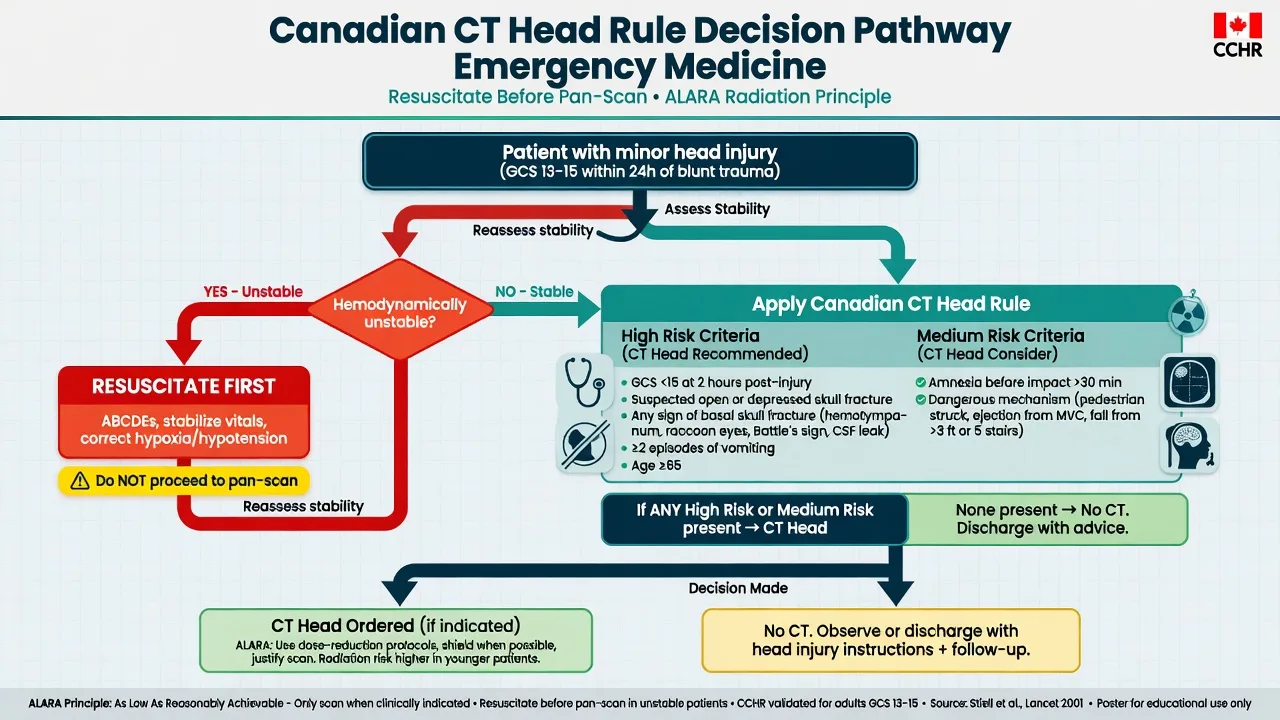
The radiation burden is the reason CT is not given freely. A typical CT phase delivers 2 to 10 mSv, which compares with about 0.02 mSv for a chest radiograph, 0.7 mSv for a mammogram, and 2 to 5 mSv for annual natural background radiation.[1] The risk is stochastic — a small increase in lifetime cancer incidence that accumulates with each scan and is largest in the young, because they have more time to develop a cancer and their dividing tissues are more radiosensitive. The principle of ALARA — as low as reasonably achievable — is applied by justifying every scan, using the minimum dose that answers the question (ultralow-dose renal colic protocols, paediatric weight-based mA reduction), and choosing a non-radiating alternative where it is equivalent.
Special populations
In children, the radiation risk is highest and CT is used selectively: weight-based dose reduction, ultralow-dose renal colic protocols, and ultrasound or magnetic resonance imaging as the first test for appendicitis. In pregnancy, ionising radiation carries a teratogenic risk in the first trimester and a small lifetime cancer risk to the fetus; a necessary CT (the pregnant trauma patient, suspected pulmonary embolism) is justified and performed with shielding and dose minimisation, but ultrasound, plain radiography, and ventilation-perfusion scanning are preferred alternatives where they answer the question. In the renal-impaired patient, contrast is avoided or preceded by hydration, and a non-contrast protocol is chosen where it is diagnostic. The unstable patient is resuscitated and stabilised before transfer, is escorted by a clinician, and is monitored throughout — the CT suite is a dangerous place to arrest. [1]
Common errors and pitfalls
The recurring errors are well described. Ordering a contrast-enhanced head CT when the question is subarachnoid blood — contrast obscures the haemorrhage, and a non-contrast head CT is the correct first scan. Accepting a normal head CT as excluding subarachnoid haemorrhage after the six-hour window, without a lumbar puncture. Calling an early ischaemic CT normal and missing the hyperdense MCA sign or a low ASPECTS. Forgetting to add CT angiography and perfusion to the stroke pathway when the patient is within an extended window. Sending an unstable patient to CT unescorted and unmonitored. Requesting a pan-scan reflexively for a low-energy, stable presentation when selective imaging with a decision rule is sufficient.[2] Over-requesting contrast CT in the young and the pregnant without weighing the cumulative radiation. And treating the radiologist's report as the end of the task when the treating clinician must look at the scan in parallel with the clinical picture.
Evidence and regional guidelines
The contemporary evidence base supports selective, question-driven imaging. The REACT-2 trial showed no survival benefit from routine total-body CT over selective imaging in major trauma.[2] The CT sensitivity for subarachnoid haemorrhage within six hours underpins the selective use of lumbar puncture.[4] The ASPECTS score quantifies the early ischaemic burden and informs reperfusion decisions.[3] CT perfusion selection extends the thrombectomy window, confirmed by DEFUSE 3 and embedded in the AHA/ASA 2019 stroke guideline.[7][10] PIOPED II validated multidetector CT pulmonary angiography as a stand-alone test for pulmonary embolism.[6] The AMACING trial re-framed the role of prophylactic hydration.[9] The radiation dose and lifetime cancer risk are established from the epidemiology reviewed by Brenner and Hall.[1] The imaging appropriateness criteria of the American College of Radiology, and the local ACEM and radiology protocols, govern the requesting pathways.
ANZ practice note. The choice between pan-scan and selective CT follows the REACT-2 evidence and the institutional major-trauma guideline; the Canadian C-spine rule is applied to cervical-spine clearance of the alert, stable patient. The stroke pathway is non-contrast head CT followed by CT angiography and CT perfusion, read in parallel with the time of onset to trigger thrombolysis or thrombectomy transfer. A normal head CT within six hours of a thunderclap headache may exclude subarachnoid haemorrhage without a lumbar puncture in selected patients; after six hours a lumbar puncture is performed to exclude xanthochromia. Contrast premedication and the decision to hydrate for contrast-induced nephropathy follow the radiology department protocol, with the AMACING evidence informing a selective rather than reflexive approach. [1]
Exam practice
SAQ — Acute ischaemic stroke: the non-contrast CT head interpretation and the reperfusion pathway
10 minutes · 10 marks
A 74-year-old man is brought to the emergency department 90 minutes after his wife found him slumped in a chair with slurred speech and a left-sided weakness. He was last known well at 08:00. On arrival his blood pressure is 176/96 mmHg, GCS 14 (E4V3M7), NIHSS 14, with a dense left hemiparesis, left facial droop and expressive dysphasia. Finger-prick glucose is 6.8 mmol/L. The non-contrast CT head shows a hyperdense right middle cerebral artery sign, loss of the right insular ribbon, obscuration of the right lentiform nucleus and effacement of the cortical sulci in the right M2 and M3 territories. There is no intracranial haemorrhage.
SAQ — CT in major blunt trauma: pan-scan versus selective imaging and the systematic abdominal read
10 minutes · 10 marks
A 42-year-old unrestrained male driver is brought to the trauma bay 35 minutes after a high-speed frontal motor-vehicle collision. He is agitated but obeys commands (GCS 14), with blood pressure 108/70 mmHg after a 1-litre crystalloid bolus, heart rate 118, respiratory rate 26, SpO2 95 per cent on 15 L oxygen. He has a distracted sternal contusion with subcutaneous emphysema, a tender abdomen with guarding in the left upper quadrant, and a clinically stable pelvis. There is no evidence of head injury, he is fully alert with no neurological deficit, and he reports neck pain. The focused assessment with sonography in trauma (FAST) is positive for free fluid in Morison pouch. The nearest trauma centre is your own.
Exam pearls
- State the clinical question before the scan — "Is there blood? Is there a stone? Is there a dissection?" The question dictates the phase.
- Stroke pathway: non-contrast head CT first to exclude haemorrhage, then CT angiography for the vessel and CT perfusion for the penumbra. Never contrast the head CT when the question is subarachnoid blood.
- Know ASPECTS. The ten MCA regions (M1 to M6, caudate, lentiform, internal capsule, insula), scored ten to zero, with 7 or below a poor-prognosis large core.[3]
- SAH CT sensitivity is approximately 98 percent within 6 hours and falls with time. A normal CT after 6 hours does not exclude it — perform a lumbar puncture.[4]
- REACT-2: routine total-body CT did not improve survival over selective CT. Justify the pan-scan, and use decision rules for selective imaging.[2]
- Know the doses: 2 to 10 mSv per CT phase, an order of magnitude above a chest radiograph — apply ALARA, especially in the young and pregnant.[1]
- CT pulmonary angiography: a filling defect in an opacified pulmonary artery is a pulmonary embolism; assess for right ventricular strain.[6]
- Contrast decision: blood or stone, no contrast; ischaemia, infection, or vessel, contrast. Premedicate the allergic; hydrate the high-risk renal patient selectively.[9]
- Never send an unstable patient to CT unescorted. Resuscitate, stabilise, monitor, and escort — the scanner is a hostile environment.
Red flags
[1]References
- [1]Brenner DJ, Hall EJ. Computed tomography--an increasing source of radiation exposure N Engl J Med, 2007.PMID 18046031
- [2]Sierink JC, Treskes K, Edwards MJR, et al. Immediate total-body CT scanning versus conventional imaging and selective CT scanning in patients with severe trauma (REACT-2): a randomised controlled trial Lancet, 2016.PMID 27371185
- [3]Barber PA, Demchuk AM, Zhang J, Buchan AM. Validity and reliability of a quantitative computed tomography score in predicting outcome of hyperacute stroke before thrombolytic therapy. ASPECTS Study Group. Alberta Stroke Programme Early CT Score Lancet, 2000.PMID 10905241
- [4]Perry JJ, Stiell IG, Sivilotti MLA, et al. Sensitivity of computed tomography performed within six hours of onset of headache for diagnosis of subarachnoid haemorrhage: prospective cohort study BMJ, 2011.PMID 21768192
- [5]Doria AS, Moineddin R, Kellenberger CJ, et al. US or CT for Diagnosis of Appendicitis in Children and Adults? A Meta-Analysis Radiology, 2006.PMID 16928974
- [6]Stein PD, Fowler SE, Goodman LR, et al. Multidetector computed tomography for acute pulmonary embolism N Engl J Med, 2006.PMID 16738268
- [7]Albers GW, Marks MP, Kemp S, et al. Thrombectomy for Stroke at 6 to 16 Hours with Selection by Perfusion Imaging N Engl J Med, 2018.PMID 29364767
- [8]Stiell IG, Wells GA, Vandemheen KL, et al. The Canadian C-spine rule for radiography in alert and stable trauma patients JAMA, 2001.PMID 11597285
- [9]Nijssen EC, Rennenberg RJ, Nelemans PJ, et al. Prophylactic hydration to protect renal function from intravascular iodinated contrast material in patients at high risk of contrast-induced nephropathy (AMACING): a prospective, randomised, phase 3, controlled, open-label, non-inferiority trial Lancet, 2017.PMID 28233565
- [10]Powers WJ, Rabinstein AA, Ackerson T, et al. Guidelines for the Early Management of Patients With Acute Ischemic Stroke: 2019 Update to the 2018 Guidelines for the Early Management of Acute Ischemic Stroke: A Guideline for Healthcare Professionals From the American Heart Association/American Stroke Association Stroke, 2019.PMID 31662037
- [11]Hoffman JR, Mower WR, Wolfson AB, et al. Validity of a set of clinical criteria to rule out injury to the cervical spine in patients with blunt trauma. National Emergency X-Radiography Utilization Study Group N Engl J Med, 2000.PMID 10891516
- [12]Stiell IG, Wells GA, Vandemheen K, et al. The Canadian CT Head Rule for patients with minor head injury Lancet, 2001.PMID 11356436
- [13]Smith RC, Verga M, McCarthy S, et al. Diagnosis of acute flank pain: value of unenhanced helical CT AJR Am J Roentgenol, 1996.PMID 8571915