EM · Procedural & diagnostic ED skills
Lumbar puncture in the emergency department
Also known as Lumbar puncture · LP · Spinal tap · Cerebrospinal fluid sampling · CSF analysis
Lumbar puncture in the ED — the indications (suspected meningitis or encephalitis, subarachnoid haemorrhage after a negative CT, demyelinating disease — multiple sclerosis, CIDP, Guillain-Barre, and the therapeutic uses — idiopathic intracranial hypertension, cryptococcal meningitis, intrathecal drug delivery), the contraindications (raised intracranial pressure with papilloedema or a focal deficit, coagulopathy with an INR over 1.4 or platelets under 50, overlying skin infection), the anatomy (the conus at L1/L2, the dural sac at S2, Tuffier's line crossing L4), the technique (L3/L4 or L4/L5, midline, the bevel parallel to the longitudinal dural fibres, the atraumatic over the cutting needle), the CSF interpretation (bacterial — high neutrophils, low glucose, high protein; viral — lymphocytes, normal glucose; tuberculous — cobweb coagulum, very low glucose, very high protein; subarachnoid — xanthochromia, uniform RBC across tubes), and the complications (post-dural puncture headache treated with caffeine and the epidural blood patch). ACEM-primary, globally tagged.
On this page & tools
Your progress
Saved locally on this device.
Target exams
Red flags
Related topics
- Meningitis and encephalitis (emergency department diagnosis and management)
- Subarachnoid haemorrhage
- Red-flag headache (approach)
- Raised intracranial pressure
- Guillain-Barré syndrome and myasthenia gravis
- Procedural sedation in the emergency department
- Local anaesthesia and topical agents
- Seizures and the first fit
Lumbar puncture is the percutaneous needle aspiration of cerebrospinal fluid (CSF) from the lumbar theca for diagnostic or therapeutic purposes. In the emergency department it is performed most often to exclude a bacterial meningitis, an encephalitis or a subarachnoid haemorrhage, and the Fellowship candidate must treat it as a structured procedural encounter: a pre-procedure screen for the contraindications, a knowledge of the anatomy and the landmarks, a sterile stepwise technique with the right needle and the right bevel orientation, an opening pressure, a four-tube collection, and a systematic interpretation of the CSF. The two questions an examiner will press are when to image before the puncture, and how to interpret the four tubes once they are drawn.[1][2]
Definition and indications
Lumbar puncture delivers a sample of CSF for analysis, a measurement of the CSF pressure, or a route for drug or contrast delivery. The diagnostic indications are suspected bacterial meningitis (the febrile patient with meningism or altered mental status), viral encephalitis (the febrile patient with confusion, seizures or a focal deficit), subarachnoid haemorrhage after a negative computed tomography (the thunderclap headache, the Ottawa-rule patient), the demyelinating and inflammatory neuropathies (multiple sclerosis, CIDP, Guillain-Barre syndrome), suspected autoimmune encephalitis, and the investigation of a CNS infection in the immunocompromised. The therapeutic indications are the idiopathic intracranial hypertension drainage and pressure measurement, the cryptococcal meningitis pressure relief, the intrathecal drug delivery (methotrexate, cytarabine, baclofen), and the spinal anaesthesia. The unifying test is that the question cannot be answered by blood or imaging alone, and the CSF will change the management.[1]
Contraindications
The contraindications are the conditions in which the puncture, performed without preparation, will harm the patient. The raised intracranial pressure from a focal mass lesion is the dangerous one: the LP releases the distal pressure and the cranial-spinal gradient herniates the brain. The patient with papilloedema, a new focal neurological deficit, a new seizure, a depressed conscious state (GCS under 13), immunocompromise, a known intracranial mass, or age over 60 must have a CT before the LP — these are the CT-first criteria. A coagulopathy — an INR over 1.4, a platelet count under 50, a recent anticoagulant dose (heparin within 4 to 6 hours, a DOAC within its half-life), or a known bleeding disorder — risks a spinal epidural haematoma and is an absolute contraindication until corrected. An overlying skin infection at the puncture site (cellulitis, an abscess) risks inoculating the CSF and is a relative contraindication — choose another interspace or defer. Critical illness, sepsis, and the uncooperative patient are relative contraindications that weigh against the urgency of the question the LP would answer.[1][2]
Relevant anatomy and landmarks
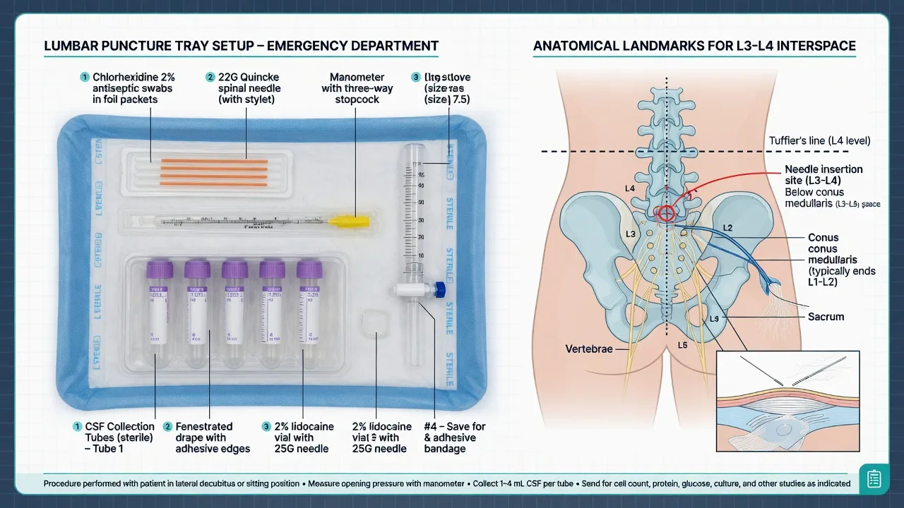
The spinal cord terminates as the conus medullaris at the level of L1/L2 in the adult (lower, at L3, in the newborn and infant), and the dural sac ends at S2 — the safe window for the puncture is therefore the L3/L4 or L4/L5 interspace, below the conus. The interspace is identified by Tuffier's line (the intercristal line), drawn between the highest points of the two iliac crests, which crosses the L4 spinous process or the L4/L5 interspace. The needle traverses, in order, the skin, subcutaneous tissue, supraspinous ligament, interspinous ligament, the ligamentum flavum (the first distinct resistance, the "pop" on piercing), the epidural space, the dura mater and the arachnoid mater, and enters the subarachnoid space where the CSF lies. The dura is composed of longitudinal fibres, and this is the anatomical basis of the bevel orientation — a bevel parallel to the fibres separates them and produces a smaller defect, a perpendicular bevel cuts them and leaves a larger CSF-leaking hole. [1]
The adult CSF volume is approximately 150 mL and is produced at roughly 500 mL per day by the choroid plexus, so the small volume taken at a diagnostic tap (10 to 15 mL) is replaced within hours. The normal opening pressure, measured in the lateral decubitus position with the legs relaxed, is 10 to 20 cmH2O in the adult (5 to 20 cmH2O in the child, lower in the newborn). [1]
Equipment
The lumbar puncture tray contains: a spinal needle with a stylet (20G or 22G in the adult, 22G or 25G in the child; the atraumatic pencil-point type — Sprotte or Whitacre — preferred over the cutting Quincke), a manometer and a three-way stopcock for the opening pressure, four numbered sterile collection tubes, sterile gloves, gown, drape, chlorhexidine or povidone-iodine skin preparation, lidocaine 1 per cent with a 25G needle for skin infiltration and a longer needle for deeper infiltration, gauze and a dressing, and a sharps container. The atraumatic needle is the contemporary standard and halves the post-dural puncture headache rate; the cutting needle is reserved for the difficult case or the operator's habit, with the trade-off accepted.[7][8]
Patient preparation and consent
The patient is consented with a discussion of the indication, the common complications (the post-LP headache in roughly 1 in 3 with a cutting needle and under 1 in 10 with the atraumatic needle, the backache, the small risk of bleeding or infection) and the alternatives. The positioning is the single most important determinant of success. The lateral decubitus position with the knees drawn to the chest and the neck flexed opens the interspinous spaces and is the position required for an accurate opening pressure. The seated position, leaning forward over a bedside table, is easier for the obese patient and may improve the landmark palpation, but the opening pressure is unreliable in this position and the patient must be repositioned to lateral for the measurement. The back is at the edge of the bed, perpendicular to it. The patient is prepped sterile from the L2 to the S1 level, and the operator is gowned and gloved. [1]
Stepwise technique — the structured encounter
The procedure is run to a sequence so that no element is missed. [1]
The lumbar puncture, in order
- Position and identify — lateral decubitus with knees to chest (or seated), the back at the edge of the bed; palpate the iliac crests and draw Tuffier's line, identifying the L4 spinous process and the L3/L4 and L4/L5 interspaces.
- Prep and consent — chlorhexidine skin preparation, sterile drape, confirm the consent and the absence of contraindications (the INR, the platelets, the CT-first criteria).
- Anaesthetise — raise a skin wheal with lidocaine 1 per cent (the maximum safe dose is 3 mg/kg plain or 7 mg/kg with adrenaline) at the chosen interspace, then infiltrate deeper along the intended needle track towards the interspinous ligament.
- Insert the needle — the spinal needle with the stylet in place, bevel parallel to the longitudinal dural fibres (towards the umbilicus in the lateral position, towards the patient's side in the seated position), in the midline, angled towards the umbilicus (approximately 15 degrees cephalad).
- Advance and feel the pops — advance slowly through the supraspinous, interspinous and the ligamentum flavum (the first resistance), then the distinctive "give" or "pop" as the needle breaches the dura and arachnoid and enters the subarachnoid space.
- Check for CSF — remove the stylet and look for CSF at the hub; if none, replace the stylet, withdraw slightly and re-advance; if bone is hit, withdraw to the subcutaneous tissue and redirect.
- Measure the opening pressure — attach the manometer and the three-way stopcock; ensure the patient's legs are partially relaxed (the legs held tight falsely raise the pressure); read the meniscus in cmH2O.
- Collect the four tubes — tube 1 for the cell count, tube 2 for the glucose and protein, tube 3 for the Gram stain, culture and the molecular testing (the polymerase chain reaction for the viral and bacterial pathogens), tube 4 for the cell count (the comparison with tube 1 distinguishes a traumatic tap) and the special tests (cytology, xanthochromia, oligoclonal bands, lactate).
- Withdraw and dress — replace the stylet (reduces the arachnoid strand uptake and the headache), withdraw the needle smoothly along the line of insertion, apply pressure and a sterile dressing.
- Document and counsel — record the interspace, the needle type and gauge, the opening pressure, the CSF appearance (clear, turbid, blood-stained, xanthochromic), the samples sent, the complications, and counsel the patient on the post-procedure care and the headache red flags. [1]
The two structural errors are to omit the opening pressure (the lateral position is required, and the legs must be relaxed) and to insert the bevel perpendicular to the longitudinal dural fibres (which cuts them and doubles the post-dural puncture headache rate). [1]
Drug doses — the local anaesthetic
The local anaesthetic infiltration is the only drug in the standard lumbar puncture. Lidocaine 1 per cent is the agent, with a maximum dose of 3 mg/kg plain (about 20 mL in the 70 kg adult) or 7 mg/kg with adrenaline (about 50 mL in the 70 kg adult). The infiltration is performed with a 25G needle for the skin wheal and a longer 21G or 23G needle for the deeper track, aspirating before each aliquot to avoid the intravascular injection. Procedural sedation is rarely required in the adult but is common in the child — ketamine 1 to 2 mg/kg intravenously or 4 to 5 mg/kg intramuscularly is the agent of choice (refer to the procedural-sedation topic for the full monitoring standard).[6]
Complications — procedure-related and disease-related
Post-dural puncture headache (PDPH) is the commonest complication, presenting as a bilateral fronto-occipital, postural headache (worse on sitting or standing, relieved by lying flat) that develops within 5 days of the puncture, with or without neck stiffness, tinnitus, hyperacusis, photophobia or nausea. The mechanism is the persistent CSF leak through the dural defect producing a low CSF pressure and a traction on the pain-sensitive meningeal and vascular structures. The incidence is up to 30 per cent with a cutting Quincke needle and under 10 per cent with the atraumatic pencil-point needle, and it is higher in the young, the female and the post-partum patient.[5][7]
Backache at the puncture site is common and self-limiting. The rare-but-serious complications are the spinal epidural haematoma (the anticoagulated patient, presenting with severe back pain and a progressive neurological deficit — a surgical emergency), the spinal epidural abscess (the delayed presentation, days later, with back pain, fever and a neurological deficit), the nerve root injury (the radicular pain on needle contact — withdraw and redirect), cerebral herniation (the patient with a raised ICP from a mass lesion in whom the LP was performed without a CT), and the sixth-nerve palsy from the prolonged intracranial hypotension (a stretch on the abducens nerve along the petrous ridge). The post-LP infection (a bacterial meningitis from a break in the sterile technique) is rare but devastating, and the sterile technique is non-negotiable.[1][6]
Pitfalls and practical tips
The pitfalls are the inverse of the structure. Skipping the CT-first screen — the raised-ICP patient in whom the LP herniates the brain. Skipping the coagulation check — the anticoagulated patient in whom the epidural haematoma paralyses. Using the cutting needle when the atraumatic is available — the avoidable post-dural puncture headache. Inserting the bevel perpendicular to the longitudinal dural fibres — the larger defect, the larger leak. Omitting the opening pressure — the missed raised pressure of a bacterial meningitis or an idiopathic intracranial hypertension, or the missed low pressure of a spontaneous CSF leak. Misreading a traumatic tap as a subarachnoid haemorrhage — the unnecessary angiogram, the unnecessary admission. Drawing fewer than four tubes — the loss of the cell-count comparison and the special tests. Forgetting the stylet on withdrawal — the arachnoid strand and the persistent leak. [1]
The practical tips are the opposite: the atraumatic needle on every patient, the opening pressure on every patient, the four tubes on every patient, the bevel parallel to the fibres, the CT-first criteria on every patient, the coagulation profile on the patient at risk, and the ultrasound-assisted localisation on the obese or the difficult landmark.[9]
CSF interpretation — the four patterns
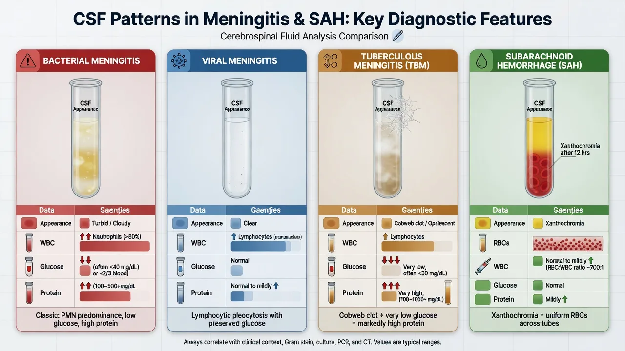
The CSF interpretation is the single most examinable block of the topic, and it is built from the appearance, the opening pressure, the cell count, the glucose, the protein and the special tests (xanthochromia, Gram stain, culture, polymerase chain reaction, oligoclonal bands). The four patterns the candidate must recognise on sight are summarised below. [1]
Bacterial meningitis
- Turbid or cloudy appearance; the opening pressure elevated
- White cell count raised (commonly over 1000), with a neutrophilic predominance
- Glucose low (under 2.2 mmol/L, or below 0.4 of the simultaneous plasma glucose)
- Protein raised (over 0.45 g/L, often over 1 g/L); Gram stain and culture positive
Viral meningitis / encephalitis
- Clear or slightly cloudy; the opening pressure normal or mildly raised
- White cell count mildly to moderately raised (10 to a few hundred), lymphocytic predominance (neutrophils early)
- Glucose normal (above 0.6 of the plasma glucose)
- Protein normal or mildly raised; the polymerase chain reaction positive (HSV, enterovirus)
Tuberculous meningitis
- Clear with a characteristic cobweb coagulum on standing; opening pressure raised
- White cell count raised (50 to 500), lymphocytic; a low neutrophil count early
- Glucose very low (under 2.2 mmol/L, often under 1.1)
- Protein markedly raised (over 1 g/L, often over 5 g/L); the PCR and culture for M. tuberculosis positive
Subarachnoid haemorrhage
- Blood-stained initially, xanthochromic (yellow) on standing or after centrifugation
- Red cell count raised and uniform across tubes 1 to 4 (no fall); white cells may rise from meningeal irritation
- Glucose normal; xanthochromia (bilirubin) present from 2 hours, peaks at 12 hours to 2 weeks
- Protein may be raised; spectrophotometry for bilirubin is the confirmatory test
The key distinctions the examiner rewards are: bacterial versus viral (the glucose and the cell type — low glucose and neutrophils are bacterial, normal glucose and lymphocytes are viral); traumatic tap versus subarachnoid (the falling RBC across tubes in the traumatic tap, the uniform RBC in the subarachnoid, confirmed by the xanthochromia on the centrifuged supernatant); and TB versus bacterial (the very low glucose, the very high protein, the cobweb coagulum and the chronic presentation in TB).[1][2]
The normal adult CSF values
Differential diagnosis — the post-LP headache and its mimics
The post-puncture headache is itself a diagnosis of exclusion, and the Fellowship candidate must distinguish it from the headaches that need an intervention. The clinical test is the postural relationship — the PDPH is relieved by lying flat and provoked by sitting or standing within 15 seconds. [1]
Post-dural puncture headache
- Postural — worse sitting, better lying flat; bilateral fronto-occipital
- Onset within 5 days of the LP; may have neck stiffness, tinnitus, photophobia, nausea
- No fever, no focal deficit, no altered consciousness
- Conservative measures first; the epidural blood patch if persistent
Bacterial meningitis (post-LP)
- Non-postural; the fever, the rigors, the photophobia, the altered conscious state
- Onset hours to days after the LP; the classic sign of a break in the sterile technique
- A medical emergency — re-sample, image, and treat empirically
- Counsel the patient on the red flags before discharge
Cerebral venous sinus thrombosis
- A progressive, severe headache; may have seizures or a focal deficit
- Risk factors — the oral contraceptive, the puerperium, the prothrombotic state
- Diagnosed on a CT venogram or an MR venogram
- Anticoagulation is the treatment
Intracranial hypotension (spontaneous)
- Identical postural headache to the PDPH but without a preceding puncture
- A spontaneous CSF leak from a dural tear or a weakness
- Diagnosed on an MRI with diffuse pachymeningeal enhancement
- Treated with the epidural blood patch
The key distinction the examiner rewards is the postural pattern: the PDPH and the spontaneous intracranial hypotension are postural (better lying, worse sitting), whereas the bacterial meningitis and the venous sinus thrombosis are not. A fever or a focal deficit after an LP is NOT a post-LP headache — re-sample, image, and treat empirically. [1]
Post-procedure care and disposition
After the LP the patient is observed briefly, with the vital signs and the puncture site checked. The traditional advice to lie flat for one to two hours is debated — the contemporary evidence does not show a clear reduction in the post-dural puncture headache, but it does no harm and is reasonable counsel. The hydration (oral or intravenous) and the oral analgesia (paracetamol, an NSAID) are the first-line measures. The CSF results are followed up and acted on (the empirical antibiotics in the suspected bacterial meningitis started before the LP if the patient is septic or the LP is delayed). The discharge advice is to return if a severe or a non-postural headache, a fever, a photophobia, a neck stiffness, a weakness or a sensory change develops in the days after the procedure.[6]
The post-dural puncture headache — management ladder
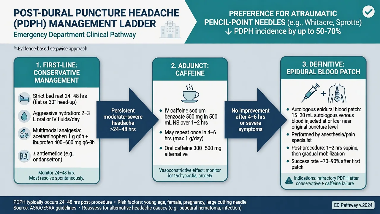
The management is a stepwise escalation from the conservative to the definitive. [1]
The post-dural puncture headache management, in order
- Conservative (first 24 to 48 hours) — rest, lie flat, hydration (oral or intravenous), oral analgesia (paracetamol, an NSAID, codeine if severe).
- Caffeine — caffeine citrate 500 mg intravenously over one hour, or caffeine 300 mg orally, repeated once; the mechanism is the cerebral vasoconstriction that offsets the compensatory vasodilation of the low CSF pressure. The evidence is short-term relief in roughly half; the headache recurs in some.
- The epidural blood patch (the definitive treatment) — for the headache persisting beyond 24 to 48 hours despite the conservative measures; 15 to 20 mL of the patient's own venous blood, drawn aseptically, is injected into the epidural space at or one space below the puncture site. The blood clots, seals the dural defect and restores the CSF pressure. The success rate is 70 to 90 per cent after one patch and higher after a second. The complications are the transient backache, the transient radiculopathy, and the rare infection or neurological injury.
- Re-image if atypical — the non-postural headache, the fever, the focal deficit, or the failure of the second blood patch mandates a re-imaging (a CT or an MRI) to exclude a subdural haematoma, a cerebral venous sinus thrombosis, an intracranial infection or a structural leak. [1]
The practical point is that the conservative measures and the caffeine are tried first, the blood patch is reserved for the persistent case, and the atypical presentation after an LP is re-imaged, not patched.[5][6]
Special populations
The child needs the puncture at the L4/L5 interspace (the conus is lower, at L3) and often a procedural sedation with ketamine 1 to 2 mg/kg intravenously. The opening pressure is lower in the young child (5 to 20 cmH2O) and the CSF values differ (a slightly higher protein in the newborn). The anticoagulated patient has the LP deferred until the INR is under 1.4 and the platelets over 50; the heparin is held 4 to 6 hours, the low-molecular-weight heparin 24 hours, and the DOAC held for 2 to 3 half-lives (refer to the anticoagulated-trauma topic for the per-drug reversal). The obese patient benefits from the ultrasound-assisted localisation of the interspace, which improves the first-attempt success and reduces the traumatic-tap rate.[9] The pregnant patient is positioned in the left lateral tilt with the fetal monitoring as indicated, and the puncture is performed by an experienced operator. The immunocompromised patient (HIV, transplant, chemotherapy) has a lower threshold for the CT-first and a broader CSF panel (cryptococcal antigen, the viral PCR, the acid-fast bacilli).
Evidence and the regional guidelines
The contemporary framework is built on the atraumatic-needle-first recommendation of the Cochrane review and the BMJ clinical practice guideline, which found that the pencil-point needle halves the post-dural puncture headache rate without a trade-off in the success rate.[7][8] The Ottawa Subarachnoid Haemorrhage Rule identifies the headache patient who needs a work-up (age over 40, a thunderclap onset, a neck pain or stiffness, a witnessed loss of consciousness, an exertional onset, a limited flexion on examination), and the 6-hour CT rule confirms that a normal non-contrast CT within 6 hours of the headache onset, in a low-risk patient, can be used to rule out the subarachnoid — but the LP and the xanthochromia remain the gold standard beyond that window.[3][4] The post-dural puncture headache diagnosis, pathophysiology and management are codified in the Bezov reviews, which established the postural pattern, the time window, the caffeine and the epidural blood patch ladder.[5][6] The bacterial meningitis CSF interpretation and the diagnostic dilemmas (the partially-treated, the immunocompromised, the atypical CSF) are reviewed in the Brouwer and van de Beek references.[1][2]
ANZ practice note. The ACEM and the Royal Australasian College of Physicians endorse the atraumatic pencil-point needle as the standard, the CT-first criteria before the LP in the high-risk patient, and the four-tube collection with the opening pressure on every patient. The post-dural puncture headache is managed with the conservative measures, the caffeine, and the epidural blood patch, performed by the anaesthetic or the pain service. The xanthochromia is the confirmatory test for the subarachnoid haemorrhage after a negative CT, with the spectrophotometry preferred over the visual inspection. [1]
Exam pearls
- L3/L4 or L4/L5 — below the conus — the cord ends at L1/L2 in the adult; Tuffier's line identifies L4.
- The bevel parallel to the longitudinal dural fibres — towards the umbilicus in the lateral position, towards the patient's side in the seated position; it spreads the fibres, not cuts them.
- The atraumatic pencil-point needle over the cutting Quincke — halves the post-dural puncture headache without a trade-off in the success.
- Measure the opening pressure on every patient — lateral position, legs relaxed; the missed raised pressure of a bacterial meningitis or an idiopathic intracranial hypertension is a classic error.
- Collect four tubes — the cell-count comparison between tubes 1 and 4 distinguishes a traumatic tap from a subarachnoid haemorrhage.
- The CT-first criteria — age over 60, immunocompromise, a known CNS disease, a new seizure, a GCS under 13, a focal neurological deficit, papilloedema; a normal CT in these patients does not exclude a subarachnoid haemorrhage.
- Xanthochromia is bilirubin — present from 2 hours, peaks at 12 hours to 2 weeks; an LP taken too early or too late can falsely exclude a subarachnoid.
- The post-LP headache is postural — a fever, a focal deficit or a non-postural pattern is NOT a post-LP headache; re-image and re-sample.
- The epidural blood patch for the persistent headache — 15 to 20 mL of the autologous blood, 70 to 90 per cent success.
- Coagulopathy first — INR under 1.4, platelets over 50, the anticoagulant held; a spinal epidural haematoma can paralyse. [1]
SAQ — Adult with suspected meningitis: LP technique and safety
10 minutes · 10 marks
A 34-year-old previously well man presents to the ED with a 24-hour history of fever, severe headache, photophobia, and neck stiffness. His GCS is 14 (confused but orientated), pupils equal, no focal neurological deficit, no papilloedema on fundoscopy, INR 1.0, platelets 220, and the Ottawa SAH rule is not met. You decide to perform a lumbar puncture.
SAQ — CSF interpretation: distinguishing meningitis, SAH and the traumatic tap
10 minutes · 10 marks
A 28-year-old woman presents 6 hours after a sudden, maximal-intensity, occipital headache with vomiting and neck stiffness. Non-contrast CT brain is normal. An LP is performed. CSF is blood-stained. Cell counts in the four tubes are: tube 1 RBC 32,000 × 10^6/L, WCC 12; tube 2 RBC 31,500, WCC 14; tube 3 RBC 31,800, WCC 13; tube 4 RBC 32,100, WCC 15. CSF glucose 3.6 mmol/L with plasma glucose 6.0. CSF protein 0.9 g/L. The supernatant is yellow on centrifugation.
Red flags
[1]References
- [1]Brouwer MC, Tunkel AR, van de Beek D. Epidemiology, diagnosis, and antimicrobial treatment of acute bacterial meningitis Clin Microbiol Rev, 2010.PMID 20610819
- [2]Brouwer MC, Tunkel AR, van de Beek D. Dilemmas in the diagnosis of acute community-acquired bacterial meningitis Lancet, 2012.PMID 23141617
- [3]Perry JJ, Sivilotti MLA, Émond M, et al. Validation of the Ottawa Subarachnoid Hemorrhage Rule in patients with acute headache CMAJ, 2017.PMID 29133539
- [4]Perry JJ, Wells JS, Sivilotti MLA, et al. Prospective Implementation of the Ottawa Subarachnoid Hemorrhage Rule and 6-Hour Computed Tomography Rule Stroke, 2020.PMID 31805846
- [5]Bezov D, Lipton RB, Ashina S, Ashina M. Post-dural puncture headache: part I diagnosis, epidemiology, etiology, and pathophysiology Headache, 2010.PMID 20533959
- [6]Bezov D, Ashina S, Lipton R, Ashina M. Post-dural puncture headache: Part II--prevention, management, and prognosis Headache, 2010.PMID 20807248
- [7]Arevalo-Rodriguez I, Pedraza OL, Cosp X, et al. Needle gauge and tip designs for preventing post-dural puncture headache (PDPH) Cochrane Database Syst Rev, 2017.PMID 28388808
- [8]Rochwerg B, Oczkowski SJ, Agoritsas T, et al. Atraumatic (pencil-point) versus conventional needles for lumbar puncture: a clinical practice guideline BMJ, 2018.PMID 29789372
- [9]Gottlieb M, Holladay D, Peksa GD. Ultrasound-assisted Lumbar Punctures: A Systematic Review and Meta-Analysis Acad Emerg Med, 2019.PMID 30129102