EM · Procedural & diagnostic ED skills
Splinting and immobilisation in the emergency department
Also known as Backslab · Plaster backslab · Plaster of Paris splint · POP cylinder · Thomas splint · Traction splint · Cervical collar · Spinal board · SAM splint · Sling and swath · Immobilisation
Splinting and immobilisation in the ED — the governing principle (immobilise the joint above and below the fracture), the device set (plaster backslab for Colles and ankle, POP cylinder for the knee, Thomas splint for the femur, sling and swath for the clavicle, rigid cervical collar and spinal board for suspected spinal injury, malleable SAM splint for the field), the layered application technique (stockinette, wool padding, plaster slabs dipped in cool water and moulded in the position of function), and the complications the Fellowship candidate must name and prevent — compartment syndrome (the 5 Ps of pain out of proportion, pallor, paraesthesia, paralysis, pulselessness), pressure sores over bony prominences, and thermal injury from the exothermic setting reaction. The central SCQ distinction is the backslab (non-circumferential, accommodates swelling, the ED acute-phase default) versus the circumferential cast (rigid, definitive, applied only after swelling resolves). ACEM Fellowship, globally tagged.
On this page & tools
Your progress
Saved locally on this device.
Target exams
Red flags
Related topics
- Fracture and dislocation reduction in the ED
- Cervical spine injury and clearance in trauma
- Paediatric trauma — the modified approach
- Wound assessment and management
- Local anaesthesia and topical agents
- Procedural sedation in the emergency department
- Acute limb ischaemia (the emergency of the 6 Ps)
- Consent, capacity and the medico-legal framework in the emergency department
Splinting and immobilisation are the procedural craft that closes almost every fracture and dislocation the emergency physician manages — the backslab for the reduced Colles, the Thomas splint for the femoral shaft, the collar and board for the suspected spinal injury, the sling for the clavicle. The Fellowship examiner tests three things together: the governing principle (immobilise the joint above and below the fracture), the layered application technique (stockinette, wool, plaster dipped in cool water and moulded in the position of function), and the complications the candidate must prevent and recognise — compartment syndrome with its 5 Ps, pressure sores over bony prominences, and thermal injury from the exothermic setting reaction. The central distinction that frames every SCQ is the backslab versus the circumferential cast: the backslab is non-circumferential, accommodates swelling, and is the ED acute-phase default; the circumferential cast is rigid, definitive, and belongs to orthopaedics, applied only after the swelling has resolved.[1][2] A candidate who has read only this topic could apply a Colles backslab in an OSCE, defend the Thomas splint in a viva, and answer any SCQ on splint choice and the cast complications.
Definition, classification and the governing principle
Immobilisation is the restriction of movement at an injury site to relieve pain, prevent further displacement and soft-tissue damage, and stabilise the limb for transport and healing. It splits into rigid (plaster of Paris, synthetic fibreglass, the circumferential cast) versus soft (sling, swath, collar, bandage), and into circumferential (the cast) versus non-circumferential (the backslab, the SAM splint). Plaster of Paris (POP) is calcium sulphate hemihydrate that sets to a rigid dihydrate when wetted — a heat-releasing (exothermic) reaction that is the mechanism of cast burns.[7][8]
The governing principle, recited aloud in every viva, is that a splint must immobilise the joint above and the joint below the fracture: a forearm fracture is splinted from the metacarpal heads to the proximal forearm (wrist and elbow controlled), a tibial fracture from the metatarsal heads to the mid-thigh (ankle and knee controlled), a femoral fracture requires traction from hip to ankle. A splint that spans only the fracture itself fails because movement at either adjacent joint displaces the fragment. [1]
Indications
The indications are the conditions in which immobilisation relieves pain, prevents secondary injury, or stabilises the limb pending definitive care: stable reduced fractures (the post-reduction Colles, the undisplaced ankle, the scaphoid suspected on clinical grounds, the buckle fracture of the child), severe soft-tissue injury (a grade II to III ankle sprain, a suspected ruptured Achilles held in equinus), pain control in the undiagnosed limb injury while imaging is obtained, prehospital extrication and transport, temporary stabilisation of a displaced fracture pending orthopaedic theatre, and post-reduction immobilisation after a joint is reduced. Splinting is also the bridge between reduction and definitive orthopaedic casting — the backslab holds the reduction through the swelling phase, and the conversion to a circumferential cast happens at fracture clinic days later, once the limb is "cold".[1][9]
Contraindications
There is no contraindication to some form of immobilisation in the injured limb — even the open fracture is splinted, in the position found, after irrigation, dressing and intravenous antibiotics. The contraindications govern the type of immobilisation and the setting.[2][3]
A suspected or established compartment syndrome contraindicates any new rigid immobilisation: any existing cast is split to skin immediately, and the limb is prepared for fasciotomy. An open fracture is irrigated, dressed and splinted in the position found, not reduced in the ED, with intravenous antibiotics and orthopaedic referral in parallel. A neurovascularly compromised limb is reduced or handed to vascular and orthopaedic surgery, not splinted and observed. A grossly contaminated wound is debrided before immobilisation. The uncooperative or unconsentable patient is sedated or the procedure deferred, with consent documented when feasible. [1]
Relevant anatomy and landmarks
The anatomy that matters for splinting is the joint above and below the injury, the bony prominences that ulcerate under pressure, and the position of function in which the limb is moulded while the plaster sets. The prominences that must be padded at every site are the malleoli, olecranon, ulnar and radial styloids, fibular head, metatarsal heads, heel and patella — an unpadded prominence under a rigid splint develops a pressure ulcer within hours.[9] The position of function varies by joint: the wrist in 15 to 20 degrees of extension with slight radial deviation, the ankle at 90 degrees neutral (never plantarflexed, which shortens the Achilles and the calf compartment), the knee in 15 to 20 degrees of flexion, the elbow at 90 degrees. Moulding the plaster in these positions while it sets is the difference between a splint that holds the reduction and one that loses it.
Equipment and the splint set
The plaster set holds a defined inventory the candidate must name at the OSCE: stockinette (the tubular cotton layer rolled onto the limb, cut to length, protecting the skin), soft wool padding (Softban, rolled cotton or synthetic padding, two to three layers, extra over the prominences), plaster slabs or POP bandages (10 to 12 layers for an adult backslab, pre-measured to the limb), crepe or conforming bandages to hold the wet plaster, adhesive tape (never circumferential tape — it constricts), a bowl of cool or tepid water (never hot), a sling, and gloves for the operator. The SAM splint is a thin sheet of aluminium sandwiched between closed-cell foam — folded into a longitudinal accordion for rigidity, versatile for the field immobilisation of a forearm, ankle or finger. The Thomas splint is a steel ring on a long side-iron, in sized sets from child to adult. The rigid cervical collar (Aspen, Malley, Stifneck) is sized to the patient by measuring the neck height from the trapezius to the chin. The spinal board is a flat rigid plank with head blocks and straps. [1]
The standard splint set — the contents at a glance
Patient preparation, consent and analgesia
Preparation has four steps stated in order. Document the pre-procedure neurovascular status — distal pulses, capillary refill, sensation and active movement — before any splint is applied, because a deficit emerging afterwards must be attributable to the injury or to the splint. Obtain consent — the procedure, the expected duration in the splint, the fracture-clinic follow-up, and the complications (compartment syndrome, pressure sores, burns) the patient must watch for and return for. Provide analgesia — a splint applied to an acutely fractured limb is itself painful, and the reduction that precedes it more so; the Fellowship answer pairs the splint with appropriate analgesia or procedural sedation. Position the limb in the position of function, with the operator seated and the limb supported by an assistant, and prepare all materials before the plaster is wetted.[2]
Stepwise technique — the layered backslab application
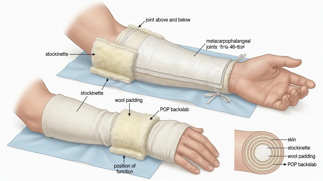
The backslab is the default ED immobilisation, and the layered sequence is the technique the OSCE examiner watches. The discipline is stockinette, wool, plaster, bandage, with the plaster moulded in the position of function while it sets, and a documented neurovascular re-check once it has hardened.[2][9]
The backslab application, in order — recite at the OSCE
- Measure and cut. Before wetting anything, measure the limb and cut the POP slab to length — for a Colles backslab, from the metacarpal heads to the proximal forearm; for an ankle, from the metatarsal heads to the mid-calf; cut 10 to 12 layers for an adult, fewer for a child. Cut the stockinette and the wool to length at the same time.
- Stockinette. Roll the stockinette onto the limb, cut a slit at any joint so it lies flat, and smooth out wrinkles (a wrinkle under plaster is a pressure sore).
- Wool padding. Roll two to three layers of soft wool over the limb, with extra padding over every bony prominence — malleoli, styloids, olecranon, fibular head, heel. The wool accommodates swelling and cushions the skin.
- Dip the plaster. Immerse the POP slab in cool or tepid water (never hot), squeeze out the excess by rolling it gently — do not wring — so the slab is wet enough to set but not dripping.
- Apply and smooth. Lay the slab on the limb (volar surface for a Colles, posterior for an ankle), smooth it against the wool so the layers bond into a single sheet with no air pockets (an air pocket is a weak spot and a pressure point).
- Mould in the position of function. Hold the limb in the correct position — wrist 15 to 20 degrees extension with radial deviation, ankle 90 degrees neutral, knee 15 to 20 degrees flexion — and mould the plaster with the palms (never the fingertips, which dent the wet plaster and create pressure points). Hold still until the plaster sets (warm and rigid, about 5 to 10 minutes).
- Bandage. Apply a conforming bandage over the plaster, secure with tape (never circumferential tape), and apply a sling for elevation.
- Re-check neurovascular. Re-test distal pulses, capillary refill, sensation and active movement against the pre-procedure record. Document. Elevate the limb. [1]
The structural errors the examiner deducts for are the hot dip-water or too-thick slab (a burn from the exothermic reaction), the unpadded prominence (an ulcer), the wrong position of function (a lost reduction — a wrist splinted in flexion loses a Colles), the fingertip dent in wet plaster (a pressure sore), the circumferential tape or bandage (constriction), and the omitted post-procedure neurovascular check (the missed compartment syndrome).[7][9]
Splint types by indication — the device set
The Fellowship candidate must match the device to the injury, and recite the rationale. The set below covers the seven splints named in the topic and the situations the SCQ trades on.[4][11]
Colles backslab
- A volar plaster slab from the metacarpal heads to the proximal forearm, the wrist held in 15 to 20 degrees of extension with radial deviation
- Indication: reduced or minimally displaced distal-radius fracture; often extended as a sugar-tong (around the radial and ulnar aspects) for rotational stability
- Pitfall: splinting the wrist in flexion loses the reduction; a too-tight slab on a swelling hand drives a median-nerve compression
- Convert to a circumferential cast at fracture clinic at 5 to 7 days once swelling has resolved
Ankle / below-knee backslab
- A posterior plaster slab from the metatarsal heads to the mid-calf, the ankle held at 90 degrees neutral, often with a U-shaped stirrup slab around the malleoli
- Indication: reduced or stable ankle fracture, severe ankle sprain, Achilles injury held in slight equinus, calcaneal injury
- Pitfall: ankle splinted in plantarflexion shortens the Achilles and raises calf compartment pressure; an unpadded malleolus ulcerates
- Non-weight-bearing, elevate, fracture clinic at 5 to 7 days
POP cylinder (above-knee)
- A circumferential-or-slab plaster from the groin to the malleoli, the knee held in 15 to 20 degrees of flexion
- Indication: patellar fracture, distal femoral fracture, knee ligament injury, post-reduction knee — immobilises knee and ankle is left free
- Pitfall: a heavy cylinder is hot and cumbersome; pressure over the peroneal nerve at the fibular head causes a foot drop
- Definitive casting is orthopaedic; the ED applies a cylinder backslab or splint and refers
Thomas splint (femur traction)
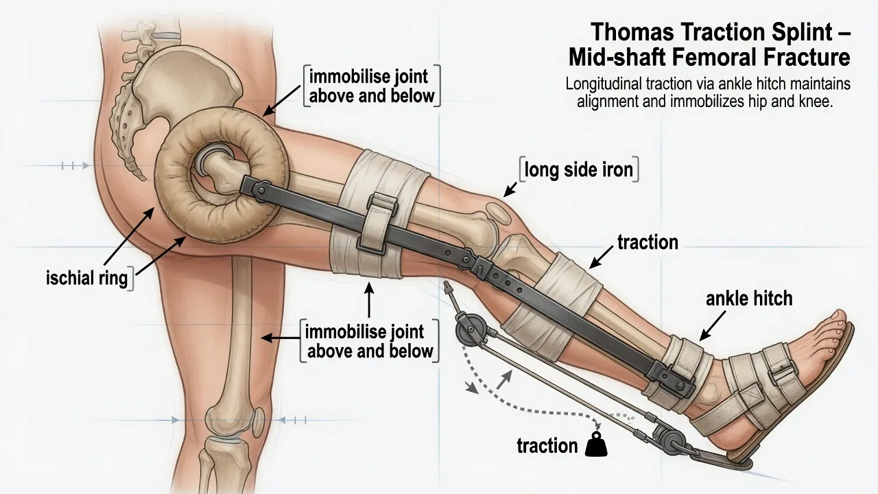
- A padded steel ring on a long side-iron — the ring seats against the ischial tuberosity in the groin, an ankle hitch applies longitudinal traction along the femur
- Indication: femoral-shaft fracture; reduces pain, blood loss and displacement by aligning the fragments and counteracting muscle spasm
- Application: pass the ring up the injured leg to the groin (the ring must pass over the flexed knee with care), apply an ankle strap and traction cord, extend the leg, tie the traction to the distal end of the side-iron — typically 10 per cent of body weight
- Pitfall: ring pressure on the perineum or sciatic nerve, ankle strap neurovascular compromise; re-check the foot pulses and sensation after traction is applied; paediatric sizes exist and the paediatric ED use is well described
Sling and swath (clavicle / shoulder)
- A broad-arm sling supports the arm and a body bandage (swath) around the arm and chest prevents rotation; together they immobilise the clavicle and the glenohumeral joint
- Indication: clavicular fracture, shoulder injury, humeral shaft fracture — the sling is the immobilisation of choice for the clavicle
- Pitfall: the figure-of-eight bandage offers no outcome benefit over a simple sling and causes more discomfort and skin problems — a sling is the modern standard
- Cochrane review of conservative management of middle-third clavicle fractures supports sling over figure-of-eight
Cervical collar
- A rigid two-piece collar sized to the patient (Aspen, Malley, Stifneck); applied to any patient with suspected cervical-spine injury after blunt trauma
- CRITICAL: a collar alone does NOT immobilise — it permits up to 30 degrees of motion; full immobilisation needs manual in-line stabilisation, head blocks and torso straps
- Pitfall: poorly sized collar (too tall flexes, too short extends), pressure sores if left for hours, raised intracranial pressure in the head-injured; remove the collar as soon as clinically cleared
- Pair with a spinal board only for extrication; log-roll off within 30 minutes
Spinal board
- A rigid flat board with head blocks and straps; for prehospital extrication and short-distance transport of the suspected spinal-injured patient
- Indication: extrication from a vehicle, transport to hospital — NOT for prolonged immobilisation
- Pitfalls: pressure sores (occiput, scapulae, sacrum, heels) within 30 to 60 minutes; respiratory restriction; pain; the board is an extrication device, not a bed
- Log-roll the patient off the board onto a trolley mattress or vacuum mattress within 30 minutes of arrival; vacuum mattress preferred for prolonged transport
SAM splint (malleable field splint)
- A thin aluminium sheet between layers of closed-cell foam, foldable and reusable; folded into a longitudinal accordion to gain rigidity, then bent to the limb
- Indication: versatile field and ED splint for the forearm, wrist, ankle and finger; lightweight and compact for prehospital and wilderness use
- Pitfall: less rigid than plaster — a temporising splint, not definitive; pad prominences as for plaster
- Pad, apply with a conforming bandage, and arrange definitive immobilisation in fracture clinic
Drug doses and local anaesthesia
Splinting itself is rarely painful enough to need local anaesthesia, but the reduction that often precedes it is, and the analgesia plan is part of the immobilisation answer. The agent set the Fellowship candidate names: nitrous oxide 50 per cent in oxygen (Entonox, self-administered) for the brief painful manoeuvre; intranasal fentanyl 1.5 mcg per kilogram for the child or the adult without intravenous access; intravenous morphine 0.1 mg per kilogram titrated or fentanyl 1 mcg per kilogram IV for the displaced fracture; procedural sedation (ketamine 1 mg per kilogram IV, propofol 1 mg per kilogram IV) to the full standard for the reduction that needs it. For a digital splint after a finger injury, a digital block with plain lidocaine 1 to 2 per cent (maximum 3 mg per kilogram plain, never adrenaline) precedes the buddy strap or aluminium foam splint. The dose is calculated before drawing up, and the patient is monitored throughout. [1]
Differential diagnosis — circumferential cast versus backslab
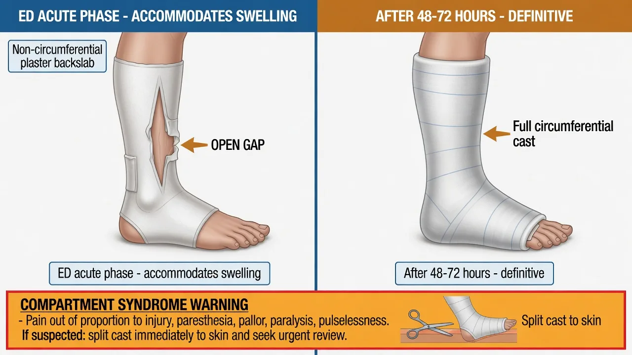
The central SCQ distinction in splinting is the backslab versus the circumferential cast, and the Fellowship answer must distinguish each by its rigidity, its accommodation of swelling, its setting, and who applies it. The discriminating question is whether the limb is still swelling — the acutely injured limb swells for 48 to 72 hours, and only a non-circumferential device is safe in that window.[1][2]
Backslab (non-circumferential)
- A plaster slab on one surface of the limb, held by a bandage — the ED default for the acute fracture
- Accommodates swelling: the slab does not encircle the limb, so a swelling limb is not strangled; the bandage can be loosened and the slab split in seconds
- Indication: every acute fracture in the first 24 to 72 hours; the bridge between reduction and definitive orthopaedic casting
- Applied by ED; converted to a circumferential cast by orthopaedics once swelling resolves
Circumferential cast
- A full tube of plaster or fibreglass encircling the limb — rigid, definitive, conforms precisely
- Does NOT accommodate swelling: a swelling limb inside a rigid tube develops compartment syndrome; this is the iatrogenic trap
- Indication: definitive orthopaedic immobilisation after the swelling phase (48 to 72 hours), in clinic or theatre; the ED almost never applies a circumferential cast to an acute fracture
- Applied by orthopaedics; the ED role is recognition that the acute limb needs a backslab, not a cast
Fibreglass (synthetic) cast
- A synthetic resin-impregnated bandage that sets lighter, stronger and more water-resistant than plaster, with less exotherm
- Used for definitive casting; lighter and more durable than POP for the weeks of immobilisation
- Pitfall: sets faster and harder to mould than POP; the lower exotherm does not abolish the burn risk if dipped in hot water
- Orthopaedic definitive; ED occasionally uses synthetic backslabs for known low-swelling injuries
The clinical rule the candidate recites: a backslab for the acute, a cast for the cold. Any limb that may swell further gets a backslab; the circumferential cast waits for the swelling to resolve. [1]
Complications — compartment syndrome, pressure sores, thermal injury
The complications the Fellowship candidate must name, prevent, and recognise divide into the three named in the topic and the secondary group. Each has a defined mechanism, prevention, and rescue.[2][3][6]
The complications — mechanism, prevention, rescue
Compartment syndrome is the limb-threatening complication of immobilisation and the one the examiner most wants recognised. The tibial fracture carries the highest risk — McQueen and Court-Brown identified the predictors as the tibial-diaphyseal fracture, the young male, and the high-energy mechanism — but any casted limb, especially the child's, can develop it.[3] The pathophysiology is a rise in pressure within a fascial compartment that exceeds capillary perfusion pressure, producing ischaemia; the cast or the swelling haematoma is often the precipitant. The classic 5 Ps are pain out of proportion (and pain on passive stretch — the earliest and most sensitive sign, present while the pulse is still palpable), pallor, paraesthesia, paralysis, and pulselessness (a late and pre-terminal sign — the absence of a pulse means the compartment is already infarcting). In the child, compartment syndrome may be silent — the child cannot report the pain and presents instead with increasing analgesia requirement, agitation, and anxiety on passive stretch; a child demanding escalating opioid analgesia in a cast is assumed to have compartment syndrome until proven otherwise.[6]
Pressure sores arise where a bony prominence presses against an unpadded rigid surface. The malleoli, the olecranon, the ulnar and radial styloids, the fibular head, the metatarsal heads and the heel are the high-risk sites. Prevention is meticulous wool padding over every prominence; recognition is localised pain under the cast (or, in the neuropathic or the child, the discovery at cast removal); treatment is relief of the pressure, local wound care, and orthopaedic review for the deep ulcer over a fracture.[9]
Thermal injury (cast burns) is a direct consequence of the exothermic setting reaction: calcium sulphate hemihydrate reacts with water to form the dihydrate and releases heat. The peak temperature rises with hotter dip-water, more plaster layers, and insulating synthetic composites, and the burn is a full-thickness injury often discovered only at cast removal. The original measurements of Gannaway (1983) and the plaster-synthetic composite work of Burghardt (2014) established that cool dip-water and a modest slab thickness keep the peak temperature below the burn threshold — the practice the candidate must recite.[7][8]
Pitfalls and practical tips
The pitfalls invert the discipline, and the Fellowship candidate must name them aloud. Circumferential casting of an acute fracture — the iatrogenic compartment syndrome. Hot dip-water or an excessive slab thickness — the exothermic burn. An unpadded prominence — the pressure ulcer. Splinting the wrist in flexion or the ankle in equinus — the lost reduction. A fingertip dent in the wet plaster — the focal pressure sore. Circumferential tape — the constriction that strangulates. A collar as sole immobilisation of the cervical spine — the missed secondary injury. Prolonged use of the spinal board — the pressure sore and the respiratory compromise. Missing compartment syndrome behind opioid sedation — the limbed patient discharged with "good analgesia". [1]
The practical tips are the mirror. Backslab, never circumferential cast, for the acute limb. Cool water, 10 to 12 layers, mould with the palms. Pad every prominence. Position of function, always. Split and spread a tight cast to skin at the first suspicion of compartment syndrome. Elevate the limb. Document neurovascular before and after, and at every review. Log-roll off the spinal board within 30 minutes. A sling, not figure-of-eight, for the clavicle. Re-image at fracture clinic at 5 to 7 days. [1]
Post-procedure care and disposition
After the splint is applied and the neurovascular check is documented, the limb is elevated (a sling for the upper limb, a pillow or footstool for the lower) to reduce swelling, and the patient is discharged with fracture-clinic follow-up at 5 to 7 days for the wrist, ankle and clavicle, sooner (24 to 48 hours) for the higher-risk paediatric forearm and the supracondylar fracture. The safety-net is explicit and written down: return immediately for increasing pain despite analgesia, swelling, numbness or tingling, cold or pale fingers or toes, burning under the cast, or an offensive discharge. The analgesia plan and the elevation instruction are reinforced at discharge, and the patient is advised to keep the splint dry and not to insert any object under it (the foreign-body and ulcer cause of "itching under the cast"). The splint that has been applied for a prehospital or transient reason (the SAM splint, the cervical collar pending clearance) is reviewed and converted or removed as the work-up proceeds. [1]
Special populations
The child is the population in whom compartment syndrome is silent — the child cannot report the pain and presents instead with analgesia-refractory agitation, anxiety on passive stretch, and a rising opioid requirement; any child in a cast demanding escalating analgesia has compartment syndrome until proven otherwise, and the threshold to split the cast is very low.[6] The paediatric forearm reduction is the commonest splinted injury, and the backslab is converted to a cast at clinic only after the swelling subsides. The elderly patient has fragile skin (the wool must be generous) and often anticoagulation, which enlarges a fracture haematoma and raises the compartment-syndrome risk; the anticoagulated limb is watched closely. The pregnant trauma patient on a spinal board is log-rolled with a left lateral tilt to relieve aortocaval compression, and immobilised with the standard devices. The polytrauma patient is splinted after the life-threats are addressed — the femoral traction splint is part of the secondary survey, not the primary, and the open fracture is dressed, splinted in position and given intravenous antibiotics in parallel. The neurologically impaired patient (the head-injured, the intubated, the intoxicated) cannot report pain under the cast, and the threshold to split and inspect is the same as for the child.
Evidence and regional guidelines
The evidence base for the cast-versus-backslab decision is Barvelink's 2024 study of redisplacement of reduced distal-radius fractures, which examined whether the type of immobilisation affects loss of reduction, and Jorgensen's 2022 assessment of casting complications in acute distal-radius fractures, which quantified the cast-related morbidity that drives the ED preference for a backslab.[1][2] The compartment-syndrome evidence rests on McQueen and Court-Brown's predictors study (the tibial fracture and the young male as the high-risk profile) and on Lee's report of silent compartment syndrome in children, which established the analgesia-refractory child as the alarm sign.[3][6] The thermal-injury evidence is the classic Gannaway study of casting-material temperatures and the Burghardt composite-cast exotherm work, which together established the cool-water, modest-thickness practice.[7][8] The Thomas-splint evidence is Chu's description of paediatric traction-splinting practice and Irajpour's comparison of simple versus traction splints showing reduced pain with traction.[4][11] The cervical-spine immobilisation evidence is Pandor's 2024 systematic review of prehospital and emergency immobilisation and the NAP7 airway-management data for the collared patient, which together support motion restriction over rigid immobilisation and the timely removal of the collar once cleared.[5][10] The clavicle evidence is Lenza's Cochrane review of conservative interventions, which found no benefit of figure-of-eight over a simple sling.[12]
ANZ practice note. In Australasian emergency departments the acute fracture is immobilised in a backslab — plaster of Paris for the common Colles and ankle, with synthetic splints in selected cases — and referred to fracture clinic for definitive casting once the swelling resolves. The Thomas splint is the standard for the femoral-shaft fracture in keeping with the Australasian trauma practice, applied with an ankle hitch and longitudinal traction and with documented post-application neurovascular review of the foot. Cervical-spine immobilisation follows the motion restriction philosophy now favoured by the ACEM-influenced prehospital services — a correctly sized rigid collar, manual in-line stabilisation during airway manoeuvres, and prompt log-roll off the spinal board within 30 minutes of arrival. Clavicular fractures are managed in a broad-arm sling, the figure-of-eight abandoned on the Cochrane evidence. Compartment syndrome in a casted limb is the audit standard: every patient with pain on passive stretch has the cast split to skin immediately and orthopaedics called for fasciotomy. [1]
SAQ — The reduced Colles fracture and the backslab technique
10 minutes · 10 marks
A 72-year-old woman has just had an emergency department reduction of a displaced distal radius fracture under a ketamine sedation. The post-reduction imaging is satisfactory and you are about to apply the backslab. The nursing student observing asks you to talk through what you are doing and why.
SAQ — The femoral shaft fracture and the cervical spine in the multi-trauma patient
10 minutes · 10 marks
A 28-year-old man is brought in by ambulance after a high-speed motorcycle crash. He has an obvious shortened and externally rotated right thigh with a femoral shaft fracture, and in the primary survey there is also a clinical suspicion of a cervical spine injury. The blood pressure is 100 over 60 and the heart rate is 110.
Exam pearls
- "Joint above and below." State the principle aloud at every splinting answer — a forearm splint controls the wrist and the elbow, a lower-limb splint controls the ankle and the knee, a femur needs traction from hip to ankle.
- "Backslab for the acute, cast for the cold." The backslab is non-circumferential, accommodates swelling, is the ED default; the circumferential cast is orthopaedic, applied only after swelling resolves at 48 to 72 hours. Applying a circumferential cast to an acute fracture is the iatrogenic cause of compartment syndrome.
- The 5 Ps, in order of onset. Pain out of proportion and pain on passive stretch (earliest, pulse still present), then pallor, paraesthesia, paralysis, pulselessness (late, pre-terminal). The pulse is a late sign — never wait for it.
- Split a tight cast to skin. On any suspicion of compartment syndrome, split the cast, the wool and the stockinette down to skin and spread it, re-check neurovascular, keep at heart level, analgesia, call orthopaedics for fasciotomy.
- Cool dip-water, 10 to 12 layers, palms not fingertips. The exotherm is controlled by cool water and a modest slab; mould with the palms because fingertips dent the wet plaster and make pressure sores.
- A collar does not immobilise alone. Pair with manual in-line stabilisation, head blocks and straps; log-roll off the spinal board within 30 minutes.
- Sling, not figure-of-eight, for the clavicle. The Cochrane evidence shows no outcome benefit and more discomfort from figure-of-eight bandaging.
- The silent child. A child in a cast demanding escalating analgesia has compartment syndrome until proven otherwise — split the cast. [1]
Red flags
[1]References
- [1]Barvelink B, van der Velde D, Sintenie JB, et al. Redisplacement of reduced distal radius fractures in adults: does the type of casting play a role? The CAST study, a multicentre cluster randomized controlled trial Bone Joint J, 2024.PMID 38945541
- [2]Jorgensen A, Tillirou A, Rousescu D, Barla M, Barut N, Hardy A, Biau DJ, Laiguillon MC, Houvet P, Massin P. Assessment of complications associated with casting of acute distal radius fractures in adults Am J Emerg Med, 2022.PMID 35397351
- [3]McQueen MM, Court-Brown CM. Predictors of Compartment Syndrome After Tibial Fracture J Orthop Trauma, 2015.PMID 25882967
- [4]Chu RS, Browne GJ, Cheng NG, Lam LT. Traction splinting of femoral shaft fractures in a paediatric emergency department: time is of the essence? Emerg Med (Fremantle), 2003.PMID 14992059
- [5]Pandor A, Horner D, Goodacre S, et al. Cervical spine immobilisation following blunt trauma in pre-hospital and emergency care: A systematic review PLoS One, 2024.PMID 38662734
- [6]Lee C, Porter K. Silent compartment syndrome in children: a report of five cases J Pediatr Orthop B, 2014.PMID 24921836
- [7]Gannaway JK, Hunter JR. Thermal effects of casting materials Clin Orthop Relat Res, 1983.PMID 6641053
- [8]Burghardt RD, Anderson JE, Heppert Z, Bronson D, Manohar S, Conrad C. Exothermic properties of plaster-synthetic composite casts J Child Orthop, 2014.PMID 24554128
- [9]Difazio R, Vesnova K, Mahan J, Connors P, Ceroni D, Copley L, Riccio AI, Andras L, Scherl S, Cooperman D, et al. Reducing the Incidence of Cast-related Skin Complications in Children Treated With Cast Immobilization J Pediatr Orthop, 2017.PMID 26683501
- [10]Wiles MD, Hanumanthappa V, Semenko T, Athanassoglou V, Pandit JJ, Hronovich M, Carey A, Smith AF, Cook TM. Airway management in patients with suspected or confirmed cervical spine injury: Guidelines from the Difficult Airway Society (DAS), Association of Anaesthetists (AoA), British Society of Orthopaedic Anaesthetists (BSOA), Intensive Care Society (ICS), Neuro Anaesthesia and Critical Care Society (NACCS), Faculty of Prehospital Care and Royal College of Emergency Medicine (RCEM) Anaesthesia, 2024.PMID 38699880
- [11]Irajpour A, Alavinia SM, Sadeghi S, Faghihzadeh S. A comparison between the effects of simple and traction splints on pain intensity in patients with femur fractures Iran J Nurs Midwifery Res, 2012.PMID 23922601
- [12]Lenza M, Buchbinder R, Johnston RV, Belloti JC, Faloppa F. Conservative interventions for treating middle third clavicle fractures in adolescents and adults Cochrane Database Syst Rev, 2016.PMID 27977849